Tag Archives: titanium

Metallic nanoparticles inside heart tissue mitochondria can cause damage

With all the focus on COVID-19, viruses , and aerosols, it’s easy to forget that there are other kinds of contaminated air too. The last time I featured work on nanoparticles and air pollution was in a May 31, 2017 posting, “Explaining the link between air pollution and heart disease?” where scientists announced they may have discovered how air pollution (nanoparticles) were making their way from lungs to the heart. Answer: the bloodstream.

A July 3, 2020 Lancaster University press release (also on EurekAlert) announces research into how air made toxic by metallic nanoparticles affects the heart in very specific ways (Note: A link has been removed),

Toxic metallic air pollution nanoparticles are getting inside the crucial, energy-producing structures within the hearts of people living in polluted cities, causing cardiac stress – a new study confirms.

The research team, led by Professors Barbara Maher of Lancaster University and Lilian Calderón-Garcidueñas of The University of Montana and the Universidad del Valle de Mexico, found the metallic nanoparticles, which included iron-rich nanoparticles and other pollution-derived metals such as titanium, inside the damaged heart cells of a 26-year-old and even a three-year-old toddler.

The hearts had belonged to people who had died in accidents and who had lived in highly-polluted Mexico City.

The findings shed new light on how air pollution can cause the development of heart disease, as the iron-rich particles were associated with damage to the cells, and increased cardiac oxidative stress, even in these very young hearts.

The repeated inhalation of these iron-rich nanoparticles, and their circulation by the bloodstream to the heart, may account for the well-established associations between exposure to particulate air pollution and increased cardiovascular disease, including heart attacks. The study indicates that heart disease can start in very early age, before progressing to full-blown cardiovascular illness later in life. This type of air pollution may thus be responsible for the ‘silent epidemic’ of heart disease, internationally. By causing pre-existing heart conditions, it may also account for some of the increased death rates from Covid-19 seen in areas with high levels of particulate air pollution.

Professor Maher said: “It’s been known for a long time that people with high exposure to particulate air pollution experience increased levels and severity of heart disease. Our new work shows that iron-rich nanoparticles from air pollution can get right inside the millions of mitochondria inside our hearts…the structures which generate the energy needed for our hearts to pump properly.

“That we found these metal particles inside the heart of even a three-year old indicates that we’re setting heart disease in train right from the earliest days, but only seeing its full, clinical effects in later life. It’s really urgent to reduce emissions of ultrafine particles from our vehicles and from industry, before we give heart disease to the next generation too.”

The researchers, using high-resolution transmission electron microscopy and energy-dispersive X-ray analysis, found that the mitochondria containing the iron-rich nanoparticles appeared to be damaged, with some cells showing deformities and others with ruptured membranes. Professor Calderón-Garcidueñas stated that increased levels of markers of cardiac oxidative stress are present in the very young cases examined.

The iron-rich nanoparticles found inside the heart cells are identical in size, shape and composition to those emitted from sources such as the exhausts, tyres and brakes of vehicles. These air pollution nanoparticles are also emitted by industrial sources as well as open fires in homes.

Some of the iron-rich nanoparticles are also strongly magnetic. This raises concerns about what might happen when people with millions of these nanoparticles in their hearts are using appliances with associated magnetic fields, such as hair dryers and mobile phones. People who work in industries that mean they are exposed to magnetic fields such as welders and power line engineers may also be at risk. This kind of exposure could potentially lead to heart electrical dysfunction and cell damage.

The findings builds on the researchers’ previous findings that show that the hearts of city dwellers contain billions of these nanoparticles and can be up to ten times more polluted than the hearts of people living in less polluted places.

The researchers say their study underlines the need for governments across the world to tackle ultrafine particulate pollution in their cities.

Professor Calderón-Garcidueñas said: “Exposure to such air pollution is a modifiable risk factor for cardiovascular disease, on a global scale, reinforcing the urgent need for individual and government actions not just to reduce PM2.5 but to monitor, regulate and reduce emissions of these specific, ultrafine components of the urban air pollution ‘cocktail’.”

Here’s a link to and a citation for the paper,

Iron-rich air pollution nanoparticles: An unrecognised environmental risk factor for myocardial mitochondrial dysfunction and cardiac oxidative stress by B.A.Maher, A.González-Maciel, R.Reynoso-Robles, R.Torres-Jardón, L.Calderón-Garcidueñas. Environmental Research Volume 188, September 2020, 109816 DOI: https://doi.org/10.1016/j.envres.2020.109816 Available online 21 June 2020

This paper appears to be open access (just keep scrolling down).
World’s smallest magnetic resonance imaging (MRI) of a single atom

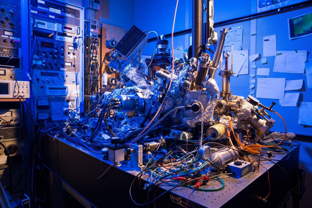
While not science’s sleekest machine, this microscope was able to capture M.R.I. scans of single atoms. Credit: IBM Research

Such a messy looking thing—it makes me feel better about my housekeeping. In any event, it’s fascinating to think this scanning tunneling microscope as seen in the above can actually act as an MRI device and create an image of a single atom.

There’s a wonderful article in the New York Times about the work but I’m starting first with a July 1, 2019 news item on Nanowerk,

Researchers at the Center for Quantum Nanoscience (QNS) within the Institute for Basic Science (IBS) at Ewha Womans University [Seoul, South Korea) have made a major scientific breakthrough by performing the world’s smallest magnetic resonance imaging (MRI). In an international collaboration with colleagues from the US, QNS scientists used their new technique to visualize the magnetic field of single atoms.

A July 2, 2019 IBS news release (also on EurekAlert but published July 1, 2019), which originated the news item, provides some insight into the research,

An MRI is routinely done in hospitals nowadays as a part of imaging for diagnostics. MRI’s detect the density of spins – the fundamental magnets in electrons and protons – in the human body. Traditionally, billions and billions of spins are required for an MRI scan. The new findings, published today [July 1, 2019] in the journal Nature Physics, show that this process is now also possible for an individual atom on a surface. To do this, the team used a Scanning Tunneling Microscope, which consists of an atomically sharp metal tip that allows researchers to image and probe single atoms by scanning the tip across the surface.

The two elements that were investigated in this work, iron and titanium, are both magnetic. Through precise preparation of the sample, the atoms were readily visible in the microscope. The researchers then used the microscope’s tip like an MRI machine to map the three-dimensional magnetic field created by the atoms with unprecedented resolution. In order to do so, they attached another spin cluster to the sharp metal tip of their microscope. Similar to everyday magnets, the two spins would attract or repel each other depending on their relative position. By sweeping the tip spin cluster over the atom on the surface, the researchers were able to map out the magnetic interaction. Lead author, Dr. Philip Willke of QNS says: “It turns out that the magnetic interaction we measured depends on the properties of both spins, the one on the tip and the one on the sample. For example, the signal that we see for iron atoms is vastly different from that for titanium atoms. This allows us to distinguish different kinds of atoms by their magnetic field signature and makes our technique very powerful.”

The researchers plan to use their single-atom MRI to map the spin distribution in more complex structures such as molecules and magnetic materials. “Many magnetic phenomena take place on the nanoscale, including the recent generation of magnetic storage devices.” says Dr. Yujeong Bae also of QNS, a co-author in this study. “We now plan to study a variety of systems using our microscopic MRI.” The ability to analyze the magnetic structure on the nanoscale can help to develop new materials and drugs. Moreover, the research team wants to use this kind of MRI to characterize and control quantum systems. These are of great interest for future computation schemes, also known as quantum computing

“I am very excited about these results. It is certainly a milestone in our field and has very promising implications for future research.” says Prof. Andreas Heinrich, Director of QNS. “The ability to map spins and their magnetic field with previously unimaginable precision, allows us to gain deeper knowledge about the structure of matter and opens new fields of basic research.”

The Center for Quantum Nanoscience, on the campus of Ewha Womans University in Seoul, South Korea, is a world-leading research center merging quantum and nanoscience to engineer the quantum future through basic research. Backed by Korea’s Institute for Basic Science, which was founded in 2011, the Center for Quantum Nanoscience draws on decades of QNS Director Andreas J. Heinrich’s (A Boy and His Atom, IBM, 2013) scientific leadership to lay the foundation for future technology by exploring the use of quantum behavior atom-by-atom on surfaces with highest precision.

You may have noticed that other than a brief mention in the first paragraph (in the Nanowerk news item excerpt), there’s no mention of the US researchers and their contribution to the work.

Interestingly, the July 1, 2019 New York Time article by Knvul Sheikh returns the favour by focusing almost entirely on US researchers while giving the Korean researchers a passing mention (Note: Links have been removed),

Different microscopy techniques allow scientists to see the nucleotide-by-nucleotide genetic sequences in cells down to the resolution of a couple atoms as seen in an atomic force microscopy image. But scientists at the IBM Almaden Research Center in San Jose, Calif., and the Institute for Basic Sciences in Seoul, have taken imaging a step further, developing a new magnetic resonance imaging technique that provides unprecedented detail, right down to the individual atoms of a sample.

When doctors want to detect tumors, measure brain function or visualize the structure of joints, they employ huge M.R.I. machines, which apply a magnetic field across the human body. This temporarily disrupts the protons spinning in the nucleus of every atom in every cell. A subsequent, brief pulse of radio-frequency energy causes the protons to spin perpendicular to the pulse. Afterward, the protons return to their normal state, releasing energy that can be measured by sensors and made into an image.

But to gather enough diagnostic data, traditional hospital M.R.I.s must scan billions and billions of protons in a person’s body, said Christopher Lutz, a physicist at IBM. So he and his colleagues decided to pack the power of an M.R.I. machine into the tip of another specialized instrument known as a scanning tunneling microscope to see if they could image individual atoms.

The tip of a scanning tunneling microscope is just a few atoms wide. And it moves along the surface of a sample, it picks up details about the size and conformation of molecules.

The researchers attached magnetized iron atoms to the tip, effectively combining scanning-tunneling microscope and M.R.I. technologies.

When the magnetized tip swept over a metal wafer of iron and titanium, it applied a magnetic field to the sample, disrupting the electrons (rather than the protons, as a typical M.R.I. would) within each atom. Then the researchers quickly turned a radio-frequency pulse on and off, so that the electrons would emit energy that could be visualized. …

Here’s a link to and a citation for the paper,

Magnetic resonance imaging of single atoms on a surface by Philip Willke, Kai Yang, Yujeong Bae, Andreas J. Heinrich & Christopher P. Lutz. Nature Physics (2019) DOI: https://doi.org/10.1038/s41567-019-0573-x Published 01 July 2019

This paper is behind a paywall.

‘Superhemophobic’ medical implants

Counterintuitively, repelling blood is the concept behind a new type of medical implant according to a Jan. 18, 2017 news item on ScienceDaily,

Medical implants like stents, catheters and tubing introduce risk for blood clotting and infection — a perpetual problem for many patients.

Colorado State University engineers offer a potential solution: A specially grown, “superhemophobic” titanium surface that’s extremely repellent to blood. The material could form the basis for surgical implants with lower risk of rejection by the body.

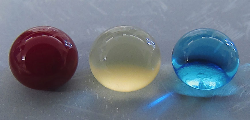
Blood, plasma and water droplets beading on a superomniphobic surface. CSU researchers have created a superhemophobic titanium surface, repellent to blood, that has potential applications for biocompatible medical devices. Courtesy: Colorado State University

A Jan. 18, 2017 Colorado State University news release by Anne Ju Manning, which originated the news item, explains more,

t’s an outside-the-box innovation achieved at the intersection of two disciplines: biomedical engineering and materials science. The work, recently published in Advanced Healthcare Materials, is a collaboration between the labs of Arun Kota, assistant professor of mechanical engineering and biomedical engineering; and Ketul Popat, associate professor in the same departments.

Kota, an expert in novel, “superomniphobic” materials that repel virtually any liquid, joined forces with Popat, an innovator in tissue engineering and bio-compatible materials. Starting with sheets of titanium, commonly used for medical devices, their labs grew chemically altered surfaces that act as perfect barriers between the titanium and blood. Their teams conducted experiments showing very low levels of platelet adhesion, a biological process that leads to blood clotting and eventual rejection of a foreign material.

Chemical compatibility

A material “phobic” (repellent) to blood might seem counterintuitive, the researchers say, as often biomedical scientists use materials “philic” (with affinity) to blood to make them biologically compatible. “What we are doing is the exact opposite,” Kota said. “We are taking a material that blood hates to come in contact with, in order to make it compatible with blood.” The key innovation is that the surface is so repellent, that blood is tricked into believing there’s virtually no foreign material there at all.

The undesirable interaction of blood with foreign materials is an ongoing problem in medical research, Popat said. Over time, stents can form clots, obstructions, and lead to heart attacks or embolisms. Often patients need blood-thinning medications for the rest of their lives – and the drugs aren’t foolproof.

“The reason blood clots is because it finds cells in the blood to go to and attach,” Popat said. “Normally, blood flows in vessels. If we can design materials where blood barely contacts the surface, there is virtually no chance of clotting, which is a coordinated set of events. Here, we’re targeting the prevention of the first set of events.”

nanotubes

Fluorinated nanotubes provided the best superhemophobic surface in the researchers’ experiments.

The researchers analyzed variations of titanium surfaces, including different textures and chemistries, and they compared the extent of platelet adhesion and activation. Fluorinated nanotubes offered the best protection against clotting, and they plan to conduct follow-up experiments.

Growing a surface and testing it in the lab is only the beginning, the researchers say. They want to continue examining other clotting factors, and eventually, to test real medical devices.

Here’s a link to and a citation for the paper,

Hemocompatibility of Superhemophobic Titania Surfaces by Sanli Movafaghi, Victoria Leszczak, Wei Wang, Jonathan A. Sorkin, Lakshmi P. Dasi, Ketul C. Popat, and Arun K. Kota. Advanced Healthcare Materials DOI: 10.1002/adhm.201600717 Version of Record online: 21 DEC 2016

© 2016 WILEY-VCH Verlag GmbH & Co. KGaA, Weinheim

Fireworks for fuel?

Scientists are attempting to harness the power in fireworks for use as fuel according to a Jan. 18, 2017 news item on Nanowerk,

The world relies heavily on gasoline and other hydrocarbons to power its cars and trucks. In search of an alternative fuel type, some researchers are turning to the stuff of fireworks and explosives: metal powders. And now one team is reporting a method to produce a metal nanopowder fuel with high energy content that is stable in air and doesn’t go boom until ignited.

A Jan. 18, 2017 American Chemical Society (ACS) news release, which originated the news item, expands on the theme,

Hydrocarbon fuels are liquid at room temperature, are simple to store, and their energy can be used easily in cars and trucks. Metal powders, which can contain large amounts of energy, have long been used as a fuel in explosives, propellants and pyrotechnics. It might seem counterintuitive to develop them as a fuel for vehicles, but some researchers have proposed to do just that. A major challenge is that high-energy metal nanopowder fuels tend to be unstable and ignite on contact with air. Albert Epshteyn and colleagues wanted to find a way to harness and control them, producing a fuel with both high energy content and good air stability.

The researchers developed a method using an ultrasound-mediated chemical process to combine the metals titanium, aluminum and boron with a sprinkle of hydrogen in a mixed-metal nanopowder fuel. The resulting material was both more stable and had a higher energy content than the standard nano-aluminum fuels. With an energy density of at least 89 kilojoules/milliliter, which is significantly superior to hydrocarbons’ 33 kilojoules/milliliter, this new titanium-aluminum-boron nanopowder packs a big punch in a small package.

Here’s a link to and a citation for the paper,

Optimization of a High Energy Ti-Al-B Nanopowder Fuel by Albert Epshteyn, Michael Raymond Weismiller, Zachary John Huba, Emily L. Maling, and Adam S. Chaimowitz. Energy Fuels, DOI: 10.1021/acs.energyfuels.6b02321 Publication Date (Web): December 30, 2016

Copyright © 2016 American Chemical Society

This paper is behind a paywall.

Inspiration from the sea for titanium implants (mussels) and adhesive panels for flexible sensors (octopuses/octopi/octopodes)

I have two sea-inspired news bits both of which concern adhesion.

Mussels and titanium implants

A July 8, 2016 news item on ScienceDaily features some mussel-inspired research from Japan into how to make better titanium implants,

Titanium is used medically in applications such as artificial joints and dental implants. While it is strong and is not harmful to tissues, the metal lacks some of the beneficial biological properties of natural tissues such as bones and natural teeth. Now, based on insights from mussels–which are able to attach themselves very tightly to even metallic surfaces due to special proteins found in their byssal threads–scientists from RIKEN have successfully attached a biologically active molecule to a titanium surface, paving the way for implants that can be more biologically beneficial.

A July 11, 2016 RIKEN press release (also on EurekAlert but dated July 8, 2016), which originated the news item, provides more information,

The work began from earlier discoveries that mussels can attach to smooth surfaces so effectively thanks to a protein, L-DOPA, which is known to be able to bind very strongly to smooth surfaces such as rocks, ceramics, or metals (…). Interestingly, the same protein functions in humans as a precursor to dopamine, and is used as a treatment for Parkinson’s disease.

According to Chen Zhang of the RIKEN Nano Medical Engineering Laboratory, the first author of the paper published in Angewandte Chemie, “We thought it would be interesting to try to use various techniques to attach a biologically active protein—in our case we chose insulin-like growth factor-1, a promoter of cell proliferation—to a titanium surface like those used in implants” (…).

Using a combination of recombinant DNA technology and treatment with tyrosinase, they were able to create a hybrid protein that contained active parts of both the growth factor and L-DOPA. Tests showed that the proteins were able to fold normally, and further experiments in cell cultures demonstrated that the IGF-1 was still functioning normally. Thanks to the incorporation of the L-DOPA, the team was able to confirm that the proteins bound strongly to the titanium surface, and remained attached even when the metal was washed with phosphate-buffered saline, a water-based solution. Zhang says, “This is similar to the powerful properties of mussel adhesive, which can remain fixed to metallic materials even underwater.”

According to Yoshihiro Ito, Team Leader of the Emergent Bioengineering Research Team of the RIKEN Center for Emergent Matter Science, “We are very excited by this finding, because the modification process is a universal one that could be used with other proteins. It could allow us to prepare new cell-growth enhancing materials, with potential applications in cell culture systems and regenerative medicine. And it is particularly interesting that this is an example of biomimetics, where nature can teach us new ways to do things. The mussel has given us insights that could be used to allow us to live healthier lives.”

The work was done by RIKEN researchers in collaboration with Professor Peibiao Zhang of the Changchun Institute of Applied Chemistry, Chinese Academy of Sciences, and Professor Yi Wang of the School of Pharmaceutical Sciences, Jilin University. The work was partially supported by the Japan Society for the Promotion of Science KAKENHI (Grant Number 15H01810 and 22220009), CAS-JSPS joint fund (GJHZ1519), and RIKEN MOST joint project.

Here’s a link to and a citation for the paper,

A Bioorthogonal Approach for the Preparation of a Titanium-Binding Insulin-like Growth-Factor-1 Derivative by using Tyrosinase by Chen Zhang, Hideyuki Miyatake, Yu Wang, Takehiko Inaba, Yi Wang, Peibiao Zhang, and Prof. Yoshihiro Ito. Angewandte Chemie International Edition DOI: 10.1002/anie.201603155 Version of Record online: 6 JUL 2016

© 2016 WILEY-VCH Verlag GmbH & Co. KGaA, Weinheim

This paper is behind a paywall.

Octopuses/octopi/octopodes and adhesive panels

Before launching into the science part of this news bit, here’s some grammar (from the Octopus Wikipedia entry; Note: Links have been removed),

The standard pluralized form of “octopus” in the English language is “octopuses” /ˈɒktəpʊsɪz/,[10] although the Ancient Greek plural “octopodes” /ɒkˈtɒpədiːz/, has also been used historically.[9] The alternative plural “octopi” — which misguidedly assumes it is a Latin “-us”-word — is considered grammatically incorrect.[11][12][13][14] It is nevertheless used enough to make it notable, and was formally acknowledged by the descriptivist Merriam-Webster 11th Collegiate Dictionary and Webster’s New World College Dictionary. The Oxford English Dictionary (2008 Draft Revision)[15] lists “octopuses”, “octopi”, and “octopodes”, in that order, labelling “octopodes” as rare and noting that “octopi” derives from the apprehension that octōpus comes from Latin.[16] In contrast, New Oxford American Dictionary (3rd Edition 2010) lists “octopuses” as the only acceptable pluralization, with a usage note indicating “octopodes” as being still occasionally used but “octopi” as being incorrect.[17]

Now the news. A July 12, 2016 news item on Nanowerk highlights some research into adhesives and octopuses,

With increased study of bio-adhesives, a significant effort has been made in search for novel adhesives that will combine reversibility, repeated usage, stronger bonds and faster bonding time, non-toxic, and more importantly be effective in wet and other extreme conditions.

A team of Korean scientists-made up of scientists from Korea Institute of Science and Technology (KIST) and UNIST has recently found a way to make building flexible pressure sensors easier–by mimicking the suction cups on octopus’s tentacles.

A July 5, 2016 UNIST (Ulsan National Institute of Science and Technology) press release, which originated the news item, provides more information,

According to the research team, “Although flexible pressure sensors might give future prosthetics and robots a better sense of touch, building them requires a lot of laborious transferring of nano- and microribbons of inorganic semiconductor materials onto polymer sheets.”

In search of an easier way to process this transfer printing, Prof. Hyunhyub Ko (School of Energy and Chemical Engineering, UNIST) and his colleagues turned to the octopus suction cups for inspiration.

An octopus uses its tentacles to move to a new location and uses suction cups underneath each tentacle to grab onto something. Each suction cup contains a cavity whose pressure is controlled by surrounding muscles. These can be made thinner or thicker on demand, increasing or decreasing air pressure inside the cup, allowing for sucking and releasing as desired.

By mimicking muscle actuation to control cavity-pressure-induced adhesion of octopus suckers, Prof. Ko and his team engineered octopus-inspired smart adhesive pads. They used the rubbery material polydimethylsiloxane (PDMS) to create an array of microscale suckers, which included pores that are coated with a thermally responsive polymer to create sucker-like walls.

The team discovered that the best way to replicate organic nature of muscle contractions would be through applied heat. Indeed, at room temperature, the walls of each pit sit in an ‘open’ state, but when the mat is heated to 32°C, the walls contract, creating suction, therby allowing the entire mate to adhere to a material (mimicking the suction function of an octopus). The adhesive strength also spiked from .32 kilopascals to 94 kilopascals at high temperature.

The team reports that the mat worked as envisioned—they made some indium gallium arsenide transistors that sat on a flexible substrate and also used it to move some nanomaterials to a different type of flexible material.

Prof. Ko and his team expect that their smart adhesive pads can be used as the substrate for wearable health sensors, such as Band-Aids or sensors that stick to the skin at normal body temperatures but fall off when rinsed under cold water.

Here’s a link to and a citation for the paper,

Octopus-Inspired Smart Adhesive Pads for Transfer Printing of Semiconducting Nanomembranes by Hochan Lee, Doo-Seung Um, Youngsu Lee, Seongdong Lim, Hyung-jun Kim,  and Hyunhyub Ko. Advanced Materials DOI: 10.1002/adma.201601407 Version of Record online: 20 JUN 2016

© 2016 WILEY-VCH Verlag GmbH & Co. KGaA, Weinheim

This paper is behind a paywall.

X-rays reveal memristor workings

A June 14, 2016 news item on ScienceDaily focuses on memristors. (It’s been about two months since my last memristor posting on April 22, 2016 regarding electronic synapses and neural networks). This piece announces new insight into how memristors function at the atomic scale,

In experiments at two Department of Energy national labs — SLAC National Accelerator Laboratory and Lawrence Berkeley National Laboratory — scientists at Hewlett Packard Enterprise (HPE) [also referred to as HP Labs or Hewlett Packard Laboratories] have experimentally confirmed critical aspects of how a new type of microelectronic device, the memristor, works at an atomic scale.

This result is an important step in designing these solid-state devices for use in future computer memories that operate much faster, last longer and use less energy than today’s flash memory. …

“We need information like this to be able to design memristors that will succeed commercially,” said Suhas Kumar, an HPE scientist and first author on the group’s technical paper.

A June 13, 2016 SLAC news release, which originated the news item, offers a brief history according to HPE and provides details about the latest work,

The memristor was proposed theoretically [by Dr. Leon Chua] in 1971 as the fourth basic electrical device element alongside the resistor, capacitor and inductor. At its heart is a tiny piece of a transition metal oxide sandwiched between two electrodes. Applying a positive or negative voltage pulse dramatically increases or decreases the memristor’s electrical resistance. This behavior makes it suitable for use as a “non-volatile” computer memory that, like flash memory, can retain its state without being refreshed with additional power.

Over the past decade, an HPE group led by senior fellow R. Stanley Williams has explored memristor designs, materials and behavior in detail. Since 2009 they have used intense synchrotron X-rays to reveal the movements of atoms in memristors during switching. Despite advances in understanding the nature of this switching, critical details that would be important in designing commercially successful circuits  remained controversial. For example, the forces that move the atoms, resulting in dramatic resistance changes during switching, remain under debate.

In recent years, the group examined memristors made with oxides of titanium, tantalum and vanadium. Initial experiments revealed that switching in the tantalum oxide devices could be controlled most easily, so it was chosen for further exploration at two DOE Office of Science User Facilities – SLAC’s Stanford Synchrotron Radiation Lightsource (SSRL) and Berkeley Lab’s Advanced Light Source (ALS).

At ALS, the HPE researchers mapped the positions of oxygen atoms before and after switching. For this, they used a scanning transmission X-ray microscope and an apparatus they built to precisely control the position of their sample and the timing and intensity of the 500-electronvolt ALS X-rays, which were tuned to see oxygen.

The experiments revealed that even weak voltage pulses create a thin conductive path through the memristor. During the pulse the path heats up, which creates a force that pushes oxygen atoms away from the path, making it even more conductive. Reversing the voltage pulse resets the memristor by sucking some of oxygen atoms back into the conducting path, thereby increasing the device’s resistance. The memristor’s resistance changes between 10-fold and 1 million-fold, depending on operating parameters like the voltage-pulse amplitude. This resistance change is dramatic enough to exploit commercially.

To be sure of their conclusion, the researchers also needed to understand if the tantalum atoms were moving along with the oxygen during switching. Imaging tantalum required higher-energy, 10,000-electronvolt X-rays, which they obtained at SSRL’s Beam Line 6-2. In a single session there, they determined that the tantalum remained stationary.

“That sealed the deal, convincing us that our hypothesis was correct,” said HPE scientist Catherine Graves, who had worked at SSRL as a Stanford graduate student. She added that discussions with SLAC experts were critical in guiding the HPE team toward the X-ray techniques that would allow them to see the tantalum accurately.

Kumar said the most promising aspect of the tantalum oxide results was that the scientists saw no degradation in switching over more than a billion voltage pulses of a magnitude suitable for commercial use. He added that this knowledge helped his group build memristors that lasted nearly a billion switching cycles, about a thousand-fold improvement.

“This is much longer endurance than is possible with today’s flash memory devices,” Kumar said. “In addition, we also used much higher voltage pulses to accelerate and observe memristor failures, which is also important in understanding how these devices work. Failures occurred when oxygen atoms were forced so far away that they did not return to their initial positions.”

Beyond memory chips, Kumar says memristors’ rapid switching speed and small size could make them suitable for use in logic circuits. Additional memristor characteristics may also be beneficial in the emerging class of brain-inspired neuromorphic computing circuits.

“Transistors are big and bulky compared to memristors,” he said. “Memristors are also much better suited for creating the neuron-like voltage spikes that characterize neuromorphic circuits.”

The researchers have provided an animation illustrating how memristors can fail,

This animation shows how millions of high-voltage switching cycles can cause memristors to fail. The high-voltage switching eventually creates regions that are permanently rich (blue pits) or deficient (red peaks) in oxygen and cannot be switched back. Switching at lower voltages that would be suitable for commercial devices did not show this performance degradation. These observations allowed the researchers to develop materials processing and operating conditions that improved the memristors’ endurance by nearly a thousand times. (Suhas Kumar) Courtesy: SLAC

This animation shows how millions of high-voltage switching cycles can cause memristors to fail. The high-voltage switching eventually creates regions that are permanently rich (blue pits) or deficient (red peaks) in oxygen and cannot be switched back. Switching at lower voltages that would be suitable for commercial devices did not show this performance degradation. These observations allowed the researchers to develop materials processing and operating conditions that improved the memristors’ endurance by nearly a thousand times. (Suhas Kumar) Courtesy: SLAC

Here’s a link to and a citation for the paper,

Direct Observation of Localized Radial Oxygen Migration in Functioning Tantalum Oxide Memristors by Suhas Kumar, Catherine E. Graves, John Paul Strachan, Emmanuelle Merced Grafals, Arthur L. David Kilcoyne3, Tolek Tyliszczak, Johanna Nelson Weker, Yoshio Nishi, and R. Stanley Williams. Advanced Materials, First published: 2 February 2016; Print: Volume 28, Issue 14 April 13, 2016 Pages 2772–2776 DOI: 10.1002/adma.201505435

This paper is behind a paywall.

Some of the ‘memristor story’ is contested and you can find a brief overview of the discussion in this Wikipedia memristor entry in the section on ‘definition and criticism’. There is also a history of the memristor which dates back to the 19th century featured in my May 22, 2012 posting.