Tag Archives: metallic nanoparticles

Use AI to reduce worries about nanoparticles in food

A June 16, 2021 news item on ScienceDaily announces research into the impact that engineered metallic nanoparticles used in agricultural practices have on food,

While crop yield has achieved a substantial boost from nanotechnology in recent years, alarms over the health risks posed by nanoparticles within fresh produce and grains have also increased. In particular, nanoparticles entering the soil through irrigation, fertilizers and other sources have raised concerns about whether plants absorb these minute particles enough to cause toxicity.

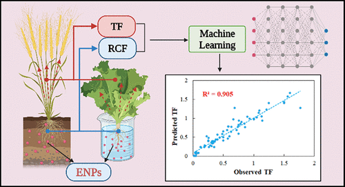
In a new study published online in the journal Environmental Science and Technology, researchers at Texas A&M University have used machine learning [a form of artificial intelligence {AI}] to evaluate the salient properties of metallic nanoparticles that make them more susceptible for plant uptake. The researchers said their algorithm could indicate how much plants accumulate nanoparticles in their roots and shoots.

A June 16, 2021 Texas A&M University news release (also on EurekAlert), which originated the news item, describes the research, which employed two different machine learning algorithms, in more detail,

Nanoparticles are a burgeoning trend in several fields, including medicine, consumer products and agriculture. Depending on the type of nanoparticle, some have favorable surface properties, charge and magnetism, among other features. These qualities make them ideal for a number of applications. For example, in agriculture, nanoparticles may be used as antimicrobials to protect plants from pathogens. Alternatively, they can be used to bind to fertilizers or insecticides and then programmed for slow release to increase plant absorption.

These agricultural practices and others, like irrigation, can cause nanoparticles to accumulate in the soil. However, with the different types of nanoparticles that could exist in the ground and a staggeringly large number of terrestrial plant species, including food crops, it is not clearly known if certain properties of nanoparticles make them more likely to be absorbed by some plant species than others.

“As you can imagine, if we have to test the presence of each nanoparticle for every plant species, it is a huge number of experiments, which is very time-consuming and expensive,” said Xingmao “Samuel” Ma, associate professor in the Zachry Department of Civil and Environmental Engineering. “To give you an idea, silver nanoparticles alone can have hundreds of different sizes, shapes and surface coatings, and so, experimentally testing each one, even for a single plant species, is impractical.”

Instead, for their study, the researchers chose two different machine learning algorithms, an artificial neural network and gene-expression programming. They first trained these algorithms on a database created from past research on different metallic nanoparticles and the specific plants in which they accumulated. In particular, their database contained the size, shape and other characteristics of different nanoparticles, along with information on how much of these particles were absorbed from soil or nutrient-enriched water into the plant body.

Once trained, their machine learning algorithms could correctly predict the likelihood of a given metallic nanoparticle to accumulate in a plant species. Also, their algorithms revealed that when plants are in a nutrient-enriched or hydroponic solution, the chemical makeup of the metallic nanoparticle determines the propensity of accumulation in the roots and shoots. But if plants are grown in soil, the contents of organic matter and the clay in soil are key to nanoparticle uptake.

Ma said that while the machine learning algorithms could make predictions for most food crops and terrestrial plants, they might not yet be ready for aquatic plants. He also noted that the next step in his research would be to investigate if the machine learning algorithms could predict nanoparticle uptake from leaves rather than through the roots.

“It is quite understandable that people are concerned about the presence of nanoparticles in their fruits, vegetables and grains,” said Ma. “But instead of not using nanotechnology altogether, we would like farmers to reap the many benefits provided by this technology but avoid the potential food safety concerns.”

This image accompanies the paper’s research abstract,

[downloaded frm https://pubs.acs.org/doi/full/10.1021/acs.est.1c01603]

Here’s a link to and a citation for the paper,

Prediction of Plant Uptake and Translocation of Engineered Metallic Nanoparticles by Machine Learning by Xiaoxuan Wang, Liwei Liu, Weilan Zhang, and Xingmao Ma. Environ. Sci. Technol. 2021, 55, 11, 7491–7500 DOI: https://doi.org/10.1021/acs.est.1c01603 Publication Date:May 17, 2021 Copyright © 2021 American Chemical Society

This paper is behind a paywall.

Metallic nanoparticles inside heart tissue mitochondria can cause damage

With all the focus on COVID-19, viruses , and aerosols, it’s easy to forget that there are other kinds of contaminated air too. The last time I featured work on nanoparticles and air pollution was in a May 31, 2017 posting, “Explaining the link between air pollution and heart disease?” where scientists announced they may have discovered how air pollution (nanoparticles) were making their way from lungs to the heart. Answer: the bloodstream.

A July 3, 2020 Lancaster University press release (also on EurekAlert) announces research into how air made toxic by metallic nanoparticles affects the heart in very specific ways (Note: A link has been removed),

Toxic metallic air pollution nanoparticles are getting inside the crucial, energy-producing structures within the hearts of people living in polluted cities, causing cardiac stress – a new study confirms.

The research team, led by Professors Barbara Maher of Lancaster University and Lilian Calderón-Garcidueñas of The University of Montana and the Universidad del Valle de Mexico, found the metallic nanoparticles, which included iron-rich nanoparticles and other pollution-derived metals such as titanium, inside the damaged heart cells of a 26-year-old and even a three-year-old toddler.

The hearts had belonged to people who had died in accidents and who had lived in highly-polluted Mexico City.

The findings shed new light on how air pollution can cause the development of heart disease, as the iron-rich particles were associated with damage to the cells, and increased cardiac oxidative stress, even in these very young hearts.

The repeated inhalation of these iron-rich nanoparticles, and their circulation by the bloodstream to the heart, may account for the well-established associations between exposure to particulate air pollution and increased cardiovascular disease, including heart attacks. The study indicates that heart disease can start in very early age, before progressing to full-blown cardiovascular illness later in life. This type of air pollution may thus be responsible for the ‘silent epidemic’ of heart disease, internationally. By causing pre-existing heart conditions, it may also account for some of the increased death rates from Covid-19 seen in areas with high levels of particulate air pollution.

Professor Maher said: “It’s been known for a long time that people with high exposure to particulate air pollution experience increased levels and severity of heart disease. Our new work shows that iron-rich nanoparticles from air pollution can get right inside the millions of mitochondria inside our hearts…the structures which generate the energy needed for our hearts to pump properly.

“That we found these metal particles inside the heart of even a three-year old indicates that we’re setting heart disease in train right from the earliest days, but only seeing its full, clinical effects in later life. It’s really urgent to reduce emissions of ultrafine particles from our vehicles and from industry, before we give heart disease to the next generation too.”

The researchers, using high-resolution transmission electron microscopy and energy-dispersive X-ray analysis, found that the mitochondria containing the iron-rich nanoparticles appeared to be damaged, with some cells showing deformities and others with ruptured membranes. Professor Calderón-Garcidueñas stated that increased levels of markers of cardiac oxidative stress are present in the very young cases examined.

The iron-rich nanoparticles found inside the heart cells are identical in size, shape and composition to those emitted from sources such as the exhausts, tyres and brakes of vehicles. These air pollution nanoparticles are also emitted by industrial sources as well as open fires in homes.

Some of the iron-rich nanoparticles are also strongly magnetic. This raises concerns about what might happen when people with millions of these nanoparticles in their hearts are using appliances with associated magnetic fields, such as hair dryers and mobile phones. People who work in industries that mean they are exposed to magnetic fields such as welders and power line engineers may also be at risk. This kind of exposure could potentially lead to heart electrical dysfunction and cell damage.

The findings builds on the researchers’ previous findings that show that the hearts of city dwellers contain billions of these nanoparticles and can be up to ten times more polluted than the hearts of people living in less polluted places.

The researchers say their study underlines the need for governments across the world to tackle ultrafine particulate pollution in their cities.

Professor Calderón-Garcidueñas said: “Exposure to such air pollution is a modifiable risk factor for cardiovascular disease, on a global scale, reinforcing the urgent need for individual and government actions not just to reduce PM2.5 but to monitor, regulate and reduce emissions of these specific, ultrafine components of the urban air pollution ‘cocktail’.”

Here’s a link to and a citation for the paper,

Iron-rich air pollution nanoparticles: An unrecognised environmental risk factor for myocardial mitochondrial dysfunction and cardiac oxidative stress by B.A.Maher, A.González-Maciel, R.Reynoso-Robles, R.Torres-Jardón, L.Calderón-Garcidueñas. Environmental Research Volume 188, September 2020, 109816 DOI: https://doi.org/10.1016/j.envres.2020.109816 Available online 21 June 2020

This paper appears to be open access (just keep scrolling down).

Algae outbreaks (dead zones) in wetlands and waterways

It’s been over seven years since I first started writing about Duke University’s  Center for the Environmental Implications of Nanotechnology and mesocosms (miniature ecosystems) and the impact that nanoparticles may have on plants and water (see August 11, 2011 posting). Since then, their focus has shifted from silver nanoparticles and their impact on plants, fish, bacteria, etc. to a more general examination of metallic nanoparticles and water. A June 25, 2018 news item on ScienceDaily announces some of their latest work,

The last 10 years have seen a surge in the use of tiny substances called nanomaterials in agrochemicals like pesticides and fungicides. The idea is to provide more disease protection and better yields for crops, while decreasing the amount of toxins sprayed on agricultural fields.

But when combined with nutrient runoff from fertilized cropland and manure-filled pastures, these “nanopesticides” could also mean more toxic algae outbreaks for nearby streams, lakes and wetlands, a new study finds.

A June 25, 2018 Duke University news release (also on EurekAlert) by Robin A. Smith, which originated the news item, provides more detail,

Too small to see with all but the most powerful microscopes, engineered nanomaterials are substances manufactured to be less than 100 nanometers in diameter, many times smaller than a hair’s breadth.

Their nano-scale gives them different chemical and physical properties from their bulk counterparts, including more surface area for reactions and interactions.

Those interactions could intensify harmful algal blooms in wetlands, according to experiments led by Marie Simonin, a postdoctoral associate with biology professor Emily Bernhardt at Duke University.

Carbon nanotubes and teeny tiny particles of silver, titanium dioxide and other metals are already added to hundreds of commercial products to make everything from faster, lighter electronics, self-cleaning fabrics, and smarter food packaging that can monitor food for spoilage. They are also used on farms for slow- or controlled-release plant fertilizers and pesticides and more targeted delivery, and because they are effective at lower doses than conventional products.

These and other applications have generated tremendous interest and investment in nanomaterials. However the potential risks to human health or the environment aren’t fully understood, Simonin said.

Most of the 260,000 to 309,000 metric tons of nanomaterials produced worldwide each year are eventually disposed in landfills, according to a previous study. But of the remainder, up to 80,400 metric tons per year are released into soils, and up to 29,200 metric tons end up in natural bodies of water.

“And these emerging contaminants don’t end up in water bodies alone,” Simonin said. “They probably co-occur with nutrient runoff. There are likely multiple stressors interacting.”

Algae outbreaks already plague polluted waters worldwide, said Steven Anderson, a research analyst in the Bernhardt Lab at Duke and one of the authors of the research.

Nitrogen and phosphorous pollution makes its way into wetlands and waterways in the form of agricultural runoff and untreated wastewater. The excessive nutrients cause algae to grow out of control, creating a thick mat of green scum or slime on the surface of the water that blocks sunlight from reaching other plants.

These nutrient-fueled “blooms” eventually reduce oxygen levels to the point where fish and other organisms can’t survive, creating dead zones in the water. Some algal blooms also release toxins that can make pets and people who swallow them sick.

To find out how the combined effects of nutrient runoff and nanoparticle contamination would affect this process, called eutrophication, the researchers set up 18 separate 250-liter tanks with sandy sloped bottoms to mimic small wetlands.

Each open-air tank was filled with water, soil and a variety of wetland plants and animals such as waterweed and mosquitofish.

Over the course of the nine-month experiment, some tanks got a weekly dose of algae-promoting nitrates and phosphates like those found in fertilizers, some tanks got nanoparticles — either copper or gold — and some tanks got both.

Along the way the researchers monitored water chemistry, plant and algae growth and metabolism, and nanoparticle accumulation in plant tissues.

“The results were surprising,” Simonin said. The nanoparticles had tiny effects individually, but when added together with nutrients, even low concentrations of gold and copper nanoparticles used in fungicides and other products turned the once-clear water a murky pea soup color, its surface covered with bright green smelly mats of floating algae.

Over the course of the experiment, big algal blooms were more than three times more frequent and more persistent in tanks where nanoparticles and nutrients were added together than where nutrients were added alone. The algae overgrowths also reduced dissolved oxygen in the water.

It’s not clear yet how nanoparticle exposure shifts the delicate balance between plants and algae as they compete for nutrients and other resources. But the results suggest that nanoparticles and other “metal-based synthetic chemicals may be playing an under-appreciated role in the global trends of increasing eutrophication,” the researchers said.

Here’s a link to and a citation for the paper,

Engineered nanoparticles interact with nutrients to intensify eutrophication in a wetland ecosystem experiment by Marie Simonin, Benjamin P. Colman, Steven M. Anderson, Ryan S. King, Matthew T. Ruis, Astrid Avellan, Christina M. Bergemann, Brittany G. Perrotta, Nicholas K. Geitner, Mengchi Ho, Belen de la Barrera, Jason M. Unrine, Gregory V. Lowry, Curtis J. Richardson, Mark R. Wiesner, Emily S. Bernhardt. Ecological Applications, 2018; DOI: 10.1002/eap.1742 First published: 25 June 2018

This paper is behind a paywall.

Sunscreens: 2018 update

I don’t usually concern myself with SPF numbers on sunscreens as my primary focus has been on the inclusion of nanoscale metal particles (these are still considered safe). However, a recent conversation with a dental hygienist and coincidentally tripping across a June 19, 2018 posting on the blog shortly after the convo. has me reassessing my take on SPF numbers (Note: Links have been removed),

So, what’s the deal with SPF? A recent interview of Dr Steven Q Wang, M.D., chair of The Skin Cancer Foundation Photobiology Committee, finally will give us some clarity. Apparently, the SPF number, be it 15, 30, or 50, refers to the amount of UVB protection that that sunscreen provides. Rather than comparing the SPFs to each other, like we all do at the store, SPF is a reflection of the length of time it would take for the Sun’s UVB radiation to redden your skin (used exactly as directed), versus if you didn’t apply any sunscreen at all. In ideal situations (in lab settings), if you wore SPF 30, it would take 30 times longer for you to get a sunburn than if you didn’t wear any sunscreen.

What’s more, SPF 30 is not nearly half the strength of SPF 50. Rather, SPF 30 allows 3% of UVB rays to hit your skin, and SPF 50 allows about 2% of UVB rays to hit your skin. Now before you say that that is just one measly percent, it actually is much more. According to Dr Steven Q. Wang, SPF 30 allows around 1.5 times more UV radiation onto your skin than SPF 50. That’s an actual 150% difference [according to Wang’s article “… SPF 30 is allowing 50 percent more UV radiation onto your skin.”] in protection.

(author of the ‘eponymous’ blog) offers a good overview of the topic in a friendly, informative fashion albeit I found the ‘percentage’ to be a bit confusing. (S)he also provides a link to a previous posting about the ingredients in sunscreens (I do have one point of disagreement with regarding oxybenzone) as well as links to Dr. Steven Q. Wang’s May 24, 2018 Ask the Expert article about sunscreens and SPF numbers on skincancer.org. You can find the percentage under the ‘What Does the SPF Number Mean?’ subsection, in the second paragraph.

Ingredients: metallic nanoparticles and oxybenzone

The use of metallic nanoparticles  (usually zinc oxide and/or (titanium dioxide) in sunscreens was loathed by civil society groups, in particular Friends of the Earth (FOE) who campaigned relentlessly against their use in sunscreens. The nadir for FOE was in February 2012 when the Australian government published a survey showing that 13% of the respondents were not using any sunscreens due to their fear of nanoparticles. For those who don’t know, Australia has the highest rate of skin cancer in the world. (You can read about the debacle in my Feb. 9, 2012 posting.)

At the time, the only civil society group which supported the use of metallic nanoparticles in sunscreens was the Environmental Working Group (EWG).  After an examination of the research they, to their own surprise, came out in favour (grudgingly) of metallic nanoparticles. (The EWG were more concerned about the use of oxybenzone in sunscreens.)

Over time, the EWG’s perspective has been adopted by other groups to the point where sunscreens with metallic nanoparticles are commonplace in ‘natural’ or ‘organic’ sunscreens.

As for oxybenzones, in a May 23, 2018 posting about sunscreen ingredients notes this (Note: Links have been removed),

Oxybenzone – Chemical sunscreen, protects from UV damage. Oxybenzone belongs to the chemical family Benzophenone, which are persistent (difficult to get rid of), bioaccumulative (builds up in your body over time), and toxic, or PBT [or: Persistent, bioaccumulative and toxic substances (PBTs)]. They are a possible carcinogen (cancer-causing agent), endocrine disrupter; however, this is debatable. Also could cause developmental and reproductive toxicity, could cause organ system toxicity, as well as could cause irritation and potentially toxic to the environment.

It seems that the tide is turning against the use of oxybenzones (from a July 3, 2018 article by Adam Bluestein for Fast Company; Note: Links have been removed),

On July 3 [2018], Hawaii’s Governor, David Ig, will sign into law the first statewide ban on the sale of sunscreens containing chemicals that scientists say are damaging the Earth’s coral reefs. Passed by state legislators on May 1 [2018], the bill targets two chemicals, oxybenzone and octinoxate, which are found in thousands of sunscreens and other skincare products. Studies published over the past 10 years have found that these UV-filtering chemicals–called benzophenones–are highly toxic to juvenile corals and other marine life and contribute to the fatal bleaching of coral reefs (along with global warming and runoff pollutants from land). (A 2008 study by European researchers estimated that 4,000 to 6,000 tons of sunblock accumulates in coral reefs every year.) Also, though both substances are FDA-approved for use in sunscreens, the nonprofit Environmental Working Group notes numerous studies linking oxybenzone to hormone disruption and cell damage that may lead to skin cancer. In its 2018 annual sunscreen guide, the EWG found oxybenzone in two-thirds of the 650 products it reviewed.

The Hawaii ban won’t take effect until January 2021, but it’s already causing a wave of disruption that’s affecting sunscreen manufacturers, retailers, and the medical community.

For starters, several other municipalities have already or could soon join Hawaii’s effort. In May [2018], the Caribbean island of Bonaire announced a ban on chemicals sunscreens, and nonprofits such as the Sierra Club and Surfrider Foundation, along with dive industry and certain resort groups, are urging legislation to stop sunscreen pollution in California, Colorado, Florida, and the U.S. Virgin Islands. Marine nature reserves in Mexico already prohibit oxybenzone-containing sunscreens, and the U.S. National Park Service website for South Florida, Hawaii, U.S. Virgin Islands, and American Samoa recommends the use of “reef safe” sunscreens, which use natural mineral ingredients–zinc oxide or titanium oxide–to protect skin.

Makers of “eco,” “organic,” and “natural” sunscreens that already meet the new standards are seizing on the news from Hawaii to boost their visibility among the islands’ tourists–and to expand their footprint on the shelves of mainland retailers. This past spring, for example, Miami-based Raw Elements partnered with Hawaiian Airlines, Honolulu’s Waikiki Aquarium, the Aqua-Aston hotel group (Hawaii’s largest), and the Sheraton Maui Resort & Spa to get samples of its reef-safe zinc-oxide-based sunscreens to their guests. “These partnerships have had a tremendous impact raising awareness about this issue,” says founder and CEO Brian Guadagno, who notes that inquiries and sales have increased this year.

As Bluestein notes there are some concerns about this and other potential bans,

“Eliminating the use of sunscreen ingredients considered to be safe and effective by the FDA with a long history of use not only restricts consumer choice, but is also at odds with skin cancer prevention efforts […],” says Bayer, owner of the Coppertone brand, in a statement to Fast Company. Bayer disputes the validity of studies used to support the ban, which were published by scientists from U.S. National Oceanic & Atmospheric Administration, the nonprofit Haereticus Environmental Laboratory, Tel Aviv University, the University of Hawaii, and elsewhere. “Oxybenzone in sunscreen has not been scientifically proven to have an effect on the environment. We take this issue seriously and, along with the industry, have supported additional research to confirm that there is no effect.”

Johnson & Johnson, which markets Neutrogena sunscreens, is taking a similar stance, worrying that “the recent efforts in Hawaii to ban sunscreens that contain oxybenzone may actually adversely affect public health,” according to a company spokesperson. “Science shows that sunscreens are a key factor in preventing skin cancer, and our scientific assessment of the lab studies done to date in Hawaii show the methods were questionable and the data insufficient to draw factual conclusions about any impact on coral reefs.”

Terrified (and rightly so) about anything scaring people away from using sunblock, The American Academy of Dermatology, also opposes Hawaii’s ban. Suzanne M. Olbricht, president of the AADA, has issued a statement that the organization “is concerned that the public’s risk of developing skin cancer could increase due to potential new restrictions in Hawaii that impact access to sunscreens with ingredients necessary for broad-spectrum protection, as well as the potential stigma around sunscreen use that could develop as a result of these restrictions.”

The fact is that there are currently a large number of widely available reef-safe products on the market that provide “full spectrum” protection up to SPF50–meaning they protect against both UVB rays that cause sunburns as well as UVA radiation, which causes deeper skin damage. SPFs higher than 50 are largely a marketing gimmick, say advocates of chemical-free products: According to the Environmental Working Group, properly applied SPF 50 sunscreen blocks 98% of UVB rays; SPF 100 blocks 99%. And a sunscreen lotion’s SPF rating has little to do with its ability to shield skin from UVA rays.

I notice neither Bayer nor Johnson & Johnson nor the American Academy of Dermatology make mention of oxybenzone’s possible role as a hormone disruptor.

Given the importance that coral reefs have to the environment we all share, I’m inclined to support the oxybenzone ban based on that alone. Of course, it’s conceivable that metallic nanoparticles may also have a deleterious effect on coral reefs as their use increases. It’s to be hoped that’s not the case but if it is, then I’ll make my decisions accordingly and hope we have a viable alternative.

As for your sunscreen questions and needs, the Environment Working Group (EWG) has extensive information including a product guide on this page (scroll down to EWG’s Sunscreen Guide) and a discussion of ‘high’ SPF numbers I found useful for my decision-making.

Magic nano ink

Colour changes © Nature Communications 2017 / MPI [Max Planck Institute] for Intelligent Systems

A March 1, 2017 news item on Nanowerk helps to explain the image seen above (Note: A link has been removed),

Plasmonic printing produces resolutions several times greater than conventional printing methods. In plasmonic printing, colours are formed on the surfaces of tiny metallic particles when light excites their electrons to oscillate. Researchers at the Max Planck Institute for Intelligent Systems in Stuttgart have now shown how the colours of such metallic particles can be altered with hydrogen (Nature Communications, “Dynamic plasmonic colour display”).

The technique could open the way for animating ultra-high-resolution images and for developing extremely sharp displays. At the same time, it provides new approaches for encrypting information and detecting counterfeits.

A March 1, 2017 Max Planck Institute press release, which originated the news item, provides more  history and more detail about the research,

Glass artisans in medieval times exploited the effect long before it was even known. They coloured the magnificent windows of gothic cathedrals with nanoparticles of gold, which glowed red in the light. It was not until the middle of the 20th century that the underlying physical phenomenon was given a name: plasmons. These collective oscillations of free electrons are stimulated by the absorption of incident electromagnetic radiation. The smaller the metallic particles, the shorter the wavelength of the absorbed radiation. In some cases, the resonance frequency, i.e., the absorption maximum, falls within the visible light spectrum. The unabsorbed part of the spectrum is then scattered or reflected, creating an impression of colour. The metallic particles, which usually appear silvery, copper-coloured or golden, then take on entirely new colours.

A resolution of 100,000 dots per inch

Researchers are also taking advantage of the effect to develop plasmonic printing, in which tailor-made square metal particles are arranged in specific patterns on a substrate. The edge length of the particles is in the order of less than 100 nanometres (100 billionths of a metre). This allows a resolution of 100,000 dots per inch – several times greater than what today’s printers and displays can achieve.

For metallic particles measuring several 100 nanometres across, the resonance frequency of the plasmons lies within the visible light spectrum. When white light falls on such particles, they appear in a specific colour, for example red or blue. The colour of the metal in question is determined by the size of the particles and their distance from each other. These adjustment parameters therefore serve the same purpose in plasmonic printing as the palette of colours in painting.

The trick with the chemical reaction

The Smart Nanoplasmonics Research Group at the Max Planck Institute for Intelligent Systems in Stuttgart also makes use of this colour variability. They are currently working on making dynamic plasmonic printing. They have now presented an approach that allows them to alter the colours of the pixels predictably – even after an image has been printed. “The trick is to use magnesium. It can undergo a reversible chemical reaction in which the metallic character of the element is lost,” explains Laura Na Liu, who leads the Stuttgart research group. “Magnesium can absorb up to 7.6% of hydrogen by weight to form magnesium hydride, or MgH2”, Liu continues. The researchers coat the magnesium with palladium, which acts as a catalyst in the reaction.

During the continuous transition of metallic magnesium into non-metallic MgH2, the colour of some of the pixels changes several times. The colour change and the speed of the rate at which it proceeds follow a clear pattern. This is determined both by the size of and the distance between the individual magnesium particles as well as by the amount of hydrogen present.

In the case of total hydrogen saturation, the colour disappears completely, and the pixels reflect all the white light that falls on them. This is because the magnesium is no longer present in metallic form but only as MgH2. Hence, there are also no free metal electrons that can be made to oscillate.

Minerva’s vanishing act

The scientists demonstrated the effect of such dynamic colour behaviour on a plasmonic print of Minerva, the Roman goddess of wisdom, which also bore the logo of the Max Planck Society. They chose the size of their magnesium particles so that Minerva’s hair first appeared reddish, the head covering yellow, the feather crest red and the laurel wreath and outline of her face blue. They then washed the micro-print with hydrogen. A time-lapse film shows how the individual colours change. Yellow turns red, red turns blue, and blue turns white. After a few minutes all the colours disappear, revealing a white surface instead of Minerva.

The scientists also showed that this process is reversible by replacing the hydrogen stream with a stream of oxygen. The oxygen reacts with the hydrogen in the magnesium hydride to form water, so that the magnesium particles become metallic again. The pixels then change back in reverse order, and in the end Minerva appears in her original colours.

In a similar manner the researchers first made the micro image of a famous Van Gogh painting disappear and then reappear. They also produced complex animations that give the impression of fireworks.

The principle of a new encryption technique

Laura Na Liu can imagine using this principle in a new encryption technology. To demonstrate this, the group formed various letters with magnesium pixels. The addition of hydrogen then caused some letters to disappear over time, like the image of Minerva. “As for the rest of the letters, a thin oxide layer formed on the magnesium particles after exposing the sample in air for a short time before palladium deposition,” Liu explains. This layer is impermeable to hydrogen. The magnesium lying under the oxide layer therefore remains metallic − and visible − because light is able to excite the plasmons in the magnesium.

In this way it is possible to conceal a message, for example by mixing real and nonsensical information. Only the intended recipient is able to make the nonsensical information disappear and filter out the real message. For example, after decoding the message “Hartford” with hydrogen, only the words “art or” would remain visible. To make it more difficult to crack such encrypted messages, the group is currently working on a process that would require a precisely adjusted hydrogen concentration for deciphering.

Liu believes that the technology could also be used some day in the fight against counterfeiting. “For example, plasmonic security features could be printed on banknotes or pharmaceutical packs, which could later be checked or read only under specific conditions unknown to counterfeiters.”

It doesn’t necessarily have to be hydrogen

Laura Na Liu knows that the use of hydrogen makes some applications difficult and impractical for everyday use such as in mobile displays. “We see our work as a starting shot for a new principle: the use of chemical reactions for dynamic printing,” the Stuttgart physicist says. It is certainly conceivable that the research will soon lead to the discovery of chemical reactions for colour changes other than the phase transition between magnesium and magnesium dihydride, for example, reactions that require no gaseous reactants.

Here’s a link to and a citation for the paper,

Dynamic plasmonic colour display by Xiaoyang Duan, Simon Kamin, & Na Liu. Nature Communications 8, Article number: 14606 (2017) doi:10.1038/ncomms14606 Published online: 24 February 2017

This paper is open access.

Metallic nanoflowers produce neuron-like fractals

I was a bit surprised to find that this University of Oregon story was about a patent. Here’s more from a July 28, 2015 news item on Azonano,

Richard Taylor’s vision of using artificial fractal-based implants to restore sight to the blind — part of a far-reaching concept that won an innovation award this year from the White House — is now covered under a broad U.S. patent.

The patent goes far beyond efforts to use the emerging technology to restore eyesight. It covers all fractal-designed electronic implants that link signaling activity with nerves for any purpose in animal and human biology.

Fractals are objects with irregular curves or shapes. “They are a trademark building block of nature,” said Taylor, a professor of physics and director of the Materials Science Institute at the University of Oregon [UO]. “In math, that property is self-similarity. Trees, clouds, rivers, galaxies, lungs and neurons are fractals. What we hope to do is adapt the technology to nature’s geometry.”

Named in U.S. patent 9079017 are Taylor, the UO, Taylor’s research collaborator Simon Brown, and Brown’s home institution, the University of Canterbury in New Zealand.

A July 28, 2015 University of Oregon news release (also on EurekAlert) by Jim Barlow, which originated the news item, continues the patent celebration,

“We’re very delighted,” Taylor said. “The U.S. Patent and Trademark Office has recognized the novelty and utility of our general concept, but there is a lot to do. We want to get all of the fundamental science sorted out. We’re looking at least another couple of years of basic science before moving forward.”

The patent solidifies the relationship between the two universities, said Charles Williams, associate vice president for innovation at the UO. “This is still in the very early days. This project has attracted national attention, awards and grants.

“We hope to engage the right set of partners to develop the technology over time as the concept moves into potentially vast forms of medical applications,” Williams added. “Dr. Taylor’s interdisciplinary science is a hallmark of the creativity at the University of Oregon and a great example of the international research collaborations that our faculty engage in every day.”

Here’s an image illustrating the ‘fractal neurons’,

FractalImplant

Caption: Retinal neurons, outlined in yellow, attach to and follows branches of a fractal interconnect. Such connections, says University of Oregon physicist Richard Taylor, could some day help to treat eye diseases such as macular degeneration. Credit: Courtesy of Richard Taylor

The news release goes on to describe the ‘fractal approach’ to eye implants which is markedly different from the implants entering the marketplace,

Taylor raised the idea of a fractal-based approach to treat eye diseases in a 2011 article in Physics World, writing that it could overcome problems associated with efforts to insert photodiodes behind the eyes. Current chip technology doesn’t allow sufficient connections with neurons.

“The wiring — the neurons — in the retina is fractal, but the chips are not fractal,” Taylor said. His vision, based on research with Brown, is to grow nanoflowers seeded from nanoparticles of metals that self assemble in a natural process, producing fractals that mimic and communicate with neurons.

It is conceivable, Taylor said, that fractal interconnects — as the implants are called in the patent — could be shaped so they network with like-shaped neurons to address narrow needs, such as a feedback loop for the sensation of touch from a prosthetic arm or leg to the brain.

Such implants would overcome the biological rejection of implants with smooth surfaces or those randomly patterned that have been developed in a trial-and-error approach to link to neurons.

Once perfected, he said, the implants would generate an electrical field that would fool a sea of glial cells that insulate and protect neurons from foreign invaders. Fractal interconnects would allow electrical signals to operate in “a safety zone biologically” that avoids toxicity issues.

“The patent covers any generic interface for connecting any electronics to any nerve,” Taylor said, adding that fractal interconnects are not electrodes. “Our interface is multifunctional. The primary thing is to get the electrical field into the system so that reaches the neurons and induces the signal.”

Taylor’s proposal for using fractal-based technology earned the top prize in a contest held by the innovation company InnoCentive. Taylor was honored in April [2015] at a meeting of the White House Office of Science and Technology Policy.

The competition was sponsored by a collaboration of science philanthropies including the Research Corporation for Science Advancement, the Gordon and Betty Moore Foundation, the W.M. Keck Foundation, the Kavli Foundation, the Templeton Foundation and the Burroughs Wellcome Fund.

You can find out more about InnoCentive here. As for other types of artificial eye implants, the latest here is a June 30, 2015 post titled, Clinical trial for bionic eye (artificial retinal implant) shows encouraging results (safety and efficacy).

Metallic nanoparticles: measuring their discrete quantum states

I tend to forget how new nanotechnology is and unconsciously take for granted stunning feats such as measuring a metallic nanoparticle’s electronic properties. A June 15, 2015 news item on Nanowerk provides a reminder with its description of the difficulties and a new technique to make it easier (Note:  A link has been removed),

How do you measure the electronic properties of individual nanoparticles or molecules that are only a few nanometers in size? Conventional methods using electron transport spectroscopy rely on contacting a material with two contacts, a source and a drain electrode. By applying a small potential difference over the electrodes and monitoring the resulting current, valuable information about the electronic properties are extracted. For example if a material is metallic or semiconducting.
But this becomes quite a challenge if the material is only a few nm in size. Even the most sophisticated fabrication tools such as electron-beam lithography have a resolution of about 10 nm at best, which is not precise enough. Scientists have developed workarounds such as creating small gaps in narrow metallic wires in which a nanoparticle can be trapped if it matches the gap size. However, even though there have been some notable successes using this approach, this method has a low yield and is not very reproducible.

Now an international collaboration including researchers in Japan, the university [sic] of Cambridge and the LCN [London Centre for Nanotechnology] in the UK have approached this in a different way as described in a paper in Nature’s Scientific Reports (“Radio-frequency capacitance spectroscopy of metallic nanoparticles”). Their method only requires a single electrode to be in direct contact with a nanoparticle or molecule, thus significantly simplifying fabrication.

A June 15, 2015 (?) LCN press release, which originated the news item, describes the achievement,

The researchers demonstrated the potential of the radio-frequency reflectometry technique by measurements on Au nanoparticles of only 2.7 nm in diameter. For such small particles, the electronic spectrum is discrete which was indeed observed in the measurements and in very good agreement with theoretical models. The researchers now plan to extend these measurements to other nanoparticles and molecules with applications in a range of areas such as biomedicine, spintronics and quantum information processing.

Here’s a link to and a citation for the paper,

Radio-frequency capacitance spectroscopy of metallic nanoparticles by James C. Frake, Shinya Kano, Chiara Ciccarelli, Jonathan Griffiths, Masanori Sakamoto,  Toshiharu Teranishi, Yutaka Majima, Charles G. Smith & Mark R. Buitelaar. Scientific RepoRts 5:10858 DOi: 10.1038/srep10858 Published June 4, 2015

This is an open access paper.

Metal nanoparticles and gut microbiomes

What happens when you eat or drink nanoparticles, metallic or otherwise? No one really knows. Part of the problem with doing research now is there are no benchmarks for the quantity we’ve been ingesting over the centuries. Nanoparticles do occur naturally, as well, people who’ve eaten with utensils made of or coated with silver or gold have ingested silver or gold nanoparticles that were shed by those very utensils. In short, we’ve been ingesting any number of nanoparticles through our food, drink, and utensils in addition to the engineered nanoparticles that are found in consumer products. So, part of what researchers need to determine is the point at which we need to be concerned about nanoparticles. That’s trickier than it might seem since we ingest our nanoparticles and recycle them into the environment (air, water, soil) to reingest (inhale, drink, eat, etc.) them at a later date. The endeavour to understand what impact engineered nanoparticles in particular will have on us as more are used in our products is akin to assembling a puzzle.

There’s a May 5, 2015 news item on Azonano which describes research into the effects that metallic nanoparticles have on the micriobiome (bacteria) in our guts,

Exposure of a model human colon to metal oxide nanoparticles, at levels that could be present in foods, consumer goods, or treated drinking water, led to multiple, measurable differences in the normal microbial community that inhabits the human gut. The changes observed in microbial metabolism and the gut microenvironment with exposure to nanoparticles could have implications for overall human health, as discussed in an article published in Environmental Engineering Science, a peer-reviewed journal from Mary Ann Liebert, Inc., publishers. The article is available free on the Environmental Engineering Science website until June 1, 2015.

A May 4, 2015 Mary Ann Liebert publisher news release on EurekAlert, which originated the news item, describes the research in more detail (Note: A link has been removed),

Alicia Taylor, Ian Marcus, Risa Guysi, and Sharon Walker, University of California, Riverside, individually introduced three different nanoparticles–zinc oxide, cerium dioxide, and titanium dioxide–commonly used in products such as toothpastes, cosmetics, sunscreens, coatings, and paints, into a model of the human colon. The model colon mimics the normal gut environment and contains the microorganisms typically present in the human microbiome.

In the article “Metal Oxide Nanoparticles Induce Minimal Phenotypic Changes in a Model Colon Gut Microbiota” the researchers described changes in both specific characteristics of the microbial community and of the gut microenvironment after exposure to the nanoparticles.

Here’s a link to and a citation for the paper,

Metal Oxide Nanoparticles Induce Minimal Phenotypic Changes in a Model Colon Gut Microbiota by Alicia A. Taylor, Ian M. Marcus Ian, Risa L., Guysi, and Sharon L. Walker. Environmental Engineering Science. DOI:10.1089/ees.2014.0518 Online Ahead of Print: April 24, 2015

I’ve taken a quick look at the research while it’s still open access (till June 1, 2015) to highlight the bits I consider interesting. There’s this about the nanoparticle (NP) quantities used in the study (Note: Links have been removed),

Environmentally relevant NP concentrations were chosen to emulate human exposures to NPs through ingestion of both food and drinking water at 0.01 μg/L ZnO NP, 0.01 μg/L CeO2 NP, and 3 mg/L TiO2 NP (Gottschalk et al., 2009; Kiser et al., 2009, 2013; Weir et al., 2012; Keller and Lazareva, 2013). Recent work has also indicated that adults in the USA ingest 5 mg TiO2 per day, half of which is in the nano-size range (Lomer et al., 2000; Powell et al., 2010). Exposure routes and reliable dosing information of NPs that are embedded in solid matrices are difficult to predict, and this is often a limitation of analytical techniques (Nowack et al., 2012; Yang and Westerhoff, 2014). The exposure levels used in this study were predominately selected from literature values that give predictions on amount of NPs in water and food sources (Gottschalk et al., 2009; Kiser et al., 2009; Weir et al., 2012; Keller and Lazareva, 2013; Keller et al., 2013).

For anyone unfamiliar with chemical notations, ZnO NP is zinc oxide nanoparticle, 0.01 μg/L is one/one hundredth of a microgram per litre,  CeO2 is cesisum dioxide NP, and TiO2 is titanium dioxide NP while 3 mg/L, is 3 milligrams per litre.

After assuring the quantities used in the study are the same as they expect humans to be ingesting on a regular basis, the researchers describe some of the factors which may affect the interaction between the tested nanoparticles and the bacteria (Note: Links have been removed),

It is essential to note that interactions between NPs and bacteria in the intestines may be dependent on numerous factors: the surface charge of the NPs and bacteria, the chemical composition and surface charge of the digested food, and variability in diet. These factors may ultimately correlate to effects seen in humans on an individual basis. In fact, similar work has demonstrated that exposing common NPs found in food to stomach-like conditions will change their surface chemistry from negative to neutral or positive, causing the NPs to interact with negatively charged mucus proteins in the gastrointestinal tract and, in turn, affecting the transport of NPs within the intestine (McCracken et al., 2013). The purpose of this work was to measure responses of the microbial community during the NP exposures. Based on previous research, it is anticipated that the NPs altered by stomach-like conditions would also cause changes in the gut environment (McCracken et al., 2013).

Here’s some of what they discovered,

Our initial hypothesis, that NPs induce phenotypic changes in a gut microbial community, was affirmed through significant measurable effects seen in the data. Tests that supported that NPs caused changes in the phenotype included hydrophobicity, EPM, sugar content of the EPS, cell size, conductivity, and SFCA (specifically butyric acid) production. Data for cell concentration and the protein content of the EPS demonstrated no significant results. Data were inconclusive for pH. With such a complex biological system, it is very likely that the phenotypic and biochemical changes observed are combinations of responses happening in parallel. The effects seen may be attributed to both changes induced by the NPs and natural phenomena associated with microbial community activity and other metabolic processes in a multifaceted environment such as the gut. Some examples of natural processes that could also influence the phenotypic and biochemical parameters are osmolarity, active metabolites, and electrolyte concentrations (Miller and Wood, 1996; Record et al., 1998).

Here’s the concluding sentence from the abstract,

Overall, the NPs caused nonlethal, significant changes to the microbial community’s phenotype, which may be related to overall health effects. [emphasis mine]

This research like the work I featured in a June 27, 2013 posting points to some issues with researching the impact that nanoparticles may have on our bodies. There was no cause for immediate alarm in 2013 and it appears that is still the case in 2015. However, that assumes quantities being ingested don’t increase significantly.

Poopy gold, silver, platinum, and more

In the future, gold rushes could occur in sewage plants. Precious metals have been found in large quantity by researchers investigating waste and the passage of nanoparticles (gold, silver, platinum, etc.) into our water. From a Jan. 29, 2015 news article by Adele Peters for Fast Company (Note: Links have been removed),

One unlikely potential source of gold, silver, platinum, and other metals: Sewage sludge. A new study estimates that in a city of a million people, $13 million of metals could be collecting in sewage every year, or $280 per ton of sludge. There’s gold (and silver, copper, and platinum) in them thar poop.

Funded in part by a grant for “nano-prospecting,” the researchers looked at a huge sample of sewage from cities across the U.S., and then studied several specific waste treatment plants. “Initially we thought gold was at just one or two hotspots, but we find it even in smaller wastewater treatment plants,” says Paul Westerhoff, an engineering professor at Arizona State University, who led the new study.

Some of the metals likely come from a variety of sources—we may ingest tiny particles of silver, for example, when we eat with silverware or when we drink water from pipes that have silver alloys. Medical diagnostic tools often use gold or silver. …

The metallic particles Peters is describing are nanoparticles some of which are naturally occurring  as she notes but, increasingly, we are dealing with engineered nanoparticles making their way into the environment.

Engineered or naturally occurring, a shocking quantity of these metallic nanoparticles can be found in our sewage. For example, a waste treatment centre in Japan recorded 1,890 grammes of gold per tonne of ash from incinerated sludge as compared to the 20 – 40 grammes of gold per tonne of ore recovered from one of the world’s top producing gold mines (Miho Yoshikawa’s Jan. 30, 2009 article for Reuters).

While finding it is one thing, extracting it is going to be something else as Paul Westerhoff notes in Peters’ article. For the curious, here’s a link to and a citation for the research paper,

Characterization, Recovery Opportunities, and Valuation of Metals in Municipal Sludges from U.S. Wastewater Treatment Plants Nationwide by Paul Westerhoff, Sungyun Lee, Yu Yang, Gwyneth W. Gordon, Kiril Hristovski, Rolf U. Halden, and Pierre Herckes. Environ. Sci. Technol., Article ASAP DOI: 10.1021/es505329q Publication Date (Web): January 12, 2015

Copyright © 2015 American Chemical Society

This paper is behind a paywall.

On a completely other topic, this is the first time I’ve noticed this type of note prepended to an abstract,

 Note

This article published January 26, 2015 with errors throughout the text. The corrected version published January 27, 2015.

Getting back to the topic at hand, I checked into nano-prospecting and found this Sept. 19, 2013 Arizona State University news release describing the project launch,

Growing use of nanomaterials in manufactured products is heightening concerns about their potential environmental impact – particularly in water resources.

Tiny amounts of materials such as silver, titanium, silica and platinum are being used in fabrics, clothing, shampoos, toothpastes, tennis racquets and even food products to provide antibacterial protection, self-cleaning capability, food texture and other benefits.

Nanomaterials are also put into industrial polishing agents and catalysts, and are released into the environment when used.

As more of these products are used and disposed of, increasing amounts of the nanomaterials are accumulating in soils, waterways and water-systems facilities. That’s prompting efforts to devise more effective ways of monitoring the movement of the materials and assessing their potential threat to environmental safety and human health.

Three Arizona State University faculty members will lead a research project to help improve methods of gathering accurate information about the fate of the materials and predicting when, where and how they may pose a hazard.

Their “nanoprospecting” endeavor is supported by a recently awarded $300,000 grant from the National Science Foundation.

You can find out more about Paul Westerhoff and his work here.

FOE, nano, and food: part three of three (final guidance)

The first part of this food and nano ‘debate’ started off with the May 22, 2014 news item on Nanowerk announcing the Friends of the Earth (FOE) report ‘Way too little: Our Government’s failure to regulate nanomaterials in food and agriculture‘. Adding energy to FOE’s volley was a Mother Jones article written by Tom Philpott which had Dr. Andrew Maynard (Director of the University of Michigan’s Risk Science Center) replying decisively in an article published both on Nanowerk and on the Conversation.

The second part of this series focused largely on a couple of  research efforts (a June 11, 2014 news item on Nanowerk highlights a Franco-German research project, SolNanoTox) and in the US (a  June 19, 2014 news item on Azonano about research from the University of Arizona focusing on nanoscale additives for dietary supplement drinks) and noted another activist group’s (As You Sow) initiative with Dunkin’ Donuts (a July 11, 2014 article by Sarah Shemkus in a sponsored section in the UK’s Guardian newspaper0).

This final part in the series highlights the US Food and Drug Administration’s (FDA) final guidance document on nanomaterials and food issued some five weeks after the FOE’s report and an essay by a Canadian academic on the topic of nano and food.

A July 9, 2014 news item on Bloomberg BNA sums up the FDA situation,

The Food and Drug Administration June 24 [2014] announced new guidance to provide greater regulatory clarity for industry on the use of nanotechnology in FDA-regulated products, including drugs, devices, cosmetics and food.

In this final guidance, the agency said that nanotechnology “can be used in a broad array of FDA-regulated products, including medical products (e.g., to increase bioavailability of a drug), foods (e.g., to improve food packaging) and cosmetics (e.g., to affect the look and feel of cosmetics).”

Also on the agency website, the FDA said it “does not make a categorical judgment that nanotechnology is inherently safe or harmful. We intend our regulatory approach to be adaptive and flexible and to take into consideration the specific characteristics and the effects of nanomaterials in the particular biological context of each product and its intended use.”

This July 18, 2014 posting by Jeannie Perron, Miriam Guggenheimm and Allan J. Topol of Covington & Burling LLP on the National Law Review blog provides a better summary and additional insight,

On June 24, 2014, the Food and Drug Administration (FDA) released three final guidance documents addressing the agency’s general approach to nanotechnology and its use by the food and cosmetics industries, as well as a draft guidance on the use of nanomaterials in food for animals.

These guidance documents reflect FDA’s understanding of nanomaterials as an emerging technology of major importance with the potential to be used in novel ways across the entire spectrum of FDA- regulated products.

The documents suggest that FDA plans to approach nanotechnology-related issues cautiously, through an evolving regulatory structure that adapts to manufacturers’ changing uses of this technology. FDA has not established regulatory definitions of “nanotechnology,” “nanomaterial,” “nanoscale,” or other related terms. …

The notion of an “evolving regulatory structure” is very appealing in situations with emerging technologies with high levels of uncertainty. It’s surprising that more of the activist groups don’t see an opportunity with this approach. An organization that hasn’t devised a rigid regulatory structure has no investment in defending it. Activist groups can make the same arguments, albeit from a different perspective, about an emerging technology as the companies do and, theoretically, the FDA has become a neutral party with the power to require a company to prove its products’ safety.

You can find the FDA final guidance and other relevant documents here.

Finally, Sylvain Charlebois, associate dean at the College of Business and Economics at the University of Guelph, offers a rather provocative (and not from the perspective you might expect given his credentials) opinion on the topic of ‘nano and food’  in a July 18, 2014 article for TheRecord.com,

Nanotechnology and nanoparticles have been around for quite some time. In fact, consumers have been eating nanoparticles for years without being aware they are in their food.

Some varieties of Dentyne gum and Jell-O, M&M’s, Betty Crocker whipped cream frosting, Kool-Aid, Pop-Tarts, you name it, contain them. Even food packaging, such as plastic containers and beer bottles, have nanoparticles.

While consumers and interest groups alike are registering their concerns about genetically modified organisms, the growing role of nanotechnology in food and agriculture is impressive. When considering the socio-economic and ethical implications of nanotechnology, comparisons to the genetic modification debate are unavoidable.

The big picture is this. For years, capitalism has demonstrated its ability to create wealth while relying on consumers’ willingness to intrinsically trust what is being offered to them. With trans fats, genetically modified organisms and now nanoparticles, our food industry is literally playing with fire. [emphasis mine]

Most consumers may not have the knowledge to fully comprehend the essence of what nanotechnology is or what it can do. However, in an era where data access in almost constant real-time is king, the industry should at least give public education a shot.

In the end and despite their tactics, the activist groups do have a point. The food and agricultural industries need to be more frank about what they’re doing with our food. As Charlebois notes, they might want to invest in some public education, perhaps taking a leaf out of the Irish Food Board’s book and presenting the public with information both flattering and nonflattering about their efforts with our food.

Part one (an FOE report is published)

Part two (the problem with research)

ETA Aug. 22, 2014: Coincidentally, Michael Berger has written an Aug. 22, 2014 Nanowerk Spotlight article titled: How to identify nanomaterials in food.

ETA Sept. 1, 2014: Even more coincidentally, Michael Berger has written a 2nd Nanowerk Spotlight (dated Aug. 25, 2014) on the food and nano topic titled, ‘Nanotechnology in Agriculture’ based on the European Union’s Joint Research Centre’s ‘Workshop on Nanotechnology for the agricultural sector: from research to the field”, held on November 21-22 2013’.