Tag Archives: silver nanoparticles

Antimicrobial ‘safe-tea’ with silver nanoparticles and green tea

This work is not about drinking tea with silver nanoparticles in it or ingesting colloidal silver by any means, a dangerous practice as Nicole Karlis’s January 7, 2024 article for Salon highlights, Note: Links have been removed,

The HBO docuseries “Love Has Won: The Cult of Mother God” begins with a jarring image. The corpse of the cult leader, Amy Carlson, laying in a bed, wrapped in blankets and string lights. She is noticeably gaunt and her face is a very blue color. When Carlson died in 2021 at the age of 45, a coroner’s report deemed her cause of death to be “alcohol abuse, anorexia and chronic colloidal silver ingestion.”

Most medical experts advise against ingesting silver — especially in large amounts. That’s because too much of it can build up in a person’s body and lead to argyria, which is the condition that Carlson and Stan Jones both had that turned them a blue. While argyria alone isn’t a serious health condition, it doesn’t go away when a person stops ingesting silver. Plus, too much silver can be fatal. [emphasis mine]

A November 17, 2023 news item on phys.org announced research from the Polish Academy of Sciences into improving antimicrobial activity, Note: A link has been removed,

Researchers at the Institute of Physical Chemistry of the Polish Academy of Sciences (IPC PAS) have demonstrated that green tea–silver nanoparticles as a powerful tool against pathogens such as bacteria and yeast. Their work is published in Nanoscale Advances.

An undated Institute of Physical Chemistry of the Polish Academy of Sciences (IPC PAS) press release (also published on EurekAlert and dated November 17, 2023), which originated the news item, describes this work, which is intended for medical applications, in more detail,

Once upon a time, people believed to be invincible against bacterial diseases, thanks to the antibiotics. Does this sound like a fairy tale? By all means! Nothing could be further from the truth. Despite widespread access to antibiotic therapy, many lives are lost due to pathogens invisible to the eye. The ability to develop drugs that can combat resistant strains of bacteria has not kept pace with the spread of resistance. So far, innovations to defeat antimicrobial-resistant strains of bacteria are in high demand. Recently, researchers at the Institute of Physical Chemistry of the Polish Academy of Sciences (IPC PAS) demonstrated green tea-silver nanoparticles as a powerful tool against pathogens such as bacteria and yeast. Their goal was to develop an efficient method to combat bacteria that are otherwise unaffected by antimicrobial agents, such as antibiotics.

Following the discovery of antibiotics, there came a change in the curse of mankind by accelerating the development of medicine and extending human life expectancy. Their successful implementation led to the rapid development of pharmacy, providing more and more drugs against many pathogens. Nevertheless, the overuse of antibiotics has led to the emergence of resistance to these compounds, becoming one of the biggest health threats worldwide. As a result, antibiotic resistance has emerged faster than the advancement of antibiotics . The appearance of new drugs on the horizon to combat these pathogens is a short-lasting spark. Even if we seem to be on the losing end, there is still a chance to defeat an invisible enemy.

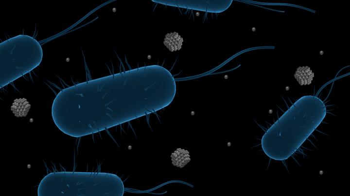
This hitch was researched by the team of scientists from the IPC PAS under the supervision of Prof. Jan Paczesny, who proposed new nanoformulations for use against widespread and challenging pathogens such as ESKAPE bacteria (Enterococcus faecium, Staphylococcus aureus, Klebsiella pneumoniae, Acinetobacter baumannii, Pseudomonas aeruginosa and Enterobacter spp.) and other problematic yeast pathogens such as Candida auris or Cryptococcus neoformans. These microorganisms, treated with commercially available antibiotics, rapidly develop antibiotic resistance. Researchers chose ESKAPE as the target group since these pathogens lead to serious diseases, from sepsis to even cancer. How? This is where the story begins.

A few months ago, Paczesny’s team decided to try combining silver nanoparticles, which are known for their antimicrobial and antifungal properties, and tea extracts rich in polyphenols additionally possessing antioxidant properties. The concept was built to enhance broad-spectrum efficacy against pathogens using green hybrid silver nanoparticles (AgNPs), which are significantly more effective than all ingredients and even more effective than certain antibiotics. Why are these hybrid particles so special? In their work, three well-known tea varieties: black tea (B-Tea), green tea (G-Tea) and Pu-erh tea (R-Tea) were used as a capping agent, which acts as a stabilizer to protect the synthesized  particles from aggregation. In this way, the particles offer a high active surface area compared to other formulations. Additionally, such synthesis is eco-friendly for the use of natural ingredients during precipitation. The structures produced vary in shape and size from 34 to 65 nm, depending on the type of tea used during synthesis, and show different reactivity towards microorganisms.

Initially, silver nanoparticles produced in the presence of tea extracts (B-TeaNPs, G-TeaNPs and R-TeaNPs) were used to treat Gram-negative (E. coli) and Gram-positive (E. faecium) bacterial strains to test the effect on strains with different cell envelope morphologies. They looked at the interactions between the manufactured nanoparticles and the pathogens to determine efficacy, comparing the results with commercially available antibiotics. The ESKAPE pathogens were then tested according to a protocol for the most effective concentration and composition of the particles, revealing up to a 25% decrease in the number of bacterial cells in E. faecium and a 90% decrease in the case of E. cloacae. Interestingly, the green silver nanoparticles also showed antifungal activity, leading to an 80% decrease in the number of viable cells of C. auris and about a 90% decrease for C. neoformans.

The first author, Sada Raza claims “What is more, the size of nanoparticles is usually related to the cytotoxic effect of nanomaterials, with smaller particles being more cytotoxic. This should favor control AgNPs and R-TeaNPs over G-TeaNPs and B-TeaNPs in our experiments. This was not the case. In most experiments, C-AgNPs and R-TeaNPs showed the lowest antimicrobial efficacy. This is in line with other studies, which demonstrated that size is not a primary factor affecting the antimicrobial activity of AgNPs.

The antibacterial and antifungal properties of silver nanoparticles made with tea extracts are greater than those of silver nanoparticles alone due to their high content of phenolic compounds, isoflavonoids (especially catechins such as epigallocatechin (EGC) and epigallocatechin gallate (EGCG)). These combinations, using biologically active tea extracts and smaller amounts of silver nanoparticles, appear to be a potential way to combat a range of infections and even replace antibiotics in some applications.

“We established that silver nanoparticles synthesized with tea extracts have higher antibacterial properties than silver nanoparticles alone. Therefore, lower dosages of TeaNPs could be used (0.1 mg mL−1). We confirmed that in some cases, the synergistic effect of tea extracts and silver nanoparticles allowed for efficacy higher than that of antibiotics (ampicillin) when tested at the same concentrations (0.1 mg mL−1) and after a relatively short exposure time of three hours.” – remarks Mateusz Wdowiak, co-author of this work.

The researchers found that the antimicrobial hybrid nanoparticles resulted in a significant reduction in bacteria compared to antibiotics or compounds separately. Although not all bacteria were killed, this is a significant improvement that could aid the treatment of superbugs using much lower doses than other commercially available compounds. The amount of hybrid silver nanoparticles needed to overcome bacteria or fungal infections is extremely low, making them cost-effective, so the key to using them well is not only functionality, but also the low cost of application.

It is an approach that can also be adapted to combat other difficult-to-treat bacterial infections. The new nanoparticles developed by researchers at the IPC PAS could bring us one step closer to effectively killing deadly drug-resistant superbugs, providing an alternative to antibiotics against Gram-negative and Gram-positive bacteria. This study also shows how much more work there is to be done in this field. Compounds used separately were much less effective than the green hybrid.

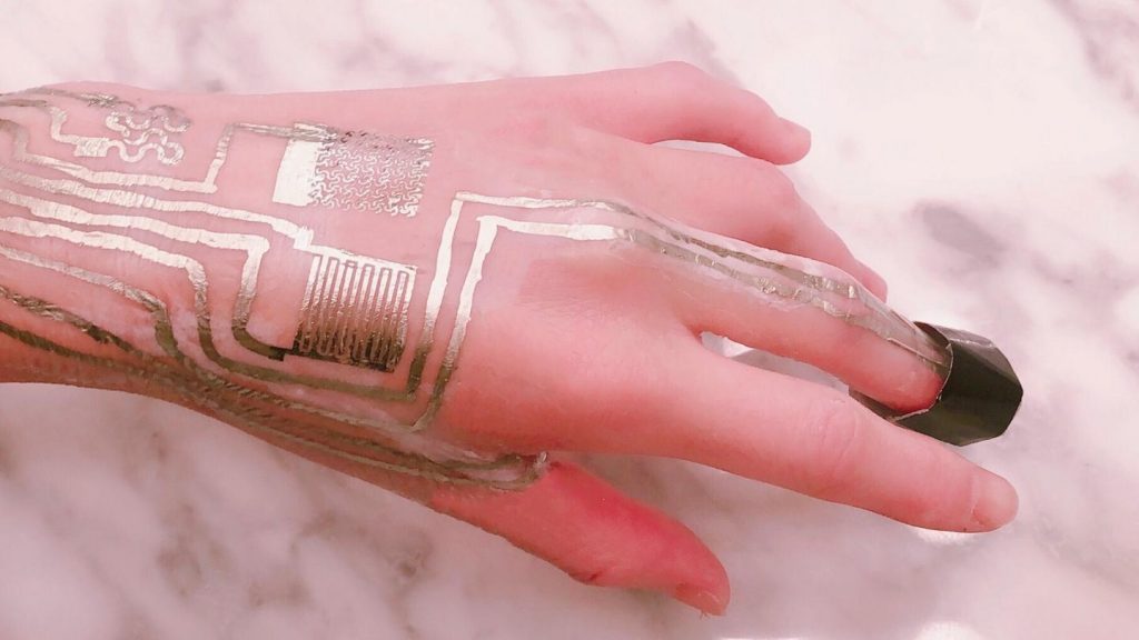
In the future, the researchers’ main goal is to use nanoparticles in everyday life, starting with agricultural applications, replacing harmful compounds used in fields to overcome infestations in plants and bring us closer to organic farming. On a larger scale, the proposed material could also be used in biomedical applications, such as an additive for wound dressings to protect against Gram-negative and Gram-positive bacteria. They hope to use nanotechnology to develop more targeted treatments for drug-resistant superbugs.

Their work was published in Nanoscale Advances journal and was financed by the National Science Centre, Poland, within the SONATA BIS grant number 2017/26/E/ST4/00041 and Foundation for Polish Science from the European Regional Development Fund within the project POIR.04.04.00-00-14D6/18-00 ‘Hybrid sensor platforms for integrated photonic systems based on ceramic and polymer materials (HYPHa)’ (TEAM-NET program).

Here’s a link to and a citation for the paper,

Enhancing the antimicrobial activity of silver nanoparticles against ESKAPE bacteria and emerging fungal pathogens by using tea extracts by Sada Raza, Mateusz Wdowiak, Mateusz Grotek, Witold Adamkiewicz, Kostiantyn Nikiforow, Pumza Mente, and Jan Paczesny. Nanoscale Adv., 2023,5, 5786-5798 DOI: https://doi.org/10.1039/D3NA00220A

This paper is licensed under a Creative Commons Attribution 3.0 Unported Licence. “You can use material from this article in other publications without requesting further permissions from the RSC [Royal Society of Chemistry], provided that the correct acknowledgement is given.” Or, consider it an open access paper.

Finally, this is not a recommendation not is it an endorsement for the ingestion of colloidal silver.

Nigeria and its nanotechnology research

Agbaje Lateef’s (Professor of Microbiology and Head of Nanotechnology Research Group (NANO+) at Ladoke Akintola University of Technology) April 20, 2022 essay on nanotechnology in Nigeria for The Conversation offers an overview and a plea, Note: Links have been removed,

Egypt, South Africa, Tunisia, Nigeria and Algeria lead the field in Africa. Since 2006, South Africa has been developing scientists, providing infrastructure, establishing centres of excellence, developing national policy and setting regulatory standards for nanotechnology. Companies such as Mintek, Nano South Africa, SabiNano and Denel Dynamics are applying the science.

In contrast, Nigeria’s nanotechnology journey, which started with a national initiative in 2006, has been slow. It has been dogged by uncertainties, poor funding and lack of proper coordination. Still, scientists in Nigeria have continued to place the country on the map through publications.

In addition, research clusters at the University of Nigeria, Nsukka, Ladoke Akintola University of Technology and others have organised conferences. Our research group also founded an open access journal, Nano Plus: Science and Technology of Nanomaterials.

To get an idea of how well Nigeria was performing in nanotechnology research and development, we turned to SCOPUS, an academic database.

Our analysis shows that research in nanotechnology takes place in 71 Nigerian institutions in collaboration with 58 countries. South Africa, Malaysia, India, the US and China are the main collaborators. Nigeria ranked fourth in research articles published from 2010 to 2020 after Egypt, South Africa and Tunisia.

Five institutions contributed 43.88% of the nation’s articles in this period. They were the University of Nigeria, Nsukka; Covenant University, Ota; Ladoke Akintola University of Technology, Ogbomoso; University of Ilorin; and University of Lagos.

The number of articles published by Nigerian researchers in the same decade was 645. Annual output grew from five articles in 2010 to 137 in the first half of 2020. South Africa published 2,597 and Egypt 5,441 from 2010 to 2020. The global total was 414,526 articles.

The figures show steady growth in Nigeria’s publications. But the performance is low in view of the fact that the country has the most universities in Africa.

The research performance is also low in relation to population and economy size. Nigeria produced 1.58 articles per 2 million people and 1.09 articles per US$3 billion of GDP in 2019. South Africa recorded 14.58 articles per 2 million people and 3.65 per US$3 billion. Egypt published 18.51 per 2 million people and 9.20 per US$3 billion in the same period.

There is no nanotechnology patent of Nigerian origin in the US patents office. Standards don’t exist for nano-based products. South Africa had 23 patents in five years, from 2016 to 2020.

Nigerian nanotechnology research is limited by a lack of sophisticated instruments for analysis. It is impossible to conduct meaningful research locally without foreign collaboration on instrumentation. The absence of national policy on nanotechnology and of dedicated funds also hinder research.

In February 2018, Nigeria’s science and technology minister unveiled a national steering committee on nanotechnology policy. But the policy is yet to be approved by the federal government. In September 2021, I presented a memorandum to the national council on science, technology and innovation to stimulate national discourse on nanotechnology.

Given that this essay is dated more than six months after Professor Lateef’s memorandum to the national council, I’m assuming that no action has been taken as of yet.

A June 2022 addition to the Nigerian nanotechnology story

Agbaje Lateef has written a June 8, 2022 essay for The Conversation about nanotechnology and the Nigerian textile industry (Note: Links have been removed),

Nigeria’s cotton production has fallen steeply in recent years. It once supported the largest textile industry in Africa. The fall is due to weak demand for cotton and to poor yields resulting from planting low-quality cottonseeds. For these reasons, farmers switched from cotton to other crops.

Nigeria’s cotton output fell from 602,400 tonnes in 2010 to 51,000 tonnes in 2020. In the 1970s and early 1980s, the country’s textile industry had 180 textile mills employing over 450,000 people, supported by about 600,000 cotton farmers. By 2019, there were 25 textile mills and 25,000 workers.

Nowadays, textiles’ properties can be greatly improved through nanotechnology – the use of extremely small materials with special properties. Nanomaterials like graphene and silver nanoparticles make textiles stronger, durable, and resistant to germs, radiation, water and fire.

Adding nanomaterials to textiles produces nanotextiles. These are often “smart” because they respond to the external environment in different ways when combined with electronics. They can be used to harvest and store energy, to release drugs, and as sensors in different applications.

Nanotextiles are increasingly used in defence and healthcare. For hospitals, they are used to produce bandages, curtains, uniforms and bedsheets with the ability to kill pathogens. The market value of nanotextiles was US$5.1 billion in 2019 and could reach US$14.8 billion in 2024.

At the moment, Nigeria is not benefiting from nanotextiles’ economic potential as it produces none. With over 216 million people, the country should be able to support its textile industry. It could also explore trading opportunities in the African Continental Free Trade Agreement to market innovative nanotextiles.

Lateef goes on to describe his research (from his June 8, 2022 essay),

Our nanotechnology research group has made the first attempt to produce nanotextiles using cotton and silk in Nigeria. We used silver and silver-titanium oxide nanoparticles produced by locust beans’ wastewater. Locust bean is a multipurpose tree legume found in Nigeria and some other parts of Africa. The seeds, the fruit pulp and the leaves are used to prepare foods and drinks.

The seeds are used to produce a local condiment called “iru” in southwest Nigeria. The processing of iru generates a large quantity of wastewater that is not useful. We used the wastewater to reduce some compounds to produce silver and silver-titanium nanoparticles in the laboratory.

Fabrics were dipped into nanoparticle solutions to make nanotextiles. Thereafter, the nanotextiles were exposed to known bacteria and fungi. The growth of the organisms was monitored to determine the ability of the nanotextiles to kill them.

The nanotextiles prevented growth of several pathogenic bacteria and black mould, making them useful as antimicrobial materials. They were active against germs even after being washed five times with detergent. Textiles without nanoparticles did not prevent the growth of microorganisms.

These studies showed that nanotextiles can kill harmful microorganisms including those that are resistant to drugs. Materials such as air filters, sportswear, nose masks, and healthcare fabrics produced from nanotextiles possess excellent antimicrobial attributes. Nanotextiles can also promote wound healing and offer resistance to radiation, water and fire.

Our studies established the value that nanotechnology can add to textiles through hygiene and disease prevention. Using nanotextiles will promote good health and well-being for sustainable development. They will assist to reduce infections that are caused by germs.

Despite these benefits, nanomaterials in textiles can have some unwanted effects on the environment, health and safety. Some nanomaterials can harm human health causing irritation when they come in contact with skin or inhaled. Also, their release to the environment in large quantities can harm lower organisms and reduce growth of plants. We recommend that the impacts of nanotextiles should be evaluated case by case before use.

Dear Professor Lateef, I hope you see some action on your suggestions soon and thank you for the update. Also, good luck with your nanotextiles.

Resisting silver’s microbial properties?

Yes, it is possible for bacteria to become resistant to silver nanoparticles. However, that yes comes with some qualifications according to a July 13, 2021 news item on ScienceDaily (Note: Links have been removed),

Antimicrobials are used to kill or slow the growth of bacteria, viruses and other microorganisms. They can be in the form of antibiotics, used to treat bodily infections, or as an additive or coating on commercial products used to keep germs at bay. These life-saving tools are essential to preventing and treating infections in humans, animals and plants, but they also pose a global threat to public health when microorganisms develop resistance to them, a concept known as antimicrobial resistance.

One of the main drivers of antimicrobial resistance is the misuse and overuse of antimicrobial agents, which includes silver nanoparticles, [emphases mine] an advanced material with well-documented antimicrobial properties. It is increasingly used in commercial products that boast enhanced germ-killing performance — it has been woven into textiles, coated onto toothbrushes, and even mixed into cosmetics as a preservative.

The Gilbertson Group at the University of Pittsburgh [Pennsylvania, US} Swanson School of Engineering used laboratory strains of E.coli to better understand bacterial resistance to silver nanoparticles and attempt to get ahead of the potential misuse of this material. The team recently published their results in Nature Nanotechnology.

Caption: A depiction of hyper-motile E.coli, a strain of bacteria found to resist silver nanoparticles’ antimicrobial properties after repeated exposure. Credit: Lisa Stabryla/University of Pittsburgh.

A July 13, 2021 University of Pittsburgh news release (also on EurekAlert), which originated the news item, provides more insight into the research,

“Bacterial resistance to silver nanoparticles is understudied, so our group looked at the mechanisms behind this event,” said Lisa Stabryla, lead author on the paper and a recent civil and environmental PhD graduate at Pitt. “This is a promising innovation to add to our arsenal of antimicrobials, but we need to consciously study it and perhaps regulate its use to avoid decreased efficacy like we’ve seen with some common antibiotics.”

Stabryla exposed E.coli to 20 consecutive days of silver nanoparticles and monitored bacterial growth over time. Nanoparticles are roughly 50 times smaller than a bacterium.

“In the beginning, bacteria could only survive at low concentrations of silver nanoparticles, but as the experiment continued, we found that they could survive at higher doses,” Stabryla noted. “Interestingly, we found that bacteria developed resistance to the silver nanoparticles but not their released silver ions alone.”

The group sequenced the genome of the E.coli that had been exposed to silver nanoparticles and found a mutation in a gene that corresponds to an efflux pump that pushes heavy metal ions out of the cell.

“It is possible that some form of silver is getting into the cell, and when it arrives, the cell mutates to quickly pump it out,” she added. “More work is needed to determine if researchers can perhaps overcome this mechanism of resistance through particle design.”

The group then studied two different types of E.coli: a hyper-motile strain that swims through its environment more quickly than normally motile bacteria and a non-motile strain that does not have physical means for moving around. They found that only the hyper-motile strain developed resistance.

“This finding could suggest that silver nanoparticles may be a good option to target certain types of bacteria, particularly non-motile strains,” Stabryla said.

In the end, bacteria will still find a way to evolve and evade antimicrobials. The hope is that an understanding of the mechanisms that lead to this evolution and a mindful use of new antimicrobials will lessen the impact of antimicrobial resistance.

“We are the first to look at bacterial motility effects on the ability to develop resistance to silver nanoparticles,” said Leanne Gilbertson, assistant professor of civil and environmental engineering at Pitt. “The observed difference is really interesting and merits further investigation to understand it and how to link the genetic response – the efflux pump regulation – to the bacteria’s ability to move in the system.

“The results are promising for being able to tune particle properties for a desired response, such as high efficacy while avoiding resistance.”

Here’s a link to and a citation for the paper,

Role of bacterial motility in differential resistance mechanisms of silver nanoparticles and silver ions by Lisa M. Stabryla, Kathryn A. Johnston, Nathan A. Diemler, Vaughn S. Cooper, Jill E. Millstone, Sarah-Jane Haig & Leanne M. Gilbertson. Nature Nanotechnology (2021) DOI: https://doi.org/10.1038/s41565-021-00929-w Published: 21 June 2021

This paper appears to be open access.

Nanomaterial shapes and forms affect passage through blood brain barrier (BBB)

I meant to get this published a lot sooner.

There seems to be a lot of excitement about this research. I got an embargoed press release further in advance than usual and now the embargo is lifted, it’s everywhere except, at the time of this writing (0920 PDT July 6, 2021), on the publisher’s (Proceedings of the National Academy of Sciences [PNAS]) website.

A July 5, 2021 news item on Medical Express announces the news,

Nanomaterials found in consumer and health-care products can pass from the bloodstream to the brain side of a blood-brain barrier model with varying ease depending on their shape—creating potential neurological impacts that could be both positive and negative, a new study reveals.

A July 5, 2021 University of Birmingham press release (also on EurekAlert), which originated the news item, delves into the details,

Scientists found that metal-based nanomaterials such as silver and zinc oxide can cross an in vitro model of the ‘blood brain barrier’ (BBB) as both particles and dissolved ions – adversely affecting the health of astrocyte cells, which control neurological responses.

But the researchers also believe that their discovery will help to design safer nanomaterials and could open up new ways of targeting hard-to-reach locations when treating brain disease.

Publishing its findings today in PNAS, an international team of researchers discovered that the physiochemical properties of metallic nanomaterials influence how effective they are at penetrating the in vitro model of the blood brain barrier and their potential levels of toxicity in the brain.

Higher concentration of certain shapes of silver nanomaterials and zinc oxide may impair cell growth and cause increased permeability of the BBB, which can lead to the BBB allowing easier brain access to these compounds.

The BBB plays a vital role in brain health by restricting the passage of various chemical substances and foreign molecules into the brain from surrounding blood vessels.

Impaired BBB integrity compromises the health of the central nervous system and increased permeability to foreign substances may eventually cause damage to the brain (neurotoxicity).

Study co-author Iseult Lynch, Professor of Environmental Nanosciences at the University of Birmingham, commented: “We found that silver and zinc oxide nanomaterials, which are widely used in various daily consumer and health-care products, passed through our in vitro BBB model, in the form of both particles and dissolved ions.

“Variation in shape, size and chemical composition can dramatically influence nanomaterials penetration through the (in vitro) blood brain barrier. This is of paramount importance for tailored medical application of nanomaterials – for example targeted delivery systems, bioimaging and assessing possible risks associated with each type of metallic nanomaterial.”

The BBB is a physical barrier composed of a tightly packed layer of endothelial cells surrounding the brain which separates the blood from the cerebrospinal fluid allowing the transfer of oxygen and essential nutrients but preventing the access of most molecules.

Recent studies found nanomaterials such as zinc oxide can accumulate on the brain side of the in vitro BBB in altered states which can affect neurological activity and brain health. Inhaled, ingested, and dermally-applied nanomaterials can reach the blood stream and a small fraction of these may cross the BBB – impacting on the central nervous system.

The researchers synthesised a library of metallic nanomaterials with different particle compositions, sizes, and shapes – evaluating their ability to penetrate the BBB using an in vitro BBB model, followed by assessment of their behaviour and fate in and beyond the model BBB.

Co-author Zhiling Guo, a Research Fellow at the University of Birmingham, commented: “”Understanding these materials’ behaviour once past the blood brain barrier is vital for evaluating the neurological effects arising from their unintentional entry into the brain. Neurotoxicity potential is greater in some materials than others, due to the different ways their shapes allow them to move and be transported.”

The research team tested varied sizes of cerium oxide and iron oxide, along with zinc oxide and four different shapes of silver – spherical (Ag NS), disc-like (Ag ND), rod-shaped (Ag NR) and nanowires (Ag NW).

Zinc oxide slipped through the in vitro BBB with the greatest ease. The researchers found spherical and disc-like silver nanomaterials underwent different dissolution regimes – gradually transforming to silver-sulfur compounds within the BBB, creating ‘easier’ entry pathways.

Zinc oxide is used as a bulking agent and a colorant. In over-the-counter drug products, it is used as a skin protectant and a sunscreen – reflecting and scattering UV radiation to help reduce or prevent sunburn and premature aging of the skin. Silver is used in cosmetic and skincare products such as anti-aging creams.

There’s still a long way to go with this research. For anyone who’s unfamiliar with the term ‘in vitro’, the rough translation is ‘in glass’ meaning test tubes, petri dishes, etc. are used. Even though the research paper has been peer-reviewed (not a perfect process), once it becomes available there will be added scrutiny from scientists with regard to how the research was conducted and whether or not the conclusions drawn are reasonable. One more question should also be asked, are the results reproducible by other scientists?

Here’s a link to and a citation for the paper,

Biotransformation modulates the penetration of metallic nanomaterials across an artificial blood–brain barrier model by Zhiling Guo, Peng Zhang, Swaroop Chakraborty, Andrew J Chetwynd, Fazel Abdolahpur Monikh, Christopher Stark, Hanene Ali-Boucetta, Sandra Wilson, Iseult Lynch, and Eugenia Valsami-Jones. PNAS 118 (28) e2105245118 DOI: https://doi.org/10.1073/pnas.2105245118 Published: July 13, 2021

This paper appears to be open access.

Printing wearable circuits onto skin

It seems that this new technique for creating wearable electronics will be more like getting a permanent tattoo where the circuits are applied directly to your skin as opposed to being like a temporary tattoo where the circuits are printed onto a substrate and then applied to then, worn on your skin.

Caption: On-body sensors, such as electrodes and temperature sensors, were directly printed and sintered on the skin surface. Credit: Adapted from ACS Applied Materials & Interfaces 2020, DOI: 10.1021/acsami.0c11479

An Oct. 14, 2020 American Chemical Society (ACS) news release (also on EurekAlert) announced this latest development in wearable electronics,

Wearable electronics are getting smaller, more comfortable and increasingly capable of interfacing with the human body. To achieve a truly seamless integration, electronics could someday be printed directly on people’s skin. As a step toward this goal, researchers reporting in ACS Applied Materials & Interfaces have safely placed wearable circuits directly onto the surface of human skin to monitor health indicators, such as temperature, blood oxygen, heart rate and blood pressure.

The latest generation of wearable electronics for health monitoring combines soft on-body sensors with flexible printed circuit boards (FPCBs) for signal readout and wireless transmission to health care workers. However, before the sensor is attached to the body, it must be printed or lithographed onto a carrier material, which can involve sophisticated fabrication approaches. To simplify the process and improve the performance of the devices, Peng He, Weiwei Zhao, Huanyu Cheng and colleagues wanted to develop a room-temperature method to sinter metal nanoparticles onto paper or fabric for FPCBs and directly onto human skin for on-body sensors. Sintering — the process of fusing metal or other particles together — usually requires heat, which wouldn’t be suitable for attaching circuits directly to skin.

The researchers designed an electronic health monitoring system that consisted of sensor circuits printed directly on the back of a human hand, as well as a paper-based FPCB attached to the inside of a shirt sleeve. To make the FPCB part of the system, the researchers coated a piece of paper with a novel sintering aid and used an inkjet printer with silver nanoparticle ink to print circuits onto the coating. As solvent evaporated from the ink, the silver nanoparticles sintered at room temperature to form circuits. A commercially available chip was added to wirelessly transmit the data, and the resulting FPCB was attached to a volunteer’s sleeve. The team used the same process to sinter circuits on the volunteer’s hand, except printing was done with a polymer stamp. As a proof of concept, the researchers made a full electronic health monitoring system that sensed temperature, humidity, blood oxygen, heart rate, blood pressure and electrophysiological signals and analyzed its performance. The signals obtained by these sensors were comparable to or better than those measured by conventional commercial devices. 

Here’s a link to and a citation for the paper,

Wearable Circuits Sintered at Room Temperature Directly on the Skin Surface for Health Monitoring by Ling Zhang, Hongjun Ji, Houbing Huang, Ning Yi, Xiaoming Shi, Senpei Xie, Yaoyin Li, Ziheng Ye, Pengdong Feng, Tiesong Lin, Xiangli Liu, Xuesong Leng, Mingyu Li, Jiaheng Zhang, Xing Ma, Peng He, Weiwei Zhao, and Huanyu Cheng. ACS Appl. Mater. Interfaces 2020, 12, 40, 45504–45515 Publication Date:September 11, 2020 DOI: https://doi.org/10.1021/acsami.0c11479 Copyright © 2020 American Chemical Society

This paper is behind a paywall.

Norwegian Institute for Water Research (NIVA) releases study on silver and titanium nanomaterials in wastewater

It turns out that silver and titanium nanomaterials (e.g. silver nanoparticles washed out of athletic clothing) in wastewater may have ‘negative’ and ‘positive’ effects on freshwater and marine life depending on the species.

A November 18, 2019 news item on Nanowerk provides an introduction to the research (Note: Links have been removed),

You may not always think about it when you do your laundry or flush the toilet; but whatever you eat, wear or apply on your skin ends up in wastewater and eventually reaches the environment. The use of nanoparticles in consumer products like textiles, foods and personal care products is increasing.

What is so special about nanoparticles, is their tiny size: One nanometer is one billionth of a meter. The small size gives nanoparticles unique and novel properties compared to their bigger counterparts and may for example reach locations that bigger particles cannot reach.
Further, pristine nanoparticles behave differently from nanoparticles in the environment. In the environment, nanoparticles are transformed by interacting and forming aggregates with other particles, elements or solids, and thereby obtain other physicochemical properties.

The transformation of these tiny particles in wastewater treatment processes and their effect on freshwater and marine organisms, have largely been unknown.
Increased mortality of marine crustaceans.

In a study (“Ecotoxicological Effects of Transformed Silver and Titanium Dioxide Nanoparticles in the Effluent from a Lab-Scale Wastewater Treatment System”) conducted at the Norwegian Institute for Water Research (NIVA), Anastasia Georgantzopoulou and colleagues from NIVA and SINTEF investigated how silver and titanium dioxide nanoparticles behave in wastewater treatment plants, and how marine and freshwater organisms are affected by them.

Exposure to treated wastewater did not have any adverse effects on the freshwater crustacean Daphnia magna. (Photo: NIVA)

A November 18, 2019 NIVA press release, which originated the news item, fills in the details,

The researchers made a laboratory-scale wastewater treatment plant, using sludge from a wastewater treatment plant in Norway. They added environmentally relevant concentrations of silver (Ag) and titanium dioxide (TiO2) nanoparticles over a 5-week period and used the treated wastewater to assess the effects of transformed nanoparticles on freshwater and marine organisms, as well as on gill cells from rainbow trout.

The experiment demonstrated contrasting effects on the two crustacean species. For the marine copepod (Tisbe battagliai), mortality increased by 20-45%, whereas exposure to ttreated wastewater did not have any adverse effects on the freshwater crustacean (Daphnia magna).

“These differences are probably due, at least partly, to the two species’ different feeding habits, in combination with the fact that the nanoparticles showed a strong association to solids present in the wastewater”, Georgantzopoulou says, and explains:

“Daphnia magna is an organism that filters water for food, whereas the marine copepod feeds on bottom surfaces – like effluent solids that have settled out from the water. The bottom feeding crustacean is therefore more likely to ingest nanoparticles, and thereby be affected by solid-associated nanoparticles”. 

Effects on algal species

Nanoparticle-containing treated wastewater also affected algal growth, but the two algae species did not have a common response: The marine algae (Skeletonema pseudocostatum) responded with a 20-40 % growth inhibition, while the algal growth of the freshwater algae (Raphidocelis subcapitata) was actually stimulated, by a 40 % increase, accompanied by increased cell aggregation. The latter is probably some kind of a defense mechanism, aiming to decrease the surface area exposed to toxic particles.

“The results from our study indicate that algal responses to the treated wastewater exposure are species-dependent. This is possibly due to differences in algal cell size, surface area, and cell wall composition”, the NIVA researcher explains.

Increased permeability of fish gill cells

The researchers also found effects of silver and titanium nanoparticles on fish gill cells using an in vitro gill cell line model. As large amounts of water are passing through the gills, and they constitute a barrier to the external environment, this organ is highly exposed to water-borne contaminants, including nanoparticles.

“Exposure to nanoparticle-containing wastewater lead to an increase in reactive oxygen species, a group of molecules that can easily react with and damage cells. This was followed by increased permeability of the gill cells, leading to a compromised barrier function”, Georgantzopoulou says.

“However, the concentrations of silver and titanium nanoparticles in the treated wastewater were too low to fully account for the effects on cell permeability alone. The wastewater effluent is a complex mixture of materials, and the permeability response is probably caused by a combination of the presence of nanoparticles and other stressors”, Georgantzopoulou adds.

Wastewater treatment-transformation of nanoparticles

“We carried out this study on wastewater treatment plant-transformed nanoparticles, and compared them to pristine nanoparticles, as the former is more relevant to what is actually happening in the environment. The increased toxicity of the transformed nanomaterials observed in the study indicates that the effects cannot be predicted by assessing the effects of nanomaterials in their pristine form and highlights the importance of understanding their behavior, environmental transformation and interaction with organisms. Future studies should take nanoparticle transformation into account and focus on a more relevant experimental exposure conditions incorporating transformed nanoparticles in more long-term impact studies to provide a better understanding of their potential impacts”, Georgantzopoulou concludes.

Here’s a link to and a citation for the paper,

Ecotoxicological Effects of Transformed Silver and Titanium Dioxide Nanoparticles in the Effluent from a Lab-Scale Wastewater Treatment System by Anastasia Georgantzopoulou, Patricia Almeida Carvalho, Christian Vogelsang, Mengstab Tilahun, Kuria Ndungu, Andy M. Booth, Kevin V. Thomas, Ailbhe Macken. Environ. Sci. Technol. 2018, 52, 16, 9431-9441 DOI: https://doi.org/10.1021/acs.est.8b01663 Publication Date:July 26, 2018 Copyright © 2018 American Chemical Society

This paper is behind a paywall.

Nanocellulose sensors: 3D printed and biocompatible

I do like to keep up with nanocellulose doings, especially when there’s some Canadian involvement, and an October 8, 2019 news item on Nanowerk alerted me to a newish application for the product,

Physiological parameters in our blood can be determined without painful punctures. Empa researchers are currently working with a Canadian team to develop flexible, biocompatible nanocellulose sensors that can be attached to the skin. The 3D-printed analytic chips made of renewable raw materials will even be biodegradable in future.

The idea of measuring parameters that are relevant for our health via the skin has already taken hold in medical diagnostics. Diabetics, for example, can painlessly determine their blood sugar level with a sensor instead of having to prick their fingers.

An October 8, 2019 Empa (Swiss Federal Laboratories for Materials Science and Technology) press release, which originated the news item, provides more detail,

A transparent foil made of wood

Nanocellulose is an inexpensive, renewable raw material, which can be obtained in form of crystals and fibers, for example from wood. However, the original appearance of a tree no longer has anything to do with the gelatinous substance, which can consist of cellulose nanocrystals and cellulose nanofibers. Other sources of the material are bacteria, algae or residues from agricultural production. Thus, nanocellulose is not only relatively easy and sustainable to obtain. Its mechanical properties also make the “super pudding” an interesting product. For instance, new composite materials based on nanocellulose can be developed that could be used as surface coatings, transparent packaging films or even to produce everyday objects like beverage bottles.

Researchers at Empa’s Cellulose & Wood Materials lab and Woo Soo Kim from the Simon Fraser University [SFU] in Burnaby, Canada, are also focusing on another feature of nanocellulose: biocompatibility. Since the material is obtained from natural resources, it is particularly suitable for biomedical research.

With the aim of producing biocompatible sensors that can measure important metabolic values, the researchers used nanocellulose as an “ink” in 3D printing processes. To make the sensors electrically conductive, the ink was mixed with silver nanowires. The researchers determined the exact ratio of nanocellulose and silver threads so that a three-dimensional network could form.

Just like spaghetti – only a wee bit smaller

It turned out that cellulose nanofibers are better suited than cellulose nanocrystals to produce a cross-linked matrix with the tiny silver wires. “Cellulose nanofibers are flexible similar to cooked spaghetti, but with a diameter of only about 20 nanometers and a length of just a few micrometers,” explains Empa researcher Gilberto Siqueira.

The team finally succeeded in developing sensors that measure medically relevant metabolic parameters such as the concentration of calcium, potassium and ammonium ions. The electrochemical skin sensor sends its results wirelessly to a computer for further data processing. The tiny biochemistry lab on the skin is only half a millimeter thin.

While the tiny biochemistry lab on the skin – which is only half a millimeter thin – is capable of determining ion concentrations specifically and reliably, the researchers are already working on an updated version. “In the future, we want to replace the silver [nano] particles with another conductive material, for example on the basis of carbon compounds,” Siqueira explains. This would make the medical nanocellulose sensor not only biocompatible, but also completely biodegradable.

I like the images from Empa better than the ones from SFU,

Using a 3D printer, the nanocellulose “ink” is applied to a carrier plate. Silver particles provide the electrical conductivity of the material. Image: Empa
Empa researcher Gilberto Siqueira demonstrates the newly printed nanocellulose circuit. After a subsequent drying, the material can be further processed. Image: Empa

SFU produced a news release about this work back in February 2019. Again, I prefer what the Swiss have done because they’re explaining/communicating the science, as well as , communicating benefits. From a February 13, 2019 SFU news release (Note: Links have been removed),

Simon Fraser University and Swiss researchers are developing an eco-friendly, 3D printable solution for producing wireless Internet-of-Things (IoT) sensors that can be used and disposed of without contaminating the environment. Their research has been published as the cover story in the February issue of the journal Advanced Electronic Materials.

SFU professor Woo Soo Kim is leading the research team’s discovery, which uses a wood-derived cellulose material to replace the plastics and polymeric materials currently used in electronics.

Additionally, 3D printing can give flexibility to add or embed functions onto 3D shapes or textiles, creating greater functionality.

“Our eco-friendly, 3D-printed cellulose sensors can wirelessly transmit data during their life, and then can be disposed without concern of environmental contamination,” says Kim, a professor in the School of Mechatronic Systems Engineering. The SFU research is being carried out at PowerTech Labs in Surrey, which houses several state-of-the-art 3D printers used to advance the research.

“This development will help to advance green electronics. For example, the waste from printed circuit boards is a hazardous source of contamination to the environment. If we are able to change the plastics in PCB to cellulose composite materials, recycling of metal components on the board could be collected in a much easier way.”

Kim’s research program spans two international collaborative projects, including the latest focusing on the eco-friendly cellulose material-based chemical sensors with collaborators from the Swiss Federal Laboratories for Materials Science.

He is also collaborating with a team of South Korean researchers from the Daegu Gyeongbuk Institute of Science and Technology’s (DGIST)’s department of Robotics Engineering, and PROTEM Co Inc, a technology-based company, for the development of printable conductive ink materials.

In this second project, researchers have developed a new breakthrough in the embossing process technology, one that can freely imprint fine circuit patterns on flexible polymer substrate, a necessary component of electronic products.

Embossing technology is applied for the mass imprinting of precise patterns at a low unit cost. However, Kim says it can only imprint circuit patterns that are imprinted beforehand on the pattern stamp, and the entire, costly stamp must be changed to put in different patterns.

The team succeeded in developing a precise location control system that can imprint patterns directly resulting in a new process technology. The result will have widespread implications for use in semiconductor processes, wearable devices and the display industry.

This paper was made available online back in December 2018 and then published in print in February 2019. As to why there’d be such large gaps between the paper’s publication dates and the two institution’s news/press releases, it’s a mystery to me. In any event, here’s a link to and a citation for the paper,

3D Printed Disposable Wireless Ion Sensors with Biocompatible Cellulose Composites by Taeil Kim, Chao Bao, Michael Hausmann, Gilberto Siqueira, Tanja Zimmermann, Woo Soo Kim. Advanced Electronic Materials DOI: https://doi.org/10.1002/aelm.201970007 First published online December 19, 2018. First published in print: 08 February 2019 (Adv. Electron. Mater. 2/2109) Volume 5, Issue 2 February 2019 1970007

This paper is behind a paywall.

Nanoparticles in combination could be more toxic

It seems that one set of nanoparticles, e.g., silver nanoparticles, in combination with another material, e.g., cadmium ions, are more dangerous than either one separately according to an August 17, 2018 University of Southern Denmark press release by Birgitte Svennevig (also on EurekAlert but dated August 20, 2018),

Researchers warn that a combination of nanoparticles and contaminants may form a cocktail that is harmful to our cells. In their study, 72 pct. of cells died after exposure to a cocktail of nano-silver and cadmium ions.

Nanoparticles are becoming increasingly widespread in our environment. Thousands of products contain nanoparticles because of their unique properties. Silver nanoparticles are one example: They have an effective antibacterial effect and can be found in refrigerators, sports clothes, cosmetics, tooth brushes, water filters, etc.

There is a significant difference between how the cells react when exposed to nanosilver alone and when they are exposed to a cocktail of nanosilver and cadmium ions. Cadmium ions are naturally found everywhere around us on Earth.

In the study, 72 pct. of the cells died, when exposed to both nanosilver and cadmiun ions. When exposed to nanosilver only, 25 pct. died. When exposed to cadmium ions only, 12 pct. died.

The study was conducted on human liver cancer cells.

  • This study indicates, that we should not look at nanoparticles isolated when we investigate and discuss the effects, they may have on our health. We need to take cocktail effects into account, said Professor Frank Kjeldsen, Dept of Biochemistry and Molecular Biology, SDU, adding:
  • Products with nano particles are being developed and manufactured every day, but in most countries there are no regulations, so there is no way of knowing what and how many nanoparticles are being released into the environment. In my opinion, this should be stopped.

Other studies, led by Professor Kjeldsen have previously shown that human cells interact with metal nanoparticles.

One study showed that nano-silver leads to the formation free radicals in cells and changes in the form and amount of proteins. Many serious diseases are characterized by an overproduction of free radicals in cells. This applies to cancer and neurological diseases such as Alzheimer’s and Parkinson’s.

This is not great news but there are a few things to note about this research. First, it was conducted on cells and therefore not subject to some of the defensive systems found in complete biological organisms such as a mouse or a dandelion plant for example.

Also, since they were cancer cells one might suspect their reactions might differ from those of healthy cells. As for how the cells were exposed to the contaminants, I think (???) they were sitting in a solution of contaminants and most of us do not live in that kind of environment.. Finally, with regard to the concentrations, I have no idea if they are greater than one might expect to encounter in one’s lifecycle but it’s always worth questioning just how much exposure you might expect during yours or a mouse’s or a dandelion’s life.

These caveats aside, Professor Frank Kjeldsen’s work raises some very concerning issues and his work adds to a growing body of evidence.

Here’s a video featuring Dr. Kjeldsen talking about his work,

Here’s a link to and a citation for the paper,

Co-exposure to silver nanoparticles and cadmium induce metabolic adaptation in HepG2 cells by Renata Rank Miranda, Vladimir Gorshkov, Barbara Korzeniowska, Stefan J. Kempf, Francisco Filipak Neto, & Frank Kjeldsen. Nanotoxicology DOI: https://doi.org/10.1080/17435390.2018.1489987 Published online: 11 Jul 2018

This paper is open access.

Observing individual silver nanoparticles in real time

A new technique for better understanding how silver nanoparticles might affect the environment was announced in a July 30, 2018 news item on ScienceDaily,

Chemists at Ruhr-Universität Bochum have developed a new method of observing the chemical reactions of individual silver nanoparticles, which only measure a thousandth of the thickness of a human hair, in real time. The particles are used in medicine, food and sports items because they have an antibacterial and anti-inflammatory effect. However, how they react and degrade in ecological and biological systems is so far barely understood. The team in the Research Group for Electrochemistry and Nanoscale Materials showed that the nanoparticles transform into poorly soluble silver chloride particles under certain conditions. The group led by Prof Dr Kristina Tschulik reports on the results in the Journal of the American Chemical Society from July 11, 2018.

A July 30,2018 Ruhr-University Bochum (RUB) press release (also on EurekAlert) by Julia Weiler, which originated the news item, provides more information,

Even under well-defined laboratory conditions, current research has yielded different, sometimes contradictory, results on the reaction of silver nanoparticles. “In every batch of nanoparticles, the individual properties of the particles, such as size and shape, vary,” says Kristina Tschulik, a member of the Cluster of Excellence Ruhr Explores Solvation. “With previous procedures, a myriad of particles was generally investigated at the same time, meaning that the effects of these variations could not be recorded. Or the measurements took place in a high vacuum, not under natural conditions in an aqueous solution.”

The team led by Kristina Tschulik thus developed a method that enables individual silver particles to be investigated in a natural environment. “Our aim is to be able to record the reactivity of individual particles,” explains the researcher. This requires a combination of electrochemical and spectroscopic methods. With optical and hyperspectral dark-field microscopy, the group was able to observe individual nanoparticles as visible and coloured pixels. Using the change in the colour of the pixels, or more precisely their spectral information, the researchers were able to follow what was happening in an electrochemical experiment in real time.

Degradation of the particles slowed down

In the experiment, the team replicated the oxidation of silver in the presence of chloride ions, which often takes place in ecological and biological systems. “Until now, it was generally assumed that the silver particles dissolve in the form of silver ions,” describes Kristina Tschulik. However, poorly soluble silver chloride was formed in the experiment – even if only a few chloride ions were present in the solution.

“This extends the lifespan of the nanoparticles to an extreme extent and their breakdown is slowed down in an unexpectedly drastic manner,” summarises Tschulik. “This is equally important for bodies of water and for living beings because this mechanism could cause the heavy metal silver to accumulate locally, which can be toxic for many organisms.”

Further development planned

The Bochum-based group now wants to further improve its technology for analysing individual nanoparticles in order to better understand the ageing mechanisms of such particles. The researchers thus want to obtain more information about the biocompatibility of the silver particles and the lifespan and ageing of catalytically active nanoparticles in the future.

Here’s a link to and a citation for the paper,

Simultaneous Opto- and Spectro-Electrochemistry: Reactions of Individual Nanoparticles Uncovered by Dark-Field Microscopy by Kevin Wonner, Mathies V. Evers, and Kristina Tschulik. J. Am. Chem. Soc., Article ASAP DOI: 10.1021/jacs.8b02367 Publication Date (Web): July 11, 2018

Copyright © 2018 American Chemical Society

This paper is behind a paywall.

Algae outbreaks (dead zones) in wetlands and waterways

It’s been over seven years since I first started writing about Duke University’s  Center for the Environmental Implications of Nanotechnology and mesocosms (miniature ecosystems) and the impact that nanoparticles may have on plants and water (see August 11, 2011 posting). Since then, their focus has shifted from silver nanoparticles and their impact on plants, fish, bacteria, etc. to a more general examination of metallic nanoparticles and water. A June 25, 2018 news item on ScienceDaily announces some of their latest work,

The last 10 years have seen a surge in the use of tiny substances called nanomaterials in agrochemicals like pesticides and fungicides. The idea is to provide more disease protection and better yields for crops, while decreasing the amount of toxins sprayed on agricultural fields.

But when combined with nutrient runoff from fertilized cropland and manure-filled pastures, these “nanopesticides” could also mean more toxic algae outbreaks for nearby streams, lakes and wetlands, a new study finds.

A June 25, 2018 Duke University news release (also on EurekAlert) by Robin A. Smith, which originated the news item, provides more detail,

Too small to see with all but the most powerful microscopes, engineered nanomaterials are substances manufactured to be less than 100 nanometers in diameter, many times smaller than a hair’s breadth.

Their nano-scale gives them different chemical and physical properties from their bulk counterparts, including more surface area for reactions and interactions.

Those interactions could intensify harmful algal blooms in wetlands, according to experiments led by Marie Simonin, a postdoctoral associate with biology professor Emily Bernhardt at Duke University.

Carbon nanotubes and teeny tiny particles of silver, titanium dioxide and other metals are already added to hundreds of commercial products to make everything from faster, lighter electronics, self-cleaning fabrics, and smarter food packaging that can monitor food for spoilage. They are also used on farms for slow- or controlled-release plant fertilizers and pesticides and more targeted delivery, and because they are effective at lower doses than conventional products.

These and other applications have generated tremendous interest and investment in nanomaterials. However the potential risks to human health or the environment aren’t fully understood, Simonin said.

Most of the 260,000 to 309,000 metric tons of nanomaterials produced worldwide each year are eventually disposed in landfills, according to a previous study. But of the remainder, up to 80,400 metric tons per year are released into soils, and up to 29,200 metric tons end up in natural bodies of water.

“And these emerging contaminants don’t end up in water bodies alone,” Simonin said. “They probably co-occur with nutrient runoff. There are likely multiple stressors interacting.”

Algae outbreaks already plague polluted waters worldwide, said Steven Anderson, a research analyst in the Bernhardt Lab at Duke and one of the authors of the research.

Nitrogen and phosphorous pollution makes its way into wetlands and waterways in the form of agricultural runoff and untreated wastewater. The excessive nutrients cause algae to grow out of control, creating a thick mat of green scum or slime on the surface of the water that blocks sunlight from reaching other plants.

These nutrient-fueled “blooms” eventually reduce oxygen levels to the point where fish and other organisms can’t survive, creating dead zones in the water. Some algal blooms also release toxins that can make pets and people who swallow them sick.

To find out how the combined effects of nutrient runoff and nanoparticle contamination would affect this process, called eutrophication, the researchers set up 18 separate 250-liter tanks with sandy sloped bottoms to mimic small wetlands.

Each open-air tank was filled with water, soil and a variety of wetland plants and animals such as waterweed and mosquitofish.

Over the course of the nine-month experiment, some tanks got a weekly dose of algae-promoting nitrates and phosphates like those found in fertilizers, some tanks got nanoparticles — either copper or gold — and some tanks got both.

Along the way the researchers monitored water chemistry, plant and algae growth and metabolism, and nanoparticle accumulation in plant tissues.

“The results were surprising,” Simonin said. The nanoparticles had tiny effects individually, but when added together with nutrients, even low concentrations of gold and copper nanoparticles used in fungicides and other products turned the once-clear water a murky pea soup color, its surface covered with bright green smelly mats of floating algae.

Over the course of the experiment, big algal blooms were more than three times more frequent and more persistent in tanks where nanoparticles and nutrients were added together than where nutrients were added alone. The algae overgrowths also reduced dissolved oxygen in the water.

It’s not clear yet how nanoparticle exposure shifts the delicate balance between plants and algae as they compete for nutrients and other resources. But the results suggest that nanoparticles and other “metal-based synthetic chemicals may be playing an under-appreciated role in the global trends of increasing eutrophication,” the researchers said.

Here’s a link to and a citation for the paper,

Engineered nanoparticles interact with nutrients to intensify eutrophication in a wetland ecosystem experiment by Marie Simonin, Benjamin P. Colman, Steven M. Anderson, Ryan S. King, Matthew T. Ruis, Astrid Avellan, Christina M. Bergemann, Brittany G. Perrotta, Nicholas K. Geitner, Mengchi Ho, Belen de la Barrera, Jason M. Unrine, Gregory V. Lowry, Curtis J. Richardson, Mark R. Wiesner, Emily S. Bernhardt. Ecological Applications, 2018; DOI: 10.1002/eap.1742 First published: 25 June 2018

This paper is behind a paywall.