Tag Archives: contrast agents

New iron oxide nanoparticle as an MRI (magnetic resonance imaging) contrast agent

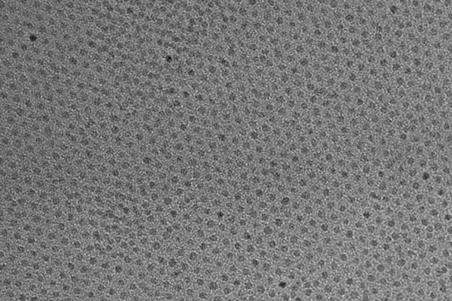
This high-resolution transmission electron micrograph of particles made by the research team shows the particles’ highly uniform size and shape. These are iron oxide particles just 3 nanometers across, coated with a zwitterion layer. Their small size means they can easily be cleared through the kidneys after injection. Courtesy of the researchers

A Feb. 14, 2017 news item on ScienceDaily announces a new MRI (magnetic resonance imaging) contrast agent,

A new, specially coated iron oxide nanoparticle developed by a team at MIT [Massachusetts Institute of Technology] and elsewhere could provide an alternative to conventional gadolinium-based contrast agents used for magnetic resonance imaging (MRI) procedures. In rare cases, the currently used gadolinium agents have been found to produce adverse effects in patients with impaired kidney function.

A Feb. 14, 2017 MIT news release (also on EurekAlert), which originated the news item, provides more technical detail,


The advent of MRI technology, which is used to observe details of specific organs or blood vessels, has been an enormous boon to medical diagnostics over the last few decades. About a third of the 60 million MRI procedures done annually worldwide use contrast-enhancing agents, mostly containing the element gadolinium. While these contrast agents have mostly proven safe over many years of use, some rare but significant side effects have shown up in a very small subset of patients. There may soon be a safer substitute thanks to this new research.

In place of gadolinium-based contrast agents, the researchers have found that they can produce similar MRI contrast with tiny nanoparticles of iron oxide that have been treated with a zwitterion coating. (Zwitterions are molecules that have areas of both positive and negative electrical charges, which cancel out to make them neutral overall.) The findings are being published this week in the Proceedings of the National Academy of Sciences, in a paper by Moungi Bawendi, the Lester Wolfe Professor of Chemistry at MIT; He Wei, an MIT postdoc; Oliver Bruns, an MIT research scientist; Michael Kaul at the University Medical Center Hamburg-Eppendorf in Germany; and 15 others.

Contrast agents, injected into the patient during an MRI procedure and designed to be quickly cleared from the body by the kidneys afterwards, are needed to make fine details of organ structures, blood vessels, and other specific tissues clearly visible in the images. Some agents produce dark areas in the resulting image, while others produce light areas. The primary agents for producing light areas contain gadolinium.

Iron oxide particles have been largely used as negative (dark) contrast agents, but radiologists vastly prefer positive (light) contrast agents such as gadolinium-based agents, as negative contrast can sometimes be difficult to distinguish from certain imaging artifacts and internal bleeding. But while the gadolinium-based agents have become the standard, evidence shows that in some very rare cases they can lead to an untreatable condition called nephrogenic systemic fibrosis, which can be fatal. In addition, evidence now shows that the gadolinium can build up in the brain, and although no effects of this buildup have yet been demonstrated, the FDA is investigating it for potential harm.

“Over the last decade, more and more side effects have come to light” from the gadolinium agents, Bruns says, so that led the research team to search for alternatives. “None of these issues exist for iron oxide,” at least none that have yet been detected, he says.

The key new finding by this team was to combine two existing techniques: making very tiny particles of iron oxide, and attaching certain molecules (called surface ligands) to the outsides of these particles to optimize their characteristics. The iron oxide inorganic core is small enough to produce a pronounced positive contrast in MRI, and the zwitterionic surface ligand, which was recently developed by Wei and coworkers in the Bawendi research group, makes the iron oxide particles water-soluble, compact, and biocompatible.

The combination of a very tiny iron oxide core and an ultrathin ligand shell leads to a total hydrodynamic diameter of 4.7 nanometers, below the 5.5-nanometer renal clearance threshold. This means that the coated iron oxide should quickly clear through the kidneys and not accumulate. This renal clearance property is an important feature where the particles perform comparably to gadolinium-based contrast agents.

Now that initial tests have demonstrated the particles’ effectiveness as contrast agents, Wei and Bruns say the next step will be to do further toxicology testing to show the particles’ safety, and to continue to improve the characteristics of the material. “It’s not perfect. We have more work to do,” Bruns says. But because iron oxide has been used for so long and in so many ways, even as an iron supplement, any negative effects could likely be treated by well-established protocols, the researchers say. If all goes well, the team is considering setting up a startup company to bring the material to production.

For some patients who are currently excluded from getting MRIs because of potential side effects of gadolinium, the new agents “could allow those patients to be eligible again” for the procedure, Bruns says. And, if it does turn out that the accumulation of gadolinium in the brain has negative effects, an overall phase-out of gadolinium for such uses could be needed. “If that turned out to be the case, this could potentially be a complete replacement,” he says.

Ralph Weissleder, a physician at Massachusetts General Hospital who was not involved in this work, says, “The work is of high interest, given the limitations of gadolinium-based contrast agents, which typically have short vascular half-lives and may be contraindicated in renally compromised patients.”

The research team included researchers in MIT’s chemistry, biological engineering, nuclear science and engineering, brain and cognitive sciences, and materials science and engineering departments and its program in Health Sciences and Technology; and at the University Medical Center Hamburg-Eppendorf; Brown University; and the Massachusetts General Hospital. It was supported by the MIT-Harvard NIH Center for Cancer Nanotechnology, the Army Research Office through MIT’s Institute for Soldier Nanotechnologies, the NIH-funded Laser Biomedical Research Center, the MIT Deshpande Center, and the European Union Seventh Framework Program.

Here’s a link to and a citation for the paper,

Exceedingly small iron oxide nanoparticles as positive MRI contrast agents by He Wei, Oliver T. Bruns, Michael G. Kaul, Eric C. Hansen, Mariya Barch, Agata Wiśniowsk, Ou Chen, Yue Chen, Nan Li, Satoshi Okada, Jose M. Cordero, Markus Heine, Christian T. Farrar, Daniel M. Montana, Gerhard Adam, Harald Ittrich, Alan Jasanoff, Peter Nielsen, and Moungi G. Bawendi. PNAS February 13, 2017 doi: 10.1073/pnas.1620145114 Published online before print February 13, 2017

This paper is behind a paywall.

Better contrast agents for magnetic resonance imaging with nanoparticles

I wonder what’s going on in the field of magnetic resonance imaging. This is the third news item I’ve stumbled across related to the topic in the last couple of months. (Links to the other two posts follow at the end of this post.) By comparison, that’s the more than in the previous seven years (2008 – 2015) combined.

The latest research concerns a new and better contrast agent. From an Aug. 3, 2016 news item on Nanowerk,

Scientists at the University of Basel [Switzerland] have developed nanoparticles which can serve as efficient contrast agents for magnetic resonance imaging. This new type of nanoparticles [sic] produce around ten times more contrast than the actual contrast agents and are responsive to specific environments.

An Aug. 3, 2016 University of Basel press release (also on EurekAlert), which originated the news item, explains further,

Contrast agents are usually based on the metal Gadolinium, which is injected and serves for an improved imaging of various organs in an MRI. Gadolinium ions should be bound with a carrier compound to avoid the toxicity to the human body of the free ions. Therefore, highly efficient contrast agents requiring lower Gadolinium concentrations represent an important step for advancing diagnosis and improving patient health prognosis.

Smart nanoparticles as contrast agents

The research groups of Prof. Cornelia Palivan and Prof. Wolfgang Meier from the Department of Chemistry at the University of Basel have introduced a new type of nanoparticles [sic], which combine multiple properties required for contrast agents: an increased MRI contrast for lower concentration, a potential for long blood circulation and responsiveness to different biochemical environments. These nanoparticles were obtained by co-assembly of heparin-functionalized polymers with trapped gadolinium ions and stimuli-responsive peptides.

The study shows, that the nanoparticles have the capacity of enhancing the MRI signal tenfold higher than the current agents. In addition, they have an enhanced efficacy in reductive milieu, characteristic for specific regions, such as cancerous tissues. These nanoparticles fulfill numerous key criteria for further development, such as absence of cellular toxicity, no apparent anticoagulation property, and high shelf stability. The concept developed by the researchers at the University of Basel to produce better contrast agents based on nanoparticles highlights a new direction in the design of MRI contrast agents, and supports their implementation for future applications.

Here’s a link to and a citation for the paper,

Nanoparticle-based highly sensitive MRI contrast agents with enhanced relaxivity in reductive milieu by
Severin J. Sigg, Francesco Santini, Adrian Najer, Pascal U. Richard, Wolfgang P. Meier, and Cornelia G. Palivan. Chem. Commun., 2016,52, 9937-9940 DOI: 10.1039/C6CC03396B First published online 13 Jul 2016

This paper is behind a paywall.

The other two MRI items featured here are in a June 10, 2016 posting (pH dependent nanoparticle-based contrast agent for MRIs [magnetic resonance images]) and in an Aug. 1, 2016 posting (Nuclear magnetic resonance microscope breaks records).

pH dependent nanoparticle-based contrast agent for MRIs (magnetic resonance images)

This news about a safer and more effective contrast agent for MRIs (magnetic resonance images) developed by Japanese scientists come from a June 6, 2016 article by Heather Zeiger on phys.org. First some explanations,

Magnetic resonance imaging relies on the excitation and subsequent relaxation of protons. In clinical MRI studies, the signal is determined by the relaxation time of the hydrogen protons in water. To get a stronger signal, scientists can use contrast agents to shorten the relaxation time of the protons.

MRI is non-invasive and does not involve radiation, making it a safe diagnostic tool. However, its weak signal makes tumor detection difficult. The ideal contrast agent would select for malignant tumors, making its location and diagnosis much more obvious.

Nanoparticle contrast agents have been of interested because nanoparticles can be functionalized and, as in this study, can contain various metals. Researchers have attempted to functionalize nanoparticles with ligands that attach to chemical factors on the surface of cancer cells. However, cancer cells tend to be compositionally heterogeneous, leading some researchers to look for nanoparticles that respond to differences in pH or redox potential compared to normal cells.

Now for the research,

Researchers from the University of Tokyo, Tokyo Institute of Technology, Kawasaki Institute of Industry Promotion, and the Japan Agency for Quantum and Radiological Science and Technology have developed a contrast agent from calcium phosphate-based nanoparticles that release a manganese ion an acidic environment. …

Peng Mi, Daisuke Kokuryo, Horacio Cabral, Hailiang Wu, Yasuko Terada, Tsuneo Saga, Ichio Aoki, Nobuhiro Nishiyama, and Kazunori Kataoka developed a contrast agent that is comprised of Mn2+– doped CaP nanoparticles with a PEG shell. They reasoned that using CaP nanoparticles, which are known to be pH sensitive, would allow the targeted release of Mn2+ ions in the tumor microenvironment. The tumor microenvironment tends to have a lower pH than the normal regions to rapid cell metabolism in an oxygen-depleted environment. Manganese ions were tested because they are paramagnetic, which makes for a good contrast agent. They also bind to proteins creating a slowly rotating manganese-protein system that results in sharp contrast enhancement.

These results were promising, so Peng Mi, et al. then tested whether the CaPMnPEG contrast agent worked in solid tumors. Because Mn2+ remains confined within the nanoparticle matrix at physiological pH, CaPMnPEG demonstrate a much lower toxicity [emphasis mine] compared to MnCl2. MRI studies showed a tumor-to-normal contrast of 131% after 30 minute, which is much higher than Gd-DTPA [emphasis mine], a clinically approved contrast agent. After an hour, the tumor-to-normal ratio was 160% and remained around 170% for several hours.

Three-dimensional MRI studies of solid tumors showed that without the addition of CaPMnPEG, only blood vessels were visible. However, upon adding CaPMnPEG, the tumor was easily distinguishable. Additionally, there is evidence that excess Mn2+ leaves the plasma after an hour. The contrast signal remained strong for several hours indicating that protein binding rather than Mn2+ concentration is important for signal enhancement.

Finally, tests with metastatic tumors in the liver (C26 colon cancer cells) showed that CaPMnPEG works well in solid organ analysis and is highly sensitive to detecting millimeter-sized micrometastasis [emphasis mine]. Unlike other contrast agents used in the clinic, CaPMnPEG provided a contrast signal that lasted for several hours after injection. After an hour, the signal was enhanced by 25% and after two hours, the signal was enhanced by 39%.

This is exciting stuff. Bravo to the researchers!

Here’s a link to and citation for the paper,

A pH-activatable nanoparticle with signal-amplification capabilities for non-invasive imaging of tumour malignancy by Peng Mi, Daisuke Kokuryo, Horacio Cabral, Hailiang Wu, Yasuko Terada, Tsuneo Saga, Ichio Aoki, Nobuhiro Nishiyama, & Kazunori Kataoka. Nature Nanotechnology (2016) doi:10.1038/nnano.2016.72 Published online 16 May 2016

This paper is behind a paywall.