Tag Archives: electrospinning

Adding nanofiber membranes results in cloth masks’ with 99% efficacy

An Oct. 22, 2020 news item on Nanowerk heralds a simple, inexpensive method for making your mask more protective,

The cloth masks many are sporting these days offer some protection against COVID-19. However, they typically provide much less than the professional N95 masks used by healthcare workers.

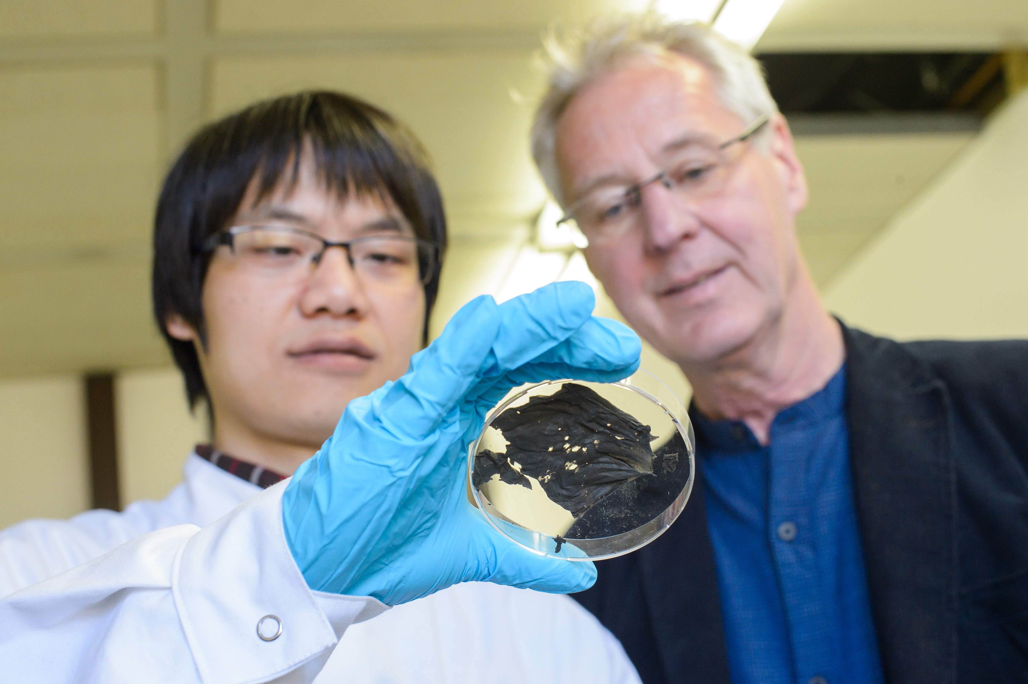
That may soon change. Recently, students from BYU’s [Brigham Young University; Utah, US] College of Engineering teamed up with Nanos Foundation [emphasis mine] to develop a nanofiber membrane that can be sandwiched between the cloth pieces in a homemade mask.

A few questions and a video

There is a video but you might find it helpful to know that when one of the students refers to OSHA she means the US Occupational Safety and Health Agency (OSHA). As for the ‘electrospinning’ I’m not sure how accessible that kind of equipment is, which calls into question how inexpensive and easy it would be to adopt this new mask insert. Fingers crossed that this will be as easy and effective as they seem to be suggesting,

The rest of the news

An October 21, 2020 BYU news release by Christie Allen, which originated the news item, delves into the technical details,

While today’s typical cloth mask might block fewer than 50% of virus particles, the membrane — which can be made using simple, inexpensive materials — will be able to block 90 to 99% of particles, increasing effectiveness [emphasis mine] while preserving breathability.

The membranes are made through a process called “electrospinning,” which involves dissolving a polymer plastic in a solution and then using an electrical current to move a droplet of the polymer downward through a needle. As the droplet accelerates, it stretches into a very small fiber that retains a static charge.

“Those nanofibers randomly land on a collector to create a sort of non-woven mesh,” said Katie Varela, a BYU mechanical engineering senior on the project team.

The remaining charge in the fibers is beneficial, she explained, because virus particles also have a static charge. “When they come close to your mask, they will be statically attracted to the mask and will not be able to go through it, and so it prevents you from inhaling viruses.”

In addition to the dramatic improvement in efficacy [emphasis mine], another key benefit of the nanofiber masks is that unlike traditional N95 masks, which have a reputation for being hot and stuffy, they allow for the circulation of (filtered) air, water and heat.

“Not only is it hard to find an N95 mask these days, but the best mask is useless if you won’t wear it,” said Will Vahle, director at Nanos Foundation. “Our nanofiber membranes are six times easier to breathe through than existing N95 masks, making them cooler, drier, and more comfortable.”

The group plans to make the instructions for creating the membranes open source. They hope that non-profit organizations will use the instructions to set up local sites where people can bring in their masks to be fitted with a membrane. They also hope other engineers will use their work as a springboard to produce more effective filters.

“We had our own proprietary nanofiber production process,” said Vahle of the project’s origins, “but we realized, hey, we have some expertise in this — why don’t we get this together and release a version that anybody can do?”

When Vahle and his colleagues approached BYU to collaborate on the project, BYU “jumped at the opportunity,” Vahle said. In addition to providing funding and facilities, the university connected the company with “fantastic students, who’ve really demonstrated an incredible work ethic and a drive to help people in need.”

Using cutting-edge science to make an immediate positive impact has also been highly valuable for the BYU students on the project.

“This experience makes things very real,” said Varela. “I’m really glad that I’m able to help with this fight against COVID-19 to help people all around the world and in my community.”

I’ve highlighted ‘effectiveness’ and ‘efficacy’, which are not synonyms although they’re often used that way. I can recall being quite surprised on discovering they were not, since I had, up to that point, confused them for many, many years. There’s a good description of the differences in a November 17, 2018 posting on the Public Health Notes website,

[downloaded from https://www.publichealthnotes.com/efficiency-vs-effectiveness-how-do-they-differ/]

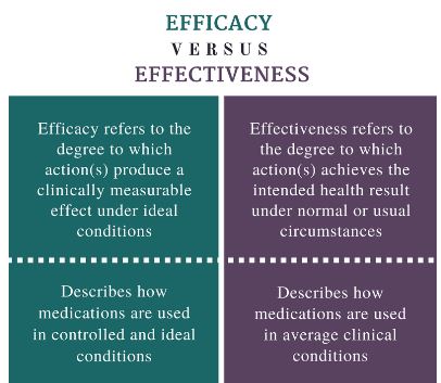
So, the difference is between controlled environments for efficacy and real life for effectiveness, in this case, a mask.

Can you get one of these improved masks?

The Nanos Foundation has a dedicated 95+ Mask webpage answering that question,

Worldwide Accessibility

Current technology has not been updated since the 1970s. The Nanos technology is inexpensive, portable and accessible.

Our Open-Source process turns common plastics into highly effective respiratory PPE [personal protective equipment].

‘Electrospinning’ nanofibers onto common cloth turns the cloth into a filter → sew the cloth into a mask to produce an effective top notch respirator.

You can use our designs, or bring your own design – 95+ is about the nanofiber membrane that turns the ‘cloth face covering’ into a respirator. Just make sure to use a design that creates a good seal or fit against the face.

The 95+ Process Requires Only A Few Simple Things

The kinds of things that can be easily found, like an old television, paint thinner & recycled plastics

I didn’t find any instructions for how to ‘electrospin’ with an old television, paint thinner, and plastics to make the nanofiber membrane. Perhaps one is required to donate before receiving instructions.

Interestingly, Nanos Foundation has three locations:

  • Greenville, AL, USA
  • Providence, RI, USA
  • Montreal, Canada

I was not expecting a Canadian connection.

Efficectiveness?

While this ‘easy to produce’ plastic insert seems very useful, it’s not clear to me what happens when the mask has to be washed or cleaned in some fashion. How long these nanofiber membranes active? Do we have to keep replacing the nanofiber membranes thereby adding more plastic to the environment?

Israeli startup (Nanomedic) and a ‘ray’ gun that shoots wound-healing skin

[downloaded from https://uploads.neatorama.com/images/posts/967/107/107967/Spray-on-Nanofiber-Skin-May-Improve-Burn-and-Wound-Care_0-x.jpg?v=10727]

Where I see a ‘ray’ gun, Rina Raphael, author of a July 6, 2019 article for Fast tCompany, sees a water pistol (Note: Links have been removed),

Imagine if bandaging looked a little more like, well, a water gun?

Israeli startup Nanomedic Technologies Ltd., a subsidiary of medical device company Nicast, has invented a new mechanical contraption to treat burns, wounds, and surgical injuries by mimicking human tissue. Shaped like a children’s toy, the lightweight SpinCare emits a proprietary nanofiber “second skin” that completely covers the area that needs to heal.

All one needs to do is aim, squeeze the two triggers, and fire off an electrospun polymer material that attaches to the skin.

The Nanomedic spray method avoids any need to come into direct contact with the wound. In that sense, it completely sidesteps painful routine bandage dressings. The transient skin then fully develops into a secure physical barrier with tough adherence. Once new skin is regenerated, usually between two to three weeks (depending on the individual’s heal time), the layer naturally peels off.

“You don’t replace it,” explains Nanomedic CEO Dr. Chen Barak. “You put it only once—on the day of application—and it remains there until it feels the new layer of skin healed.”

“It’s the same model as an espresso machine,” says Barak.

The SpinCare holds single-use ampoules containing Nanomedic’s polymer formulation. Once the capsule is firmly in place, one activates the device roughly eight inches towards the wound. Pressing the trigger activates the electron-spinning process, which sprays a web-like a layer of nano fibers directly on the wound.

The solution adjusts to the morphology of the wound, thereby creating a transient skin layer that imitates the skin structure’s human tissue. It’s a transparent, protective film that then allows the patient and doctor to monitor progress. Once the wound has healed and developed a new layer of skin, the SpinCare “bandage” falls off on its own.

The product is already being tested in hospitals. In the coming year, following FDA clearance, Nanomedic plans to expand to emergency rooms, ambulances, military use, and disaster relief response like fire truck companies. The global wound healing market is expected to hit $35 billion by 2025, according to a report by Transparency Market Research.

Nanomedic joins other researchers attempting to reimagine the wound healing process. Engineers at the University of Wisconsin-Madison, for example, created a new kind of protective bandage that sends a mild electrical stimulation, thereby “dramatically” reducing the time deep surgical wounds take to heal.

As for the the playful (yet functional) design, it resembles other medical tools utilizing the point-and-shoot feature. Researchers at the Technion-Israel Institute of Technology and Boston Children’s Hospital recently revealed a “hot-glue gun” that melds torn human tissues together. The medical glue is meant to replace painful and often scarring stitches and staples.

Down the line, Nanomedic plans to enter the in-home care market, where it believes it can better assist caretakers for treatment of chronic wounds, such as pressure ulcers. The chronic wounds segment is projected to hold the dominant share in the wound healing market due to aging populations.

But a bigger opportunity lies in the multiple uses the SpinCare can ultimately provide. It is, in essence, a platform technology that could benefit multiple categories, not just medical wound care. Currently, the SpinCare’s capsules do not contain any active ingredients.

Nanomedic is already researching how to add different additives, such as antibacterial compliments, collagen, silicone, cannabinoids—and, eventually, stem cells and cellular treatments.

Such advancements would propel the device to new markets, like plastic surgery, aesthetics, and dermatology. The latter, for example, spans “burns” caused by deep, cosmetic laser peels.

“Because it is a solution, we can combine additives inside,” explains Katz. “By that, we are transforming the transient skin into a drug delivery system and slow release system.”

Nanomedic is still at the premarket phase, [emphasis mine] having concluded one clinical trial related to the treatment of split graft donor site wounds and currently engaged in two ongoing burn studies. Barak anticipates FDA approval will take between nine to 12 months, during which the company will focus on building manufacturing lines and preparing for a European launch in early 2020.

According to the startup’s estimates, the product’s final price (not yet determined) will be far more affordable than traditional dressings. Nanomedic has raised $7 million in funding to date, including a grant by the EU’s Horizon 2020 SME Instrument program.

Barak believes Nanocare [sic] brings a highly cost-effective alternative to the healthcare system, but more than anything, she’s proud that SpinCare, above all else, mitigates patient pain and hassle. Some users, the company reports, are able to return to work and physical activity right away.

The Nanomedic website can be found here. The company has also produced a video featuring SpinCare,

There’s a bit more about the technology (I’m especially interested in the electrospinning) on Nanomedic’s Technology webpage,

Electrospinning technology allows the development of a wide range of products and devices, with tailored composition, geometry and morphology.

Almost any natural or synthetic polymer can be electrospun to create a nanofibrous mat. The intrinsic structure of the electrospun products, which mimics the natural extra cellular matrix (ECM), encourages quick and efficient tissue integration and minimizes medical complications.

Raphael’s article and the Nanomedic website offer more detail to what you can see in the excerpts provided here. If you have the time, I recommend checking out both.

Creating nanofibres from your old clothing (cotton waste)

Researchers at the University of British Columbia (UBC; Canada) have discovered a way to turn cotton waste into a potentially higher value product. An October 15, 2019 UBC news release makes the announcement (Note: Links have been removed),

In the materials engineering labs at UBC, surrounded by Bunsen burners, microscopes and spinning machines, professor Frank Ko and research scientist Addie Bahi have developed a simple process for converting waste cotton into much higher-value nanofibres.

These fibres are the building blocks of advanced products like surgical implants, antibacterial wound dressings and fuel cell batteries.

“More than 28 million tonnes of cotton are produced worldwide each year, but very little of that is actually recycled after its useful life,” explains Bahi, a materials engineer who previously worked on recycling waste in the United Kingdom. “We wanted to find a viable way to break down waste cotton and convert it into a value-added product. This is one of the first successful attempts to make nanofibres from fabric scraps – previous research has focused on using a ready cellulose base to make nanofibres.”

Compared to conventional fibres, nanofibres are extremely thin (a nanofibre can be 500 times smaller than the width of the human hair) and so have a high surface-to-volume ratio. This makes them ideal for use in applications ranging from sensors and filtration (think gas sensors and water filters) to protective clothing, tissue engineering and energy storage.
Ko and Bahi developed their process in collaboration with ecologyst, a B.C.-based company that manufactures sustainable outdoor apparel, and with the participation of materials engineering student Kosuke Ayama.

They chopped down waste cotton fabric supplied by ecologyst into tiny strips and soaked it in a chemical bath to remove all additives and artificial dyes from the fabric. The resulting gossamer-thin material was then fed to an electrospinning machine to produce very fine, smooth nanofibres. These can be further processed into various finished products.

“The process itself is relatively simple, but what we’re thrilled about is that we’ve proved you can extract a high-value product from something that would normally go to landfill, where it will eventually be incinerated. It’s estimated that only a fraction of cotton clothing is recycled. The more product we can re-process, the better it will be for the environment,” said lead researcher Frank Ko, a Canada Research Chair in advanced fibrous materials in UBC’s faculty of applied science.

The process Bahi and Ko developed is lab-scale, supported by a grant from the Natural Sciences and Engineering Research Council of Canada. In the future, the pair hope to refine and scale up their process and eventually share their methods with industry partners.

“We started with cotton because it’s one of the most popular fabrics for clothing,” said Bahi. “Once we’re able to develop the process further, we can look at converting other textiles into value-added materials. Achieving zero waste [emphasis mine] for the fashion and textile industries is extremely challenging – this is simply one of the many first steps towards that goal.”

The researchers have a 30 sec. video illustrating the need to recycle cotton materials,

You can find the researchers’ industrial partner, ecologyst here.

At the mention of ‘zero waste’, I was reminded of an upcoming conference, Oct. 30 -31, 2019 in Vancouver (Canada) where UBC is located. It’s called the 2019 Zero Waste Conference and, oddly,there’s no mention of Ko or Bahi or Ayama or ecologyst on the speakers’ list. Maybe I was looking at the wrong list or the organizers didn’t have enough lead time to add more speakers.

One final comment, I wish there was a little more science (i.e., more technical details) in the news release.

Ultra-thin superconducting film for outer space

Truth in a press release? But first, there’s this April 6, 2017 news item on Nanowerk announcing research that may have applications in aerospace and other sectors,

Experimental physicists in the research group led by Professor Uwe Hartmann at Saarland University have developed a thin nanomaterial with superconducting properties. Below about -200 °C these materials conduct electricity without loss, levitate magnets and can screen magnetic fields.

The particularly interesting aspect of this work is that the research team has succeeded in creating superconducting nanowires that can be woven into an ultra-thin film that is as flexible as cling film. As a result, novel coatings for applications ranging from aerospace to medical technology are becoming possible.

The research team will be exhibiting their superconducting film at Hannover Messe from April 24th to April 28th [2017] (Hall 2, Stand B46) and are looking for commercial and industrial partners with whom they can develop their system for practical applications.

An April 6, 2017 University of Saarland press release (also on EurekAlert), which originated the news item, provides more details along with a line that rings with the truth,

A team of experimental physicists at Saarland University have developed something that – it has to be said – seems pretty unremarkable at first sight. [emphasis mine] It looks like nothing more than a charred black piece of paper. But appearances can be deceiving. This unassuming object is a superconductor. The term ‘superconductor’ is given to a material that (usually at a very low temperatures) has zero electrical resistance and can therefore conduct an electric current without loss. Put simply, the electrons in the material can flow unrestricted through the cold immobilized atomic lattice. In the absence of electrical resistance, if a magnet is brought up close to a cold superconductor, the magnet effectively ‘sees’ a mirror image of itself in the superconducting material. So if a superconductor and a magnet are placed in close proximity to one another and cooled with liquid nitrogen they will repel each another and the magnet levitates above the superconductor. The term ‘levitation’ comes from the Latin word levitas meaning lightness. It’s a bit like a low-temperature version of the hoverboard from the ‘Back to the Future’ films. If the temperature is too high, however, frictionless sliding is just not going to happen.
Many of the common superconducting materials available today are rigid, brittle and dense, which makes them heavy. The Saarbrücken physicists have now succeeded in packing superconducting properties into a thin flexible film. The material is a essentially a woven fabric of plastic fibres and high-temperature superconducting nanowires. ‘That makes the material very pliable and adaptable – like cling film (or ‘plastic wrap’ as it’s also known). Theoretically, the material can be made to any size. And we need fewer resources than are typically required to make superconducting ceramics, so our superconducting mesh is also cheaper to fabricate,’ explains Uwe Hartmann, Professor of Nanostructure Research and Nanotechnology at Saarland University.

The low weight of the film is particularly advantageous. ‘With a density of only 0.05 grams per cubic centimetre, the material is very light, weighing about a hundred times less than a conventional superconductor. This makes the material very promising for all those applications where weight is an issue, such as in space technology. There are also potential applications in medical technology,’ explains Hartmann. The material could be used as a novel coating to provide low-temperature screening from electromagnetic fields, or it could be used in flexible cables or to facilitate friction-free motion.

In order to be able to weave this new material, the experimental physicists made use of a technique known as electrospinning, which is usually used in the manufacture of polymeric fibres. ‘We force a liquid material through a very fine nozzle known as a spinneret to which a high electrical voltage has been applied. This produces nanowire filaments that are a thousand times thinner than the diameter of a human hair, typically about 300 nanometres or less. We then heat the mesh of fibres so that superconductors of the right composition are created. The superconducting material itself is typically an yttrium-barium-copper-oxide or similar compound,’ explains Dr. Michael Koblischka, one of the research scientists in Hartmann‘s group.

The research project received €100,000 in funding from the Volkswagen Foundation as part of its ‘Experiment!’ initiative. The initiative aims to encourage curiosity-driven, blue-skies research. The positive results from the Saarbrücken research team demonstrate the value of this type of funding. Since September 2016, the project has been supported by the German Research Foundation (DFG). Total funds of around €425,000 will be provided over a three-year period during which the research team will be carrying out more detailed investigations into the properties of the nanowires.

I’d say the “unremarkable but appearances can be deceiving” comments are true more often than not. I think that’s one of the hard things about science. Big advances can look nondescript.

What looks like a pretty unremarkable piece of burnt paper is in fact an ultrathin superconductor that has been developed by the team lead by Uwe Hartmann (r.) shown here with doctoral student XianLin Zeng. Courtesy: Saarland University

In any event, here’s a link to and a citation for the paper,

Preparation of granular Bi-2212 nanowires by electrospinning by Xian Lin Zeng, Michael R Koblischka, Thomas Karwoth, Thomas Hauet, and Uwe Hartmann. Superconductor Science and Technology, Volume 30, Number 3 Published 1 February 2017

© 2017 IOP Publishing Ltd

This paper is behind a paywall.

Stronger more robust nanofibers for everything from bulletproof vests to cellular scaffolds (tissue engineering)

This work on a new technique for producing nanofibers comes from Harvard University’s School of Engineering and Applied Sciences and the Wyss Institute for Biologically Inspired Engineering (also at Harvard University). From an Oct. 10, 2016 news item on phys.org,

Fibrous materials—known for their toughness, durability and pliability—are used in everything from bulletproof vests to tires, filtration systems and cellular scaffolds for tissue engineering and regenerative medicine.

The properties of these materials are such that the smaller the fibers are, the stronger and tougher they become. But making certain fibers very small has been an engineering challenge.

Now, researchers from the Harvard John A. Paulson School of Engineering and Applied Sciences (SEAS) and the Wyss Institute for Biologically Inspired Engineering at Harvard have developed a new method to make and collect nanofibers and control their size and morphology. This could lead to stronger, more durable bulletproof vests and armor and more robust cellular scaffolding for tissue repair.

An Oct. 7, 2016 Harvard University press release by Leah Burrows, which originated the news item, describes the research in more detail (Note: A link has been removed),

Nanofibers are smaller than one micrometer in diameter.  Most nanofiber production platforms rely on dissolving polymers in a solution, which then evaporates as the fiber forms.

Rotary Jet-Spinning (RJS), the technique developed by Kit Parker’s Disease Biophysics Group, works likes a cotton candy machine. Parker is Tarr Family Professor of Bioengineering and Applied Physics at SEAS and a Core Member of the Wyss Institute. A liquid polymer solution is loaded into a reservoir and pushed out through a tiny opening by centrifugal force as the device spins. As the solution leaves the reservoir, the solvent evaporates and the polymers solidify and elongate into small, thin fibers.

“This advance is important because it allows us to manufacture ballistic protection that is much lighter, more flexible and more functional than what is available today,” said Parker, who in addition to his Harvard role is a lieutenant colonel in the United States Army Reserve and was motivated by his own combat experiences in Afghanistan. “Not only could it save lives but for the warfighter, it also could help reduce the repetitive injury motions that soldiers, sailors, marines and airmen have suffered over the last 15 years of the war on terror.”

“Rotary Jet-Spinning is great for most polymer fibers you want to make,” said Grant Gonzalez, a graduate student at SEAS and first author of the paper.  “However, some fibers require a solvent that doesn’t evaporate easily. Para-aramid, the polymer used in Kevlar® for example, is dissolved in sulfuric acid, which doesn’t evaporate off. The solution just splashes against the walls of the device without forming fibers.”

Nanofibers are smaller than one micrometer in diameter.  Most nanofiber production platforms rely on dissolving polymers in a solution, which then evaporates as the fiber forms.

Rotary Jet-Spinning (RJS), the technique developed by Kit Parker’s Disease Biophysics Group, works likes a cotton candy machine. Parker is Tarr Family Professor of Bioengineering and Applied Physics at SEAS and a Core Member of the Wyss Institute. A liquid polymer solution is loaded into a reservoir and pushed out through a tiny opening by centrifugal force as the device spins. As the solution leaves the reservoir, the solvent evaporates and the polymers solidify and elongate into small, thin fibers.

“This advance is important because it allows us to manufacture ballistic protection that is much lighter, more flexible and more functional than what is available today,” said Parker, who in addition to his Harvard role is a lieutenant colonel in the United States Army Reserve and was motivated by his own combat experiences in Afghanistan. “Not only could it save lives but for the warfighter, it also could help reduce the repetitive injury motions that soldiers, sailors, marines and airmen have suffered over the last 15 years of the war on terror.”

“Rotary Jet-Spinning is great for most polymer fibers you want to make,” said Grant Gonzalez, a graduate student at SEAS and first author of the paper.  “However, some fibers require a solvent that doesn’t evaporate easily. Para-aramid, the polymer used in Kevlar® for example, is dissolved in sulfuric acid, which doesn’t evaporate off. The solution just splashes against the walls of the device without forming fibers.”

Other methods, such as electrospinning, which uses an electric field to pull the polymer into a thin fiber, also have poor results with Kevlar and other polymers such as alginate used for tissue scaffolding and DNA.

The Harvard team overcame these challenges by developing a wet-spinning platform, which uses the same principles as the RJS system but relies on precipitation rather than evaporation to separate the solvent from the polymer.

In this system, called immersion Rotary Jet-Spinning (iRJS), when the polymer solution shoots out of the reservoir, it first passes through an area of open air, where the polymers elongate and the chains align. Then the solution hits a liquid bath that removes the solvent and precipitates the polymers to form solid fibers. Since the bath is also spinning — like water in a salad spinner — the nanofibers follow the stream of the vortex and wrap around a rotating collector at the base of the device.

Using this system, the team produced Nylon, DNA, alginate and ballistic resistant para-aramid nanofibers. The team could tune the fiber’s diameter by changing the solution concentration, the rotational speed and the distance the polymer traveled from the reservoir to the bath.

“By being able to modulate fiber strength, we can create a cellular scaffold that can mimic skeleton muscle and native tissues,” said Gonzalez.  “This platform could enable us to create a wound dressing out of alginate material or seed and mature cells on scaffolding for tissue engineering.”

Because the fibers were collected by a spinning vortex, the system also produced well-aligned sheets of nanofibers, which is important for scaffolding and ballistic resistant materials.

This is the ‘candy floss’ technique at work,

Rotary Jet-Spinning (RJS) works likes a cotton candy machine. A liquid polymer solution is loaded into a reservoir and pushed out through a tiny opening by centrifugal force as the device spins. As the solution leaves the reservoir, the solvent evaporates and the polymers solidify and elongate into small, thin fibers. Courtesy: Harvard University

Rotary Jet-Spinning (RJS) works likes a cotton candy machine. A liquid polymer solution is loaded into a reservoir and pushed out through a tiny opening by centrifugal force as the device spins. As the solution leaves the reservoir, the solvent evaporates and the polymers solidify and elongate into small, thin fibers. Courtesy: Harvard University

Here’s a link to and a citation for the paper,

Production of Synthetic, Para-Aramid and Biopolymer Nanofibers by Immersion Rotary Jet-Spinning by Grant M. Gonzalez, Luke A. MacQueen, Johan U. Lind, Stacey A. Fitzgibbons, Christophe O. Chantre, Isabelle Huggler, Holly M. Golecki, Josue A. Goss, Kevin Kit Parker. Macromolecular Materials and Engineering DOI: 10.1002/mame.201600365 Version of Record online: 7 OCT 2016

© 2016 WILEY-VCH Verlag GmbH & Co. KGaA, Weinheim

This paper is behind a paywall.

Magnetospinning with an inexpensive magnet

The fridge magnet mentioned in the headline for a May 11, 2015  Nanowerk spotlight aricle by Michael Berger isn’t followed up until the penultimate paragraph but it is worth the wait,

“Our method for spinning of continuous micro- and nanofibers uses a permanent revolving magnet,” Alexander Tokarev, Ph.D., a Research Associate in the Nanostructured Materials Laboratory at the University of Georgia, tells Nanowerk. “This fabrication technique utilizes magnetic forces and hydrodynamic features of stretched threads to produce fine nanofibers.”

“The new method provides excellent control over the fiber diameter and is compatible with a range of polymeric materials and polymer composite materials including biopolymers,” notes Tokarev. “Our research showcases this new technique and demonstrates its advantages to the scientific community.”

Electrospinning is the most popular method to produce nanofibers in labs now. Owing to its simplicity and low costs, a magnetospinning set-up could be installed in any non-specialized laboratory for broader use of magnetospun nanofibers in different methods and technologies. The total cost of a laboratory electrospinning system is above $10,000. In contrast, no special equipment is needed for magnetospinning. It is possible to build a magnetospinning set-up, such as the University of Georgia team utilizes, by just using a $30 rotating motor and a $5 permanent magnet. [emphasis mine]

Berger’s article references a recent paper published by the team,

Magnetospinning of Nano- and Microfibers by Alexander Tokarev, Oleksandr Trotsenko, Ian M. Griffiths, Howard A. Stone, and Sergiy Minko. Advanced Materials First published: 8 May 2015Full publication history DOI: 10.1002/adma.201500374View/save citation

This paper is behind a paywall.

* The headline originally stated that a ‘fridge’ magnet was used. Researcher Alexander Tokarev kindly dropped by correct this misunderstanding on my part and the headline has been changed to read  ‘inexpensive magnet’ on May 14, 2015 at approximately 1400 hundred hours PDT.

Honey nanofibres tested as scaffolding for wound dressing in an Iran-Netherlands collaboration

It’s taken me a while to get to this one but I can’t resist this honey-enabled technology any longer. According to a Sept. 19, 2013 news item on Nanowerk, honey, a well known antibiotic, has been used in a new technique for wound dressings (Note: A link has been removed),

Researchers applied electrospinning process and produced a drug-carrying nanofibrous web to be used in wound dressing by using an artificial and biodegradable polymer and honey as a natural polymer (“A novel honey-based nanofibrous scaffold for wound dressing application”).

A wide range of biological and biodegradable materials have been electrospun in recent years to produce nanofibers. In this research, a drug carrying nanofibrous web was produced to be used in wound dressing by using an artificial and biodegradable polymer and a natural polymer through electrospinning method.

The Sept. 19, 2013 Iran National Nanotechnology Initiative Council (INIC) news release, which originated the news item, mentions honey’s antibiotic properties and explains how its application in this new technique for wound dressing,

Honey has antibacterial and anti-inflammation properties. Many studies have been published on the effects of honey in the treatment of infections and in prevention of the wound from being infected. Therefore, the combination of the unique properties of nanofibers and the natural properties of honey in the production of wound dressing is the most important characteristic of this research.

SEM [scanning electron microscope] and AFM [atomic force microscope] results showed that the fibers were completely homogenous with relatively smooth surface. However, spindle-like beads were observed in nanofibers containing 60% honey. As the concentration of honey increased in the mixture, a decrease was observed in the diameter of nanofibers. Drug-loaded nanofibers, too, had relatively smooth and homogenous surface, and as the amount of drug increases, the diameter of the nanofibers decreased. Drug release behavior studies demonstrated a sudden initial release. Statistical analyses showed that the presence of honey did not have significant effect on the process or on the behavior of drug release. Therefore, electrospun nanofibers that contain honey are appropriate option to be used in wound dressing.

Wounds can be dressed faster by using the achievements of this research. Honey is considered as a well-known drug in traditional medical sciences, which has been loaded with drugs in this research.

The research paper’s (a link and citation will be provided further down) abstract provides a bit more detail,

In this study, nanofiber meshes were produced from aqueous mixtures of poly(vinyl alcohol) (PVA) and honey via electrospinning. The Electrospinning process was performed at different PVAs to honey ratios (100/0, 90/10, 80/20, 70/30, and 60/40). Dexamethasone sodium phosphate was selected as an anti-inflammatory drug and incorporated in the electrospinning solutions. Its release behavior was determined. Uniform and smooth nanofibers were formed, independent of the honey content. In case honey content increased up to 40%, some spindle-like beads on the fibers were observed. The diameter of electrospun fibers decreased as the ratio of honey increased. The release characteristics of the model drug from both the PVA and PVA/honey (80/20) nanofibrous mats were studied and statistical analysis was performed. All electrospun fibers exhibited a large initial burst release at a short time after incubation. The release profile was similar for both PVA and PVA/honey (80/20) drug-loaded nanofibers. This study shows that an anti-inflammatory drug can be released during the initial stages and honey can be used as a natural antibiotic to improve the wound dressing efficiency and increase the healing rate.

Here’s a link to and a citation for paper,

A novel honey-based nanofibrous scaffold for wound dressing application by  H. Maleki, A. A. Gharehaghaji, and P. J. Dijkstra.
Journal of Applied Polymer Science, Volume 127, Issue 5, pages 4086–4092, 5 March 2013 (Article first published online: 29 MAY 2012) DOI: 10.1002/app.37601

Copyright © 2012 Wiley Periodicals, Inc.

This article is behind a paywall.

One final note, the researchers are from (Maleki and Gharehaghaji) Amirkabir University of Technology, Tehran, Iran and (Dijkstra) the University of Twente, Enschede, The Netherlands

Contraception and HIV protection in cloth*

Researchers at the University of Washington have published a study in the peer-reviewed, open access journal, Public Library of Science ONE (PLoS ONE), concerning their work to produce fibres that can deliver both contraceptives and anti-HIV drugs, according to a Nov. 30, 2012 news item on Nanowerk,

The only way to protect against HIV and unintended pregnancy today is the condom. It’s an effective technology, but not appropriate or popular in all situations.

A University of Washington team has developed a versatile platform to simultaneously offer contraception and prevent HIV. Electrically spun cloth with nanometer-sized fibers can dissolve to release drugs, providing a platform for cheap, discrete and reversible protection.

Hannah Hickey’s  Nov. 30, 2012 University of Washington news release, which originated the news item, provides details,

“Our dream is to create a product women can use to protect themselves from HIV infection and unintended pregnancy,” said corresponding author Kim Woodrow, a UW assistant professor of bioengineering. “We have the drugs to do that. It’s really about delivering them in a way that makes them more potent, and allows a woman to want to use it.”

Electrospinning uses an electric field to catapult a charged fluid jet through air to create very fine, nanometer-scale fibers. The fibers can be manipulated to control the material’s solubility, strength and even geometry. Because of this versatility, fibers may be better at delivering medicine than existing technologies such as gels, tablets or pills. No high temperatures are involved, so the method is suitable for heat-sensitive molecules. The fabric can also incorporate large molecules, such as proteins and antibodies, that are hard to deliver through other methods.

They first dissolved polymers approved by the Food and Drug Administration and antiretroviral drugs used to treat HIV to create a gooey solution that passes through a syringe. As the stream encounters the electric field it stretches to create thin fibers measuring 100 to several thousand nanometers that whip through the air and eventually stick to a collecting plate (one nanometer is about one 25-millionth of an inch). The final material is a stretchy fabric that can physically block sperm or release chemical contraceptives and antivirals.

“This method allows controlled release of multiple compounds,” Ball said. “We were able to tune the fibers to have different release properties.”

One of the fabrics they made dissolves within minutes, potentially offering users immediate, discrete protection against unwanted pregnancy and sexually transmitted diseases.

Another dissolves gradually over a few days, providing an option for sustained delivery, more like the birth-control pill,  to provide contraception and guard against HIV.

The fabric could incorporate many fibers to guard against many different sexually transmitted infections, or include more than one anti-HIV drug to protect against drug-resistant strains (and discourage drug-resistant strains from emerging). Mixed fibers could be designed to release drugs at different times to increase their potency, like the prime-boost method used in vaccines.

The electrospun cloth could be inserted directly in the body or be used as a coating on vaginal rings or other products.

Electrospinning has existed for decades, but it’s only recently been automated to make it practical for applications such as filtration and tissue engineering. This is the first study to use nanofibers for vaginal drug delivery.

While this technology is more discrete than a condom, and potentially more versatile than pills or plastic or rubber devices, researchers say there is no single right answer.

The citation and link to the article,

Drug-Eluting Fibers for HIV-1 Inhibition and Contraception by Cameron Ball, Emily Krogstad, Thanyanan Chaowanachan, Kim A. Woodrow (2012) PLoS ONE 7(11): e49792. doi:10.1371/journal.pone.0049792

Last month, the Bill and Melinda Gates Foundation awarded these researchers a $1M grant to pursue this work.

*ETA Dec.2.12: I erroneously used the word clothing in the headline. It’s now been corrected to ‘cloth’.

Printing new knee cartilage

I was reminded of the 1992 Olympics in Barcelona while reading the Nov. 22, 2012 news item on Nanowerk about printing cartilage for knees. Some years ago I knew a Canadian wrestler who’d participated in those games and he had a story about knee cartilage that featured amputation.

Apparently, wrestlers in earlier generations had knee surgeries that involved removal of cartilage for therapeutic purposes. Unfortunately, decades later, these retired wrestlers found that whatever cartilage had remained was now worn through and bones were grinding on bones causing such pain that more than one wrestler agreed to amputation. I never did check out the story but it rang true largely because I’d come across a similar story from a physiotherapist regarding  a shoulder joint and the consequences of losing cartilage in there (very, very painful).

It seems that scientists are now working on a solution for those of us unlucky enough to have damaged or worn through cartilage in our joints, from the Nov. 22, 2012 IOP science news release, (Institute of Physics) which originated the news item,

The printing of 3D tissue has taken a major step forward with the creation of a novel hybrid printer that simplifies the process of creating implantable cartilage.


The printer is a combination of two low-cost fabrication techniques: a traditional ink jet printer and an electrospinning machine. Combining these systems allowed the scientists to build a structure made from natural and synthetic materials. …

In this study, the hybrid system produced cartilage constructs with increased mechanical stability compared to those created by an ink jet printer using gel material alone. The constructs were also shown to maintain their functional characteristics in the laboratory and a real-life system.

The key to this was the use of the electrospinning machine, which uses an electrical current to generate very fine fibres from a polymer solution. Electrospinning allows the composition of polymers to be easily controlled and therefore produces porous structures that encourage cells to integrate into surrounding tissue.

In this study, flexible mats of electrospun synthetic polymer were combined, layer-by-layer, with a solution of cartilage cells from a rabbit ear that were deposited using the traditional ink jet printer. The constructs were square with a 10cm diagonal and a 0.4mm thickness.

The researchers tested their strength by loading them with variable weights and, after one week, tested to see if the cartilage cells were still alive.

The constructs were also inserted into mice for two, four and eight weeks to see how they performed in a real life system. After eight weeks of implantation, the constructs appeared to have developed the structures and properties that are typical of elastic cartilage, demonstrating their potential for insertion into a patient.

The researchers state that in a future scenario, cartilage constructs could be clinically applied by using an MRI scan of a body part, such as the knee, as a blueprint for creating a matching construct. A careful selection of scaffold material for each patient’s construct would allow the implant to withstand mechanical forces while encouraging new cartilage to organise and fill the defect.

The researchers’ article in the IOP science jouBiofrarnal, Biofabrication, is freely available for 30 days after its date of publication, Nov. 21, 2012. You do need to register with IOP science to gain access. Here’s the citation and a link,

Hybrid printing of mechanically and biologically improved constructs for cartilage tissue engineering applications by Tao Xu, Kyle W Binder, Mohammad Z Albanna, Dennis Dice, Weixin Zhao, James J Yoo and Anthony Atala in 2013 Biofabrication 5 015001 doi:10.1088/1758-5082/5/1/015001

I believe all of the scientists involved in this bioprinting project are with the Wake Forest Institute for Regenerative Medicine.

A breath-based and handheld diagnostic device

Researcher Perena Gouma and her team at Stony Brook University (New York, US) are hoping that eventually their device will be available over the counter so anyone will be able to perform a preliminary diagnostic test as casually as you take a breath. From the May 7, 2012 news item on Nanowerk,

You blow into a small valve attached to a box that is about half the size of your typical shoebox and weighs less than one pound. Once you blow into it, the lights on top of the box will give you an instant readout. A green light means you pass (and your bad breath is not indicative of an underlying disease; perhaps it’s just a result of the raw onions you ingested recently); however, a red light means you might need to take a trip to the doctor’s office to check if something more serious is an issue.

Here’s a bit more about the device and the researchers’ hopes in a video from the US National Science Foundation (NSF) featuring the NSF’s Miles O’Brien as the reporter,

O’Brien in his May 7, 2012 article for the NSF’s Science Nation online magazine describes the technology,

With support from the National Science Foundation (NSF), Professor Perena Gouma and her team at Stony Brook University in New York developed a sensor chip that you might say is the “brain” of the breathalyzer. It’s coated with tiny nanowires that look like microscopic spaghetti and are able to detect minute amounts of chemical compounds in the breath. “These nanowires enable the sensor to detect just a few molecules of the disease marker gas in a ‘sea’ of billions of molecules of other compounds that the breath consists of,” Gouma explains. This is what nanotechnology is all about.

The manufacturing process that creates the single crystal nanowires is called “electrospinning.” It starts with a liquid compound being shot from a syringe into an electrical field. The electric field crystallizes the inserted liquid into a tiny thread or “wire” that collects onto an aluminum backing. Gouma says enough nanowire can be produced in one syringe to stretch from her lab in Stony Brook, N.Y. to the moon and still be a single grain (monocrystal).

“There can be different types of nanowires, each with a tailored arrangement of metal and oxygen atoms along their configuration, so as to capture a particular compound,” explains Gouma. “For example, some nanowires might be able to capture ammonia molecules, while others capture just acetone and others just the nitric oxide. Each of these biomarkers signal a specific disease or metabolic malfunction so a distinct diagnostic breathalyzer can be designed.”

Gouma also says the nanowires can be rigged to detect infectious viruses and microbes like Salmonella, E. coli or even anthrax. “There will be so many other applications we haven’t envisioned. It’s very exciting; it’s a whole new world,” she says.

I think most (if not all) of the handheld diagnostic projects I’ve covered have been fluids-based, i.e., they need a sample of saliva, blood, urine, etc. to perform their diagnostic function. I believe this is the first breath-based project I’ve seen.