Tag Archives: Institute of Physics

Nanoscale silver unveils intrinsic self-healing abilities

A September 7, 2024 news item on phys.org offers some intriguing information about nanoscale silver and its self-healing abilities, Note: A link has been removed,

As an innovative concept in materials science and engineering, the inspiration for self-healing materials comes from living organisms that have the innate ability to self-heal. Along this line, the search for self-healing materials has been generally focused on “soft” materials like polymers and hydrogels. For solid-state metals, one may intuitively imagine that any form of self-healing will be much more difficult to achieve.

While a few past studies have showcased the self-healing behavior in metals that more or less requires the assistance of external triggers (e.g., by heating, mechanical stimulus, or electron beam irradiation), whether the autonomous self-healing can occur in metal solids without any external intervention remains a scientific curiosity.

Now in a new study published in Matter, researchers from the Institute of Physics (IOP) of the Chinese Academy of Sciences have discovered that such an intrinsic and autonomous self-healing phenomenon can occur in nanoscale silver (Ag).

An August 26, 2024 Chinese Academy of Sciences press release (also on EurekAlert but published September 6, 2024), which originated the news item, delves further into the research,

This study, which combines advanced in-situ transmission electron microscopy (TEM) with molecular dynamics (MD) simulations, reveals that nanoscale Ag can autonomously repair itself from structural damages such as nanocracks and nanopores without external intervention.

This remarkable ability is observed not only at room temperature but also at frigid temperatures as low as 173 K.

Notably, over the same damaging area, the repeated reversible self-healing cycles can also be achieved with the same level of efficiency.

The experiments were performed inside an atomic-resolution TEM by utilizing single-crystalline Ag nanosheets as testing specimens. Both nanopores and nanocracks were purposefully fabricated through in-situ drilling by TEM electron beam.

To avoid any possible intervention to the healing process, Ag nanosheet specimen was afterwards kept at a “beam-off” state all through until each moment for interval TEM imaging.

As an interesting and perhaps surprising result, the two representative kinds of structural damages were observed to undergo rapid self-healing autonomously within several to dozens of minutes, with the healed regions perfectly restoring the crystal lattice of Ag with atomically precise ordering.

Unlike Ag, gold (Au) did not show similar self-healing behavior at room temperature, despite the fact that Au is the most relevant element to Ag in the Periodic Table and they share many similarities in physical and chemical properties.

Further MD simulation results well reproduce the experimental observations, especially regarding the difference in the healing behavior between Ag and Au. What sets Ag apart from Au is its high mobility of surface diffusion, a trait not commonly found in other metal solids.

By employing TEM, the researchers were able to in-situ track the trajectories of healing process in Ag at the atomic level. With a combination of atomistic imaging and theoretical simulation results, the research highlights that self-healing is enabled by the surface-mediated self-diffusion of Ag atoms as driven by chemical potential imbalance due to Gibbs-Thomson effect.

When a nascent damage structure (either nanopore or nanocrack) begins its existence in Ag nanosheet, there will create a concave site with the negative local curvature. Due to the general curvature-dependence of chemical potential, the concave damage site will thereby have smaller chemical potential relative to the undamaged areas of the nanosheet. This built-in imbalance of chemical potential drives Ag atoms to migrate and repair the damage autonomously, showcasing a sophisticated form of material self-maintenance in negative feedback way.

The ability of Ag to autonomously self-heal nanoscale damages at room temperature and below shows promising possibility for developing damage-tolerant components and devices at the sub-micrometer length scale.

Perhaps more importantly, in a broader term, this unusual finding at a mechanistic level may provide a guiding framework for deeper understanding of the self-healing phenomena and concepts in metal solids in general.

Here’s a link to and a citation for the paper,

Direct observation of autonomous self-healing in silver by Jianlin Wang, Qiuhao Xu, Muhua Sun, Jiyu Xu, Pan Chen, Bohan Yu, Zhongqi Wu, Zitao Chen, Xudan Huang, Huacong Sun, Lei Liao, Chen Cai, Xiaomin Li, Lifen Wang, Xuezeng Tian, Zhi Xu, Sheng Meng, Wenlong Wang, Xuedong Bai. Matter DOI: 10.1016/j.matt.2024.07.009 Published online; August 21, 2024 Copyright: © 2024 Elsevier Inc

This paper is open access for the time being.

Don’t throw that cigarette butt away—use it to store energy!

I’ve read the news release and briefly skimmed the research paper and cannot find any discussion of how these scientists got the idea to ‘recycle’ used cigarette butts for energy storage (supercapacitors) although the inspiration seems to have its roots in a desire to create better supercapacitors from recycled materials. From an Aug. 5, 2014 news item on ScienceDaily,

A group of scientists from South Korea have converted used-cigarette butts into a high-performing material that could be integrated into computers, handheld devices, electrical vehicles and wind turbines to store energy.

Presenting their findings today, 5 August 2014, in IOP Publishing’s journal Nanotechnology, the researchers have demonstrated the material’s superior performance compared to commercially available carbon, graphene and carbon nanotubes.

It is hoped the material can be used to coat the electrodes of supercapacitors — electrochemical components that can store extremely large amounts of electrical energy — whilst also offering a solution to the growing environmental problem caused by used-cigarette filters.

An Aug. 5, 2014 Institute of Physics (IOP) news release (also on EurekAlert), which originated the news item, further describes the situation regarding used cigarette butts and the characteristics that could render them into supercapacitors

It is estimated that as many as 5.6 trillion cigarette butts (equivalent to 766 571 metric tons), are deposited into the environment worldwide every year.

Co-author of the study Professor Jongheop Yi, from Seoul National University, said: “Our study has shown that used cigarette filters can be transformed into a high-performing carbon-based material using a simple one-step process, which simultaneously offers a green solution to meeting the energy demands of society.

“Numerous countries are developing strict regulations to avoid the trillions of toxic and non-biodegradable used cigarette filters that are disposed of into the environment each year; our method is just one way of achieving this.”

Carbon is the most popular material that supercapacitors are composed of, due to its low cost, high surface area, high electrical conductivity and long-term stability.

Scientists around the world are currently working towards improving the characteristics of supercapacitors – such as energy density, power density and cycle stability – while also trying to reduce production costs.

In their study, the researchers demonstrated that the cellulose acetate fibres that cigarette filters are mostly composed of could be transformed into a carbon-based material using a simple, one-step burning technique called pyrolysis.

As a result of this burning process, the resulting carbon-based material contained a number of tiny pores, increasing its performance as a supercapacitive material.

“A high-performing supercapacitor material should have a large surface area, which can be achieved by incorporating a large number of small pores into the material,” continued Professor Yi.

“A combination of different pore sizes ensures that the material has high power densities, which is an essential property in a supercapacitor for the fast charging and discharging.”

Once fabricated, the carbon-based material was attached to an electrode and tested in a three-electrode system to see how well the material could adsorb electrolyte ions (charge) and then release them (discharge).

The material stored a higher amount of electrical energy than commercially available carbon and also had a higher amount of storage compared to graphene and carbon nanotubes, as reported in previous studies.

Here’s a link to and a citation for the research paper,

Preparation of energy storage material derived from a used cigarette filter for a supercapacitor electrode by Minzae Lee, Gil-Pyo Kim, Hyeon Don Song, Soomin Park, and Jongheop Yi. Nanotechnology 25 (34) 5601 doi:10.1088/0957-4484/25/34/345601

This is an open access paper.
Printing new knee cartilage

I was reminded of the 1992 Olympics in Barcelona while reading the Nov. 22, 2012 news item on Nanowerk about printing cartilage for knees. Some years ago I knew a Canadian wrestler who’d participated in those games and he had a story about knee cartilage that featured amputation.

Apparently, wrestlers in earlier generations had knee surgeries that involved removal of cartilage for therapeutic purposes. Unfortunately, decades later, these retired wrestlers found that whatever cartilage had remained was now worn through and bones were grinding on bones causing such pain that more than one wrestler agreed to amputation. I never did check out the story but it rang true largely because I’d come across a similar story from a physiotherapist regarding  a shoulder joint and the consequences of losing cartilage in there (very, very painful).

It seems that scientists are now working on a solution for those of us unlucky enough to have damaged or worn through cartilage in our joints, from the Nov. 22, 2012 IOP science news release, (Institute of Physics) which originated the news item,

The printing of 3D tissue has taken a major step forward with the creation of a novel hybrid printer that simplifies the process of creating implantable cartilage.


The printer is a combination of two low-cost fabrication techniques: a traditional ink jet printer and an electrospinning machine. Combining these systems allowed the scientists to build a structure made from natural and synthetic materials. …

In this study, the hybrid system produced cartilage constructs with increased mechanical stability compared to those created by an ink jet printer using gel material alone. The constructs were also shown to maintain their functional characteristics in the laboratory and a real-life system.

The key to this was the use of the electrospinning machine, which uses an electrical current to generate very fine fibres from a polymer solution. Electrospinning allows the composition of polymers to be easily controlled and therefore produces porous structures that encourage cells to integrate into surrounding tissue.

In this study, flexible mats of electrospun synthetic polymer were combined, layer-by-layer, with a solution of cartilage cells from a rabbit ear that were deposited using the traditional ink jet printer. The constructs were square with a 10cm diagonal and a 0.4mm thickness.

The researchers tested their strength by loading them with variable weights and, after one week, tested to see if the cartilage cells were still alive.

The constructs were also inserted into mice for two, four and eight weeks to see how they performed in a real life system. After eight weeks of implantation, the constructs appeared to have developed the structures and properties that are typical of elastic cartilage, demonstrating their potential for insertion into a patient.

The researchers state that in a future scenario, cartilage constructs could be clinically applied by using an MRI scan of a body part, such as the knee, as a blueprint for creating a matching construct. A careful selection of scaffold material for each patient’s construct would allow the implant to withstand mechanical forces while encouraging new cartilage to organise and fill the defect.

The researchers’ article in the IOP science jouBiofrarnal, Biofabrication, is freely available for 30 days after its date of publication, Nov. 21, 2012. You do need to register with IOP science to gain access. Here’s the citation and a link,

Hybrid printing of mechanically and biologically improved constructs for cartilage tissue engineering applications by Tao Xu, Kyle W Binder, Mohammad Z Albanna, Dennis Dice, Weixin Zhao, James J Yoo and Anthony Atala in 2013 Biofabrication 5 015001 doi:10.1088/1758-5082/5/1/015001

I believe all of the scientists involved in this bioprinting project are with the Wake Forest Institute for Regenerative Medicine.

Surgery with fingertip control

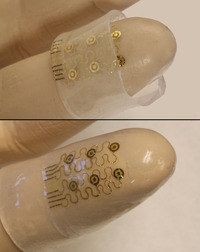
In the future, ‘surgery at your fingertips’ could be literally true. Researchers at the University of Illinois at Urbana-Champaign have created a silicon nanomembrane that can be fitted onto the fingertips and could, one day, be used in surgical procedures. From the Aug. 9, 2012 news item on ScienceDaily,

The intricate properties of the fingertips have been mimicked and recreated using semiconductor devices in what researchers hope will lead to the development of advanced surgical gloves.

The devices, shown to be capable of responding with high precision to the stresses and strains associated with touch and finger movement, are a step towards the creation of surgical gloves for use in medical procedures such as local ablations [excising or removing tissue] and ultrasound scans.

Researchers from the University of Illinois at Urbana-Champaign, Northwestern University and Dalian University of Technology have published their study August 10, in IOP [Institute of Physics] Publishing’s journal Nanotechnology.

The Aug. 10,2012 posting on the IOP website  offers this detail about the research,

The electronic circuit on the ‘skin’ is made of patterns of gold conductive lines and ultrathin sheets of silicon, integrated onto a flexible polymer called polyimide. The sheet is then etched into an open mesh geometry and transferred to a thin sheet of silicone rubber moulded into the precise shape of a finger.

This electronic ‘skin’, or finger cuff, was designed to measure the stresses and strains at the fingertip by measuring the change in capacitance – the ability to store electrical charge – of pairs of microelectrodes in the circuit.  Applied forces decreased the spacing in the skin which, in turn, increased the capacitance.

The fingertip device could also be fitted with sensors for measuring motion and temperature, with small-scale heaters as actuators for ablation and other related operations

The researchers experimented with having the electronics on the inside of the device, in contact with wearer’s skin, and also on the outside. They believe that because the device exploits materials and fabrication techniques adopted from the established semiconductor industry, the processes can be scaled for realistic use at reasonable cost.

“Perhaps the most important result is that we are able to incorporate multifunctional, silicon semiconductor device technologies into the form of soft, three-dimensional, form-fitting skins, suitable for integration not only with the fingertips but also other parts of the body,” continued Professor Rogers [John Rogers, co-author of the study].

Here’s what an image of these e-fingertips,

Virtual touch. Electronic fingertips could one day allow us to feel virtual sensations. Credit: John Rogers/University of Illinois at Urbana-Champaign

Krystnell A offers a more detailed description of the e-fingetips in an Aug. 9, 2012 story for Science NOW,

Hoping to create circuits with the flexibility of skin, materials scientist John Rogers of the University of Illinois, Urbana-Champaign, and colleagues cut up nanometer-sized strips of silicon; implanted thin, wavy strips of gold to conduct electricity; and mounted the entire circuit in a stretchable, spider web-type mesh of polymer as a support. They then embedded the circuit-polyimide structure onto a hollow tube of silicone that had been fashioned in the shape of a finger. Just like turning a sock inside out, the researchers flipped the structure so that the circuit, which was once on the outside of the tube, was on the inside where it could touch a finger placed against it.

To test the electronic fingers, the researchers put them on and pressed flat objects, such as the top of their desks. The pressure created electric currents that were transferred to the skin, which the researchers felt as mild tingling. That’s a first step in creating electrical signals that could be sent to the fingers, which could virtually recreate sensations such as heat, pressure, and texture, the team reports online today in Nanotechnology.

Rogers says another application of the technology is to custom fit the “electronic skin” around entire organs, allowing doctors to remotely monitor changes in temperature and blood flow. Electronic skin could also restore sensation to people who have lost their natural skin, he says, such as burn victims or amputees.

Here’s a link to the article which is freely accessible for 30 days after publication, from the Aug. 9, 2012 news item on ScienceDaily,

Ming Ying, Andrew P Bonifas, Nanshu Lu, Yewang Su, Rui Li, Huanyu Cheng, Abid Ameen, Yonggang Huang, John A Rogers. Silicon nanomembranes for fingertip electronics. Nanotechnology, 2012; 23 (34): 344004 DOI: 10.1088/0957-4484/23/34/344004

My best guess is that free access will no longer be available by Sept. 7 (or so) , 2012. I last wrote about John Rogers’ work in an Aug. 12, 2011 posting about electronic tattoos.

Camouflage for everyone

The Institute of Physics (IOP) journal, Bioinspiration and BIomimetics, has published an open access article on camouflage inspired by zebrafish and squid. From the IOP’s May 2, 2012 news release

Researchers from the University of Bristol have created artificial muscles that can be transformed at the flick of a switch to mimic the remarkable camouflaging abilities of organisms such as squid and zebrafish.

They demonstrate two individual transforming mechanisms that they believe could be used in ‘smart clothing’ to trigger camouflaging tricks similar to those seen in nature.

The soft, stretchy, artificial muscles are based on specialist cells called chromatophores that are found in amphibians, fish, reptiles and cephalopods, and contain pigments of colours that are responsible for the animals’ remarkable colour-changing effects.

Here’s the video mentioned in the IOP’s May 2, 2012 news release,

The lead author Jonathan Rossiter provides a description of the work (which may help you better understand what you’re seeing on the video), from the May 2, 2012 news item,

Two types of artificial chromatophores were created in the study: the first based on a mechanism adopted by a squid and the second based on a rather different mechanism adopted by zebrafish.

A typical colour-changing cell in a squid has a central sac containing granules of pigment. The sac is surrounded by a series of muscles and when the cell is ready to change colour, the brain sends a signal to the muscles and they contract. The contracting muscles make the central sacs expand, generating the optical effect which makes the squid look like it is changing colour.

The fast expansion of these muscles was mimicked using dielectric elastomers (DEs) – smart materials, usually made of a polymer, which are connected to an electric circuit and expand when a voltage is applied. They return to their original shape when they are short circuited.

In contrast, the cells in the zebrafish contain a small reservoir of black pigmented fluid that, when activated, travels to the skin surface and spreads out, much like the spilling of black ink. The natural dark spots on the surface of the zebrafish therefore appear to get bigger and the desired optical effect is achieved. The changes are usually driven by hormones.

The zebrafish cells were mimicked using two glass microscope slides sandwiching a silicone layer. Two pumps, made from flexible DEs, were positioned on both sides of the slide and were connected to the central system with silicone tubes; one pumping opaque white spirit, the other a mixture of black ink and water.

“Our artificial chromatophores are both scalable and adaptable and can be made into an artificial compliant skin which can stretch and deform, yet still operate effectively. This means they can be used in many environments where conventional ‘hard’ technologies would be dangerous, for example at the physical interface with humans, such as smart clothing,” continued Rossiter.

I wonder what these smart clothes/smart skin would feel like against your personal skin given that we are talking about ‘artificial muscles’. For example, how much movement would your clothing/smart skin have independent of you?

By independent, I mean that everything occurs externally. While we’re not ordinarily conscious of all our physical responses they are stimulated internally and part of a whole body response (even though we may notice only localized responses, e.g., a rash). In the research, there’s an external stimulus and an external response via smart clothes/smart skin.

This is just speculation as I imagine we’re several years away from any field testing of these smart clothes/smart skin, assuming that scientists are able to address all the technical hurdles between a laboratory breakthrough and developing applications.

Thanks to Nanowerk where I first came across this information (May 2, 2012 news item).

Shifting winds in the world of particle accelerators: the Fermilab

I’ve been spending more time with physicists (in my own mind, anyway) than is usual for me.  I’m sure this will pass but while I’m hot (so to speak) on the topic, here’s an item about the Fermilab in the US. From the Feb. 1, 2012 news release on EurekAlert,

In this month’s Physics World, reviews and careers editor, Margaret Harris, visits the Fermi National Accelerator Laboratory (Fermilab) to explore what future projects are in the pipeline now that the Tevatron particle accelerator has closed for good.

After 28 years of ground-breaking discoveries, the Tevatron accelerator has finally surrendered to the mighty Large Hadron Collider (LHC) at CERN [European Laboratory for Particle Physics], placing Fermilab, in some people’s mind, on the brink of disappearing into obscurity.

(I did cover some of the excitement over the Higgs Boson search at the the LHC at CERN in my Dec. 14, 2011 posting.) As for the folks at the Fermilab, they  do have plans (from the news release),

Fermilab can no longer compete with the LHC when it comes to smashing particles together at high energies, but it can look for rare interactions between particles at lower energies. In this type of experiment, the key is not a beam’s energy but its intensity: the number of particles produced per second.

Their plans include two experiments – one already being built and another in the pipeline – that will send beams of neutrinos underground to distant detectors to see how these particles change between one form and another.

More ambitious still is Project X – expected to cost between $1-2bn – which will provide intense beams of protons for experiments on neutrinos, rare decays and heavy nuclei. Outside of high-energy physics, the lab currently participates in experiments into cosmic rays, dark matter and dark energy.

One aspect,  I find particularly interesting about this news release and article is that it makes some of the positioning and jockeying for funds visible to a larger audience than is common in Canadian circles. From the news release,

One obstacle that stands in the way of Fermilab’s progression is money. With the US Congress’s budgetary process – which allocates funds one year at a time – threatening to delay projects, combined with the current economic downturn, there is cause for concern, especially for a lab currently in transition.

The other aspect I find interesting is that while the Fermilab is based in Illinois (US), the article is being published by the UK-based Institute of Physics in their Physics World journal. Is this article part of a larger public relations initiative on behalf of physicists in the UK concerned about their funding? Nassif Ghoussoub at his Piece of Mind blog notes some of the discussion currently taking place in the UK about one of its funding agencies in his Jan. 31, 2012 posting and what is sometimes called ‘basic research’.

Robert Hooke and Sir Isaac Newton; scientists and a three-century old feud

When I first came across the story, the writer was unequivocal. Sir Isaac Newton had done everything in his power to remove a rival from the history books in a campaign that persisted over years and Newton was somewhat successful.

On the recent unveiling of a Robert Hooke (Newton’s rival) portrait, the latest materials I’ve found on this topic have taken a more measured approach to Newton’s role. From the Jan. 13, 2012 news item on Science Daily,

Chroniclers of his time called him ‘despicable’, ‘mistrustful’ and ‘jealous’, and a rivalrous Isaac Newton might have had the only surviving portrait of him burnt, but, three centuries on, Robert Hooke is now regarded as one of the great Enlightenment scientists.

It was Hooke’s dispute with Isaac Newton over credit for Newton’s work on gravity that tainted more than two hundred years of historical writing about Hooke, as it is chronicled that he fought for greater credit than Newton offered for the guiding principles which were later detailed in Newton’s Principia

Hooke’s name was so thoroughly muddied his tercentenary passed unmarked. (In the UK, that’s a major affront. From what I can tell, they celebrate all historical events and important persons. Missing some of Hooke’s importance would seem unthinkable and yet, it happened.)

From the Jan. 13, 2012 news item on the European Commission CORDIS news page,

But this was only part of the story and in recent years the scientific community has woken up to the fact that Hooke was in fact one of the great Enlightenment scientists. In an effort to further correct the skewed vision of history that proliferated for so long and give Hooke credit where credit is due, the Institute of Physics (IOP) in the United Kingdom has hung a new painting of the often forgotten scientist at its London headquarters.

The painting is the work of history artist Rita Greer who started her ‘Robert Hooke project’ in 2003. Her aim was to set the record straight by chronicling the life of the scientist.

Here’s an image of the painting,

Robert Hooke painted by Rita Greer

Here are more details about Hooke from the Institute of Physics (IOP) Jan. 12, 2012 news release,

Following Hooke’s death in the early 1700s, Newton was appointed President of the Royal Society and it was during his time in this capacity that, it is thought, the only portrait of Hooke was destroyed – it is unclear whether the portrait was destroyed on Newton’s command or simply left to perish.

With no visual sources for reference, Greer has used written sources – including the chronicles of both John Aubrey and Richard Waller – to create a likeness of Hooke with details fitting to his position in the history of science.

The image set to be hung at IOP shows Hooke holding a quill and a book in his right hand and a spring in his left. The spring represents one of Hooke’s defining successes – Hooke’s law of elasticity.

Hooke’s law states that the extension of a spring is in direct proportion to the load applied to it – a law which many materials obey and which culminated in the development of a balance spring.  Balance springs subsequently enabled the development of portable timepieces – the first watches.

The history artist Rita Greer says, “Robert Hooke, brilliant, ingenious seventeenth century scientist was brushed under the carpet of history by Sir Isaac Newton and his cronies. When he had his Tercentenary there wasn’t a single memorial to him anywhere. I thought it disgraceful as Hooke did many wonderful things for science.

Sir Arnold Wolfendale FRS, a former President of the IOP and former Astronomer Royal, says, “Robert Hooke was a brilliant man of many parts of which one was physics. He was also remarkable for many advances and discoveries for which he did not receive adequate credit.

“With her fine portraits of Hooke, Rita Greer is going some way towards redressing the balance and bringing Hooke’s image to a wider audience. I think that Hooke would have been pleased with her persistence, as we are at the IOP.”

Robert Hooke was a key part of the group that went on to form the Royal Society, becoming the first Curator of Experiments for the Society in 1662.

Hooke has many physics-related credits to his name, including the construction of the vacuum pumps used in Boyle’s gas law experiments, building some of the earliest Gregorian telescopes and observing the rotations of Mars and Jupiter, deducing the wave theory of light, and being the first to suggest that matter expands when heated and that air is made of small particles.

Whether or not he intended to destroy the last portrait of Hooke (I’m inclined to think that was his intention) Newton didn’t manage to remove Hooke from the history books entirely but it certainly seems that he enjoyed three very successful centuries until Rita Greer came along.

University of Texas at Dallas lab demos cloaking device visible to naked eye

Invisibility cloaks have been everywhere lately and I’ve been getting a little blasé about them but then I saw this Oct. 4, 2011 news item on physorg.com,

Scientists have created a working cloaking device that not only takes advantage of one of nature’s most bizarre phenomenon, but also boasts unique features; it has an ‘on and off’ switch and is best used underwater.

For the first time, I was able to see an invisibility cloak in action, here’s the video,

For the curious here’s how it works (from the Oct. 4, 2011 news release on the Institute of Physics website),

This novel design, presented today, Tuesday 4 September [Tuesday 4 October?], in IOP [Institute of Physics] Publishing’s journal Nanotechnology, makes use of sheets of carbon nanotubes (CNT) – one-molecule-thick sheets of carbon wrapped up into cylindrical tubes.

CNTs have such unique properties, such as having the density of air but the strength of steel, that they have been extensively studied and put forward for numerous applications; however it is their exceptional ability to conduct heat and transfer it to surrounding areas that makes them an ideal material to exploit the so-called “mirage effect”.

The most common example of a mirage is when an observer appears to see pools of water on the ground. This occurs because the air near the ground is a lot warmer than the air higher up, causing lights rays to bend upward towards the viewer’s eye rather than bounce off the surface.

This results in an image of the sky appearing on the ground which the viewer perceives as water actually reflecting the sky; the brain sees this as a more likely occurrence.

Through electrical stimulation, the transparent sheet of highly aligned CNTs can be easily heated to high temperatures. They then have the ability to transfer that heat to its surrounding areas, causing a steep temperature gradient. Just like a mirage, this steep temperature gradient causes the light rays to bend away from the object concealed behind the device, making it appear invisible.

With this method, it is more practical to demonstrate cloaking underwater as all of the apparatus can be contained in a petri dish. It is the ease with which the CNTs can be heated that gives the device its unique ‘on and off’ feature.

Congratulations to Dr. Ali Aliev (lead author) and the rest of the University of Texas at Dallas team!

ETA Oct. 5, 2011: I added the preposition ‘of’ to the title and I’m adding a comment about invisibility cloaks.

Comment: Most of the invisibility cloaks I’ve read about are at the nanoscale which means none of us outside a laboratory could possibly observe the cloak in action. Seeing this video demonstrating an invisibility cloak in the range of visible light and at a macroscale was a dream come true, so to speak.