An adhesive that US and Chinese scientists have developed shows great promise not just for bandages but wearable robotics too. From a December 14, 2018 news item on Nanowerk,
Researchers from the Harvard John A. Paulson School of Engineering and Applied Sciences (SEAS) and Xi’an Jiaotong University in China have developed a new type of adhesive that can strongly adhere wet materials — such as hydrogel and living tissue — and be easily detached with a specific frequency of light.
The adhesives could be used to attach and painlessly detach wound dressings, transdermal drug delivery devices, and wearable robotics.
“Strong adhesion usually requires covalent bonds, physical interactions, or a combination of both,” said Yang Gao, first author of the paper and researcher at Xi’an Jiaotong University. “Adhesion through covalent bonds is hard to remove and adhesion through physical interactions usually requires solvents, which can be time-consuming and environmentally harmful. Our method of using light to trigger detachment is non-invasive and painless.”
The adhesive uses an aqueous solution of polymer chains spread between two, non-sticky materials — like jam between two slices of bread. On their own, the two materials adhere poorly together but the polymer chains act as a molecular suture, stitching the two materials together by forming a network with the two preexisting polymer networks. This process is known as topological entanglement.
When exposed to ultra-violet light, the network of stitches dissolves, separating the two materials.
The researchers, led by Zhigang Suo, the Allen E. and Marilyn M. Puckett Professor of Mechanics and Materials at SEAS, tested adhesion and detachment on a range of materials, sticking together hydrogels; hydrogels and organic tissue; elastomers; hydrogels and elastomers; and hydrogels and inorganic solids.
“Our strategy works across a range of materials and may enable broad applications,” said Kangling Wu, co-lead author and researcher at Xi’an Jiaotong University in China. While the researchers focused on using UV light to trigger detachment, their work suggests the possibility that the stitching polymer could detach with near-infrared light, a feature which could be applied to a range of new medical procedures.
“In nature, wet materials don’t like to adhere together,” said Suo. “We have discovered a general approach to overcome this challenge. Our molecular sutures can strongly adhere wet materials together. Furthermore, the strong adhesion can be made permanent, transient, or detachable on demand, in response to a cue. So, as we see it, nature is full of loopholes, waiting to be stitched.”
Here’s a link to and a citation for the paper,
Photodetachable Adhesion by Yang Gao, Kangling Wu, Zhigang Suo. https://doi.org/10.1002/adma.201806948 First published: 14 December 2018
Engineers at the University of Maryland in College Park have found a way to make wood more than ten times times stronger and tougher than before, creating a natural substance that is stronger than titanium alloy.
“This new way to treat wood makes it twelve times stronger than natural wood and ten times tougher,” said Liangbing Hu, the leader of the team that did the research, to be published on Thursday [February 7, 2018] in the journal Nature. “This could be a competitor to steel or even titanium alloys, it is so strong and durable. It’s also comparable to carbon fiber, but much less expensive.” Hu is an associate professor of materials science and engineering and a member of the Maryland Energy Innovation Institute.
“It is both strong and tough, which is a combination not usually found in nature,” said Teng Li, the co-leader of the team and the Samuel P. Langley associate professor of mechanical engineering at the University of Maryland. His team measured the dense wood’s mechanical properties. “It is as strong as steel, but six times lighter. It takes 10 times more energy to fracture than natural wood. It can even be bent and molded at the beginning of the process.”
The team’s process begins by removing the wood’s lignin, the part of the wood that makes it both rigid and brown in color. Then it is compressed under mild heat, at about 150 F. This causes the cellulose fibers to become very tightly packed. Any defects like holes or knots are crushed together. The treatment process was extended a little further with a coat of paint.
The scientists found that the wood’s fibers are pressed together so tightly that they can form strong hydrogen bonds, like a crowd of people who can’t budge – who are also holding hands. The compression makes the wood five times thinner than its original size.
The team also tested the material by shooting a bullet-like projectile at it. Unlike natural wood, which was blown straight through, the fully treated wood actually stopped the projectile partway through.
“Soft woods like pine or balsa, which grow fast and are more environmentally friendly, could replace slower-growing but denser woods like teak, in furniture or buildings,” Hu said.
“The paper provides a highly promising route to the design of light weight high performance structural materials, with tremendous potential for a broad range of applications where high strength, large toughness and superior ballistic resistance are desired, “ said Dr. Huajian Gao, a professor at Brown University, who was not involved in the study. “It is particularly exciting to note that the method is versatile for various species of wood and fairly easy to implement.”
“This kind of wood could be used in cars, airplanes, buildings – any application where steel is used,” Hu said.
“The two-step process reported in this paper achieves exceptionally high strength, much beyond what [is] reported in the literature,” said Dr. Zhigang Suo, a professor of mechanics and materials at Harvard University, also not involved with the study. “Given the abundance of wood, as well as other cellulose-rich plants, this paper inspires imagination.”
“The most outstanding observation, in my view, is the existence of a limiting concentration of lignin, the glue between wood cells, to maximize the mechanical performance of the densified wood. Too little or too much removal lower the strength compared to a maximum value achieved at intermediate or partial lignin removal. This reveals the subtle balance between hydrogen bonding and the adhesion imparted by such polyphenolic compound. Moreover, of outstanding interest, is the fact that that wood densification leads to both, increased strength and toughness, two properties that usually offset each other,” said Orlando J. Rojas, a professor at Aalto University in Finland.
Hu’s research has explored the capacities of wood’s natural nanotechnology [emphasis mine]. They previously made a range of emerging technologies out of nanocellulose related materials: (1) super clear paper for replacing plastic; (2) photonic paper for improving solar cell efficiency by 30%; (3) a battery and a supercapacitor out of wood; (4) a battery from a leaf; (5) transparent wood for energy efficient buildings; (6) solar water desalination for drinking and specifically filtering out toxic dyes. These wood-based emerging technologies are being commercialized through a UMD spinoff company, Inventwood LLC.
At a guess, “wood’s natural nanotechnology” refers to the properties of wood and other forms of cellulose at the nanoscale.
Here’s a link to and a citation for the paper,
Processing bulk natural wood into a high-performance structural material by Jianwei Song, Chaoji Chen, Shuze Zhu, Mingwei Zhu, Jiaqi Dai, Upamanyu Ray, Yiju Li, Yudi Kuang, Yongfeng Li, Nelson Quispe, Yonggang Yao, Amy Gong, Ulrich H. Leiste, Hugh A. Bruck, J. Y. Zhu, Azhar Vellore, Heng Li, Marilyn L. Minus, Zheng Jia, Ashlie Martini, Teng Li, & Liangbing Hu. Nature volume 554, pages 224–228 (08 February 2018) doi:10.1038/nature25476 Published online: 07 February 2018
Rumpelstiltskin is a fairy tale whereby a young girl is trapped by her father’s lie that she can spin straw into gold. She is forced to spin gold by the King under pain of execution when an imp offers to help in exchange for various goods. As she succeeds each time, the King demands more until finally she has nothing left to trade for the imp’s help. Well, there is one last thing: her first-born child. She agrees to the bargain and she marries the King. On the birth of their first child, the imp reappears and under pressure of her pleas makes one last bargain. She must guess his name which she does, Rumplestiltskin. (The full story along with variants is here in this Wikipedia entry.)
With this latest research, we have a reverse Rumpelstiltskin story where gold is turned into something else according to a June 13, 2016 news item on Nanowerk (Note: A link has been removed),
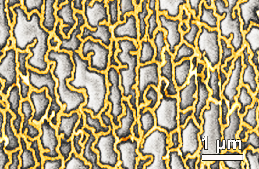
Flexible solar panels that could be rolled up for easy transport and other devices would benefit from transparent metal electrodes that can conduct electricity, are stretchable, and resist damage following repeated stretching. Researchers found that topology and the adhesion between a metal nanomesh and the underlying substrate played key roles in creating such materials. The metal nanomesh can be stretched to three times its length while maintaining a transparency comparable to similar commercial materials used in solar cells and flat panel displays. Also, nanomeshes on pre-stretched slippery substrates led to electrodes that didn’t wear out, even after being stretched 50,000 times (Proceedings of the National Academy of Sciences, “Fatigue-free, superstretchable, transparent, and biocompatible metal electrodes”).
Tuning topology and adhesion of metal nanomeshes has led to super stretchable, transparent electrodes that don’t wear out. The scanning electron microscopy image shows the structure of a gold mesh created with a special lithographic technique that controlled the dimensions of the mesh structure. Optimizing this structure and its adhesion to the substrate was key to achieving super stretchability and long lifetimes in use—nanomeshes on pre-stretched slippery substrates did not show signs of wear even after repeated stretching, up to 50,000 cycles.
Next-generation flexible electronics require highly stretchable and transparent electrodes. Fatigue, structural damage due to repeated use, is deadly in metals as it leads to poor conductivity and it commonly occurs in metals with repeated stretching—even with short elongations. However, few electronic conductors are transparent and stretchable, even fewer can be cyclically stretched to a large strain without causing fatigue. Now researchers led by the University of Houston found that optimizing topology of a metal nanomesh and its adhesion to an underlying substrate improved stretchability and eliminated fatigue, while maintaining transparency. A special lithographic technique called “grain boundary lithography” controlled the dimensions of the mesh structure. The metal nanomesh remained transparent after being stretched to three times its length. Gold nanomeshes on prestretched slippery substrates impressively showed no wear when stretched 50,000 times. The slippery surface advantageously allowed the structure of the nanomesh to reorient to relax the stress. Such electrically conductive, flexible, and transparent electrodes could lead to next-generation flexible electronics such as advanced solar cells. The nanomesh electrodes are also promising for implantable electronics because the nanomeshes are biocompatible.
There’s been a lot of talk about foldable, stretchable, and/or bendable electronics, which is exciting in itself but I find this work on developing a fatigue-free conductor particularly intriguing. After all, who hasn’t purchased something that stretches, folds, etc. only to find that it becomes ‘fatigued’ and is now ‘stretched out’.
Researchers have discovered a new stretchable, transparent conductor that can be folded or stretched and released, resulting in a large curvature or a significant strain, at least 10,000 times without showing signs of fatigue.
This is a crucial step in creating a new generation of foldable electronics – think a flat-screen television that can be rolled up for easy portability – and implantable medical devices. The work, published Monday [Sept. 21, 2015] in the Proceedings of the National Academy of Sciences, pairs gold nanomesh with a stretchable substrate made with polydimethylsiloxane, or PDMS.
The research is the result of an international collaboration including the University of Houston (US), Harvard University (US), Methodist Research Institute (US), Zhengzhou University (China), Lawrence Berkeley National Laboratory (LBNL; US).
The substrate is stretched before the gold nanomesh is placed on it – a process known as “prestretching” – and the material showed no sign of fatigue when cyclically stretched to a strain of more than 50 percent.
The gold nanomesh also proved conducive to cell growth, indicating it is a good material for implantable medical devices.
Fatigue is a common problem for researchers trying to develop a flexible, transparent conductor, making many materials that have good electrical conductivity, flexibility and transparency – all three are needed for foldable electronics – wear out too quickly to be practical, said Zhifeng Ren, a physicist at the University of Houston and principal investigator at the Texas Center for Superconductivity, who was the lead author for the paper.
The new material, produced by grain boundary lithography, solves that problem, he said.
In addition to Ren, other researchers on the project included Chuan Fei Guo and Ching-Wu “Paul” Chu, both from UH; Zhigang Suo, Qihan Liu and Yecheng Wang, all from Harvard University, and Guohui Wang and Zhengzheng Shi, both from the Houston Methodist Research Institute.
In materials science, “fatigue” is used to describe the structural damage to a material caused by repeated movement or pressure, known as “strain cycling.” Bend a material enough times, and it becomes damaged or breaks. That means the materials aren’t durable enough for consumer electronics or biomedical devices.
“Metallic materials often exhibit high cycle fatigue, and fatigue has been a deadly disease for metals,” the researchers wrote.
“We weaken the constraint of the substrate by making the interface between the Au (gold) nanomesh and PDMS slippery, and expect the Au nanomesh to achieve superstretchability and high fatigue resistance,” they wrote in the paper. “Free of fatigue here means that both the structure and the resistance do not change or have little change after many strain cycles.”
As a result, they reported, “the Au nanomesh does not exhibit strain fatigue when it is stretched to 50 percent for 10,000 cycles.”
Many applications require a less dramatic stretch – and many materials break with far less stretching – so the combination of a sufficiently large range for stretching and the ability to avoid fatigue over thousands of cycles indicates a material that would remain productive over a long period of time, Ren said.
The grain boundary lithography involved a bilayer lift-off metallization process, which included an indium oxide mask layer and a silicon oxide sacrificial layer and offers good control over the dimensions of the mesh structure.
The researchers used mouse embryonic fibroblast cells to determine biocompatibility; that, along with the fact that the stretchability of gold nanomesh on a slippery substrate resembles the bioenvironment of tissue or organ surfaces, suggest the nanomesh “might be implanted in the body as a pacemaker electrode, a connection to nerve endings or the central nervous system, a beating heart, and so on,” they wrote.
Having taught a course on bioelectronics for Simon Fraser University’s (Vancouver, Canada) Continuing Studies Program, this latest work from Harvard University (US) caught my attention. A Harvard research team has developed a technique which could allow doctors to inject us with electronics, should we need them. From a June 8, 2015 news item on phys.org,
It’s a notion that might be pulled from the pages of science-fiction novel – electronic devices that can be injected directly into the brain, or other body parts, and treat everything from neurodegenerative disorders to paralysis.
It sounds unlikely, until you visit Charles Lieber’s lab.
A team of international researchers, led by Lieber, the Mark Hyman, Jr. Professor of Chemistry, an international team of researchers developed a method for fabricating nano-scale electronic scaffolds that can be injected via syringe. Once connected to electronic devices, the scaffolds can be used to monitor neural activity, stimulate tissues and even promote regenerations of neurons. …
Here’s an image provided by the researchers,
Bright-field image showing the mesh electronics being injected through sub-100 micrometer inner diameter glass needle into aqueous solution. mage courtesy of Lieber Research Group, Harvard University
“I do feel that this has the potential to be revolutionary,” Lieber said. “This opens up a completely new frontier where we can explore the interface between electronic structures and biology. For the past thirty years, people have made incremental improvements in micro-fabrication techniques that have allowed us to make rigid probes smaller and smaller, but no one has addressed this issue – the electronics/cellular interface – at the level at which biology works.”
The idea of merging the biological with the electronic is not a new one for Lieber.
In an earlier study, scientists in Lieber’s lab demonstrated that the scaffolds could be used to create “cyborg” tissue – when cardiac or nerve cells were grown with embedded scaffolds. [emphasis mine] Researchers were then able to use the devices to record electrical signals generated by the tissues, and to measure changes in those signals as they administered cardio- or neuro-stimulating drugs.
“We were able to demonstrate that we could make this scaffold and culture cells within it, but we didn’t really have an idea how to insert that into pre-existing tissue,” Lieber said. “But if you want to study the brain or develop the tools to explore the brain-machine interface, you need to stick something into the body. When releasing the electronics scaffold completely from the fabrication substrate, we noticed that it was almost invisible and very flexible like a polymer and could literally be sucked into a glass needle or pipette. From there, we simply asked, would it be possible to deliver the mesh electronics by syringe needle injection, a process common to delivery of many species in biology and medicine – you could go to the doctor and you inject this and you’re wired up.'”
Though not the first attempts at implanting electronics into the brain – deep brain stimulation has been used to treat a variety of disorders for decades – the nano-fabricated scaffolds operate on a completely different scale.
“Existing techniques are crude relative to the way the brain is wired,” Lieber explained. “Whether it’s a silicon probe or flexible polymers…they cause inflammation in the tissue that requires periodically changing the position or the stimulation. But with our injectable electronics, it’s as if it’s not there at all. They are one million times more flexible than any state-of-the-art flexible electronics and have subcellular feature sizes. They’re what I call “neuro-philic” – they actually like to interact with neurons..”
Despite their enormous potential, the fabrication of the injectable scaffolds is surprisingly easy.
“That’s the beauty of this – it’s compatible with conventional manufacturing techniques,” Lieber said.
The process is similar to that used to etch microchips, and begins with a dissolvable layer deposited on a substrate. To create the scaffold, researchers lay out a mesh of nanowires sandwiched in layers of organic polymer. The first layer is then dissolved, leaving the flexible mesh, which can be drawn into a syringe needle and administered like any other injection.
After injection, the input/output of the mesh can be connected to standard measurement electronics so that the integrated devices can be addressed and used to stimulate or record neural activity.
“These type of things have never been done before, from both a fundamental neuroscience and medical perspective,” Lieber said. “It’s really exciting – there are a lot of potential applications.”
Going forward, Lieber said, researchers hope to better understand how the brain and other tissues react to the injectable electronics over longer periods.
Lieber’s earlier work on “cyborg tissue” was briefly mentioned here in a Feb. 20, 2014 posting.
Getting back to the most recent work, here’s a link to and a citation for the paper,
Syringe-injectable electronics by Jia Liu, Tian-Ming Fu, Zengguang Cheng, Guosong Hong, Tao Zhou, Lihua Jin, Madhavi Duvvuri, Zhe Jiang, Peter Kruskal, Chong Xie, Zhigang Suo, Ying Fang, & Charles M. Lieber. Nature Nanotechnology (2015) doi:10.1038/nnano.2015.115 Published online 08 June 2015
This paper is behind a paywall but there is a free preview via ReadCube Access.
One final note, the researchers have tested the injectable electronics (or mesh electronics) in vivo (live animals).
I have two items about implants and brains and an item about being able to exert remote control of the brain, all of which hint at a cyborg future for at least a few of us.
e-Dura, the spinal column, and the brain
The first item concerns some research, at the École Polytechnique de Lausanne (EPFL) which features flexible electronics. From a March 24, 2015 article by Ben Schiller for Fast Company (Note: Links have been removed),
Researchers at the Swiss Federal Institute of Technology, in Lausanne, have developed the e-Dura—a tiny skinlike device that attaches directly to damaged spinal cords. By sending out small electrical pulses, it stimulates the cord as if it were receiving signals from the brain, thus allowing movement.
“The purpose of the neuro-prosthesis is to excite the neurons that are on the spinal cord below the site of the injury and activate them, just like if they were receiving information from the brain,” says Stéphanie Lacour, a professor at the institute.
EPFL scientists have managed to get rats walking on their own again using a combination of electrical and chemical stimulation. But applying this method to humans would require multifunctional implants that could be installed for long periods of time on the spinal cord without causing any tissue damage. This is precisely what the teams of professors Stéphanie Lacour and Grégoire Courtine have developed. Their e-Dura implant is designed specifically for implantation on the surface of the brain or spinal cord. The small device closely imitates the mechanical properties of living tissue, and can simultaneously deliver electric impulses and pharmacological substances. The risks of rejection and/or damage to the spinal cord have been drastically reduced. An article about the implant will appear in early January [2015] in Science Magazine.
So-called “surface implants” have reached a roadblock; they cannot be applied long term to the spinal cord or brain, beneath the nervous system’s protective envelope, otherwise known as the “dura mater,” because when nerve tissues move or stretch, they rub against these rigid devices. After a while, this repeated friction causes inflammation, scar tissue buildup, and rejection.
Here’s what the implant looks like,
Courtesy: EPFL
The press release describes how the implant is placed (Note: A link has been removed),
Flexible and stretchy, the implant developed at EPFL is placed beneath the dura mater, directly onto the spinal cord. Its elasticity and its potential for deformation are almost identical to the living tissue surrounding it. This reduces friction and inflammation to a minimum. When implanted into rats, the e-Dura prototype caused neither damage nor rejection, even after two months. More rigid traditional implants would have caused significant nerve tissue damage during this period of time.
The researchers tested the device prototype by applying their rehabilitation protocol — which combines electrical and chemical stimulation – to paralyzed rats. Not only did the implant prove its biocompatibility, but it also did its job perfectly, allowing the rats to regain the ability to walk on their own again after a few weeks of training.
“Our e-Dura implant can remain for a long period of time on the spinal cord or the cortex, precisely because it has the same mechanical properties as the dura mater itself. This opens up new therapeutic possibilities for patients suffering from neurological trauma or disorders, particularly individuals who have become paralyzed following spinal cord injury,” explains Lacour, co-author of the paper, and holder of EPFL’s Bertarelli Chair in Neuroprosthetic Technology.
The press release goes on to describe the engineering achievements,
Developing the e-Dura implant was quite a feat of engineering. As flexible and stretchable as living tissue, it nonetheless includes electronic elements that stimulate the spinal cord at the point of injury. The silicon substrate is covered with cracked gold electric conducting tracks that can be pulled and stretched. The electrodes are made of an innovative composite of silicon and platinum microbeads. They can be deformed in any direction, while still ensuring optimal electrical conductivity. Finally, a fluidic microchannel enables the delivery of pharmacological substances – neurotransmitters in this case – that will reanimate the nerve cells beneath the injured tissue.
The implant can also be used to monitor electrical impulses from the brain in real time. When they did this, the scientists were able to extract with precision the animal’s motor intention before it was translated into movement.
“It’s the first neuronal surface implant designed from the start for long-term application. In order to build it, we had to combine expertise from a considerable number of areas,” explains Courtine, co-author and holder of EPFL’s IRP Chair in Spinal Cord Repair. “These include materials science, electronics, neuroscience, medicine, and algorithm programming. I don’t think there are many places in the world where one finds the level of interdisciplinary cooperation that exists in our Center for Neuroprosthetics.”
For the time being, the e-Dura implant has been primarily tested in cases of spinal cord injury in paralyzed rats. But the potential for applying these surface implants is huge – for example in epilepsy, Parkinson’s disease and pain management. The scientists are planning to move towards clinical trials in humans, and to develop their prototype in preparation for commercialization.
EPFL has provided a video of researcher Stéphanie Lacour describing e-Dura and expressing hopes for its commercialization,
Here’s a link to and a citation for the paper,
Electronic dura mater for long-term multimodal neural interfaces by Ivan R. Minev, Pavel Musienko, Arthur Hirsch, Quentin Barraud, Nikolaus Wenger, Eduardo Martin Moraud, Jérôme Gandar, Marco Capogrosso, Tomislav Milekovic, Léonie Asboth, Rafael Fajardo Torres, Nicolas Vachicouras, Qihan Liu, Natalia Pavlova, Simone Duis, Alexandre Larmagnac, Janos Vörös, Silvestro Micera, Zhigang Suo, Grégoire Courtine, Stéphanie P. Lacour. Science 9 January 2015: Vol. 347 no. 6218 pp. 159-163 DOI: 10.1126/science.1260318
This paper is behind a paywall.
Carbon nanotube fibres could connect to the brain
Researchers at Rice University (Texas, US) are excited about the possibilities that carbon nanotube fibres offer in the field of implantable electronics for the brain. From a March 25, 2015 news item on Nanowerk,
Carbon nanotube fibers invented at Rice University may provide the best way to communicate directly with the brain.
The fibers have proven superior to metal electrodes for deep brain stimulation and to read signals from a neuronal network. Because they provide a two-way connection, they show promise for treating patients with neurological disorders while monitoring the real-time response of neural circuits in areas that control movement, mood and bodily functions.
New experiments at Rice demonstrated the biocompatible fibers are ideal candidates for small, safe electrodes that interact with the brain’s neuronal system, according to the researchers. They could replace much larger electrodes currently used in devices for deep brain stimulation therapies in Parkinson’s disease patients.
They may also advance technologies to restore sensory or motor functions and brain-machine interfaces as well as deep brain stimulation therapies for other neurological disorders, including dystonia and depression, the researchers wrote.
The fibers created by the Rice lab of chemist and chemical engineer Matteo Pasquali consist of bundles of long nanotubes originally intended for aerospace applications where strength, weight and conductivity are paramount.
The individual nanotubes measure only a few nanometers across, but when millions are bundled in a process called wet spinning, they become thread-like fibers about a quarter the width of a human hair.
“We developed these fibers as high-strength, high-conductivity materials,” Pasquali said. “Yet, once we had them in our hand, we realized that they had an unexpected property: They are really soft, much like a thread of silk. Their unique combination of strength, conductivity and softness makes them ideal for interfacing with the electrical function of the human body.”
The simultaneous arrival in 2012 of Caleb Kemere, a Rice assistant professor who brought expertise in animal models of Parkinson’s disease, and lead author Flavia Vitale, a research scientist in Pasquali’s lab with degrees in chemical and biomedical engineering, prompted the investigation.
“The brain is basically the consistency of pudding and doesn’t interact well with stiff metal electrodes,” Kemere said. “The dream is to have electrodes with the same consistency, and that’s why we’re really excited about these flexible carbon nanotube fibers and their long-term biocompatibility.”
Weeks-long tests on cells and then in rats with Parkinson’s symptoms proved the fibers are stable and as efficient as commercial platinum electrodes at only a fraction of the size. The soft fibers caused little inflammation, which helped maintain strong electrical connections to neurons by preventing the body’s defenses from scarring and encapsulating the site of the injury.
The highly conductive carbon nanotube fibers also show much more favorable impedance – the quality of the electrical connection — than state-of-the-art metal electrodes, making for better contact at lower voltages over long periods, Kemere said.
The working end of the fiber is the exposed tip, which is about the width of a neuron. The rest is encased with a three-micron layer of a flexible, biocompatible polymer with excellent insulating properties.
The challenge is in placing the tips. “That’s really just a matter of having a brain atlas, and during the experiment adjusting the electrodes very delicately and putting them into the right place,” said Kemere, whose lab studies ways to connect signal-processing systems and the brain’s memory and cognitive centers.
Doctors who implant deep brain stimulation devices start with a recording probe able to “listen” to neurons that emit characteristic signals depending on their functions, Kemere said. Once a surgeon finds the right spot, the probe is removed and the stimulating electrode gently inserted. Rice carbon nanotube fibers that send and receive signals would simplify implantation, Vitale said.
The fibers could lead to self-regulating therapeutic devices for Parkinson’s and other patients. Current devices include an implant that sends electrical signals to the brain to calm the tremors that afflict Parkinson’s patients.
“But our technology enables the ability to record while stimulating,” Vitale said. “Current electrodes can only stimulate tissue. They’re too big to detect any spiking activity, so basically the clinical devices send continuous pulses regardless of the response of the brain.”
Kemere foresees a closed-loop system that can read neuronal signals and adapt stimulation therapy in real time. He anticipates building a device with many electrodes that can be addressed individually to gain fine control over stimulation and monitoring from a small, implantable device.
“Interestingly, conductivity is not the most important electrical property of the nanotube fibers,” Pasquali said. “These fibers are intrinsically porous and extremely stable, which are both great advantages over metal electrodes for sensing electrochemical signals and maintaining performance over long periods of time.”
The paper is open access provided you register on the website.
Remote control for stimulation of the brain
Mo Costandi, neuroscientist and freelance science writer, has written a March 24, 2015 post for the Guardian science blog network focusing on neuronal remote control,
Two teams of scientists have developed new ways of stimulating neurons with nanoparticles, allowing them to activate brain cells remotely using light or magnetic fields. The new methods are quicker and far less invasive than other hi-tech methods available, so could be more suitable for potential new treatments for human diseases.
Researchers have various methods for manipulating brain cell activity, arguably the most powerful being optogenetics, which enables them to switch specific brain cells on or off with unprecedented precision, and simultaneously record their behaviour, using pulses of light.
This is very useful for probing neural circuits and behaviour, but involves first creating genetically engineered mice with light-sensitive neurons, and then inserting the optical fibres that deliver light into the brain, so there are major technical and ethical barriers to its use in humans.
Nanomedicine could get around this. Francisco Bezanilla of the University of Chicago and his colleagues knew that gold nanoparticles can absorb light and convert it into heat, and several years ago they discovered that infrared light can make neurons fire nervous impulses by heating up their cell membranes.
…
Polina Anikeeva’s team at the Massachusetts Institute of Technology adopted a slightly different approach, using spherical iron oxide particles that give off heat when exposed to an alternating magnetic field.
…
Although still in the experimental stages, research like this may eventually allow for wireless and minimally invasive deep brain stimulation of the human brain. Bezanilla’s group aim to apply their method to develop treatments for macular degeneration and other conditions that kill off light-sensitive cells in the retina. This would involve injecting nanoparticles into the eye so that they bind to other retinal cells, allowing natural light to excite them into firing impulses to the optic nerve.
Costandi’s article is intended for an audience that either understands the science or can deal with the uncertainty of not understanding absolutely everything. Provided you fall into either of those categories, the article is well written and it provides links and citations to the papers for both research teams being featured.
Taken together, the research at EPFL, Rice University, University of Chicago, and Massachusetts Institute of Technology provides a clue as to how much money and intellectual power is being directed at the brain.
A multi-institutional research team has developed a method for embedding networks of biocompatible nanoscale wires within engineered tissues. These networks—which mark the first time that electronics and tissue have been truly merged in 3D—allow direct tissue sensing and potentially stimulation, a potential boon for development of engineered tissues that incorporate capabilities for monitoring and stimulation, and of devices for screening new drugs.
The Aug. 27, 2012 news item on Nanowerk provides more detail about integration of the cells and electronics,
Until now, the only cellular platforms that incorporated electronic sensors consisted of flat layers of cells grown on planar metal electrodes or transistors. Those two-dimensional systems do not accurately replicate natural tissue, so the research team set out to design a 3-D scaffold that could monitor electrical activity, allowing them to see how cells inside the structure would respond to specific drugs.
The researchers built their new scaffold out of epoxy, a nontoxic material that can take on a porous, 3-D structure. Silicon nanowires embedded in the scaffold carry electrical signals to and from cells grown within the structure.
“The scaffold is not just a mechanical support for cells, it contains multiple sensors. We seed cells into the scaffold and eventually it becomes a 3-D engineered tissue,” Tian says [Bozhi Tian, a former postdoc at MIT {Massachusetts Institute of Technology} and Children’s Hospital and a lead author of the paper ].
The team chose silicon nanowires for electronic sensors because they are small, stable, can be safely implanted into living tissue and are more electrically sensitive than metal electrodes. The nanowires, which range in diameter from 30 to 80 nanometers (about 1,000 times smaller than a human hair), can detect voltages less than one-thousandth of a watt, which is the level of electricity that might be seen in a cell.
“The current methods we have for monitoring or interacting with living systems are limited,” said Lieber [Charles M. Lieber, the Mark Hyman, Jr. Professor of Chemistry at Harvard and one of the study’s team leaders]. “We can use electrodes to measure activity in cells or tissue, but that damages them. With this technology, for the first time, we can work at the same scale as the unit of biological system without interrupting it. Ultimately, this is about merging tissue with electronics in a way that it becomes difficult to determine where the tissue ends and the electronics begin.”
The research addresses a concern that has long been associated with work on bioengineered tissue – how to create systems capable of sensing chemical or electrical changes in the tissue after it has been grown and implanted. The system might also represent a solution to researchers’ struggles in developing methods to directly stimulate engineered tissues and measure cellular reactions.
“In the body, the autonomic nervous system keeps track of pH, chemistry, oxygen and other factors, and triggers responses as needed,” Kohane [Daniel Kohane, a Harvard Medical School professor in the Department of Anesthesia at Children’s Hospital Boston and a team leader] explained. “We need to be able to mimic the kind of intrinsic feedback loops the body has evolved in order to maintain fine control at the cellular and tissue level.”
Here’s a citation and a link to the paper (which is behind a paywall),
Macroporous nanowire nanoelectronic scaffolds for synthetic tissues by Bozhi Tian, Jia Lin, Tal Dvir, Lihua Jin, Jonathan H. Tsui, Quan Qing, Zhigang Suo, Robert Langer, Daniel S. Kohane, and Charles M. Lieber in Nature Materials (2012) doi:10.1038/nmat3404 Published onlin26 August 2012.
This is the image MIT included with its Aug 27, 2012 news release (which originated the news item on Nanowerk),
A 3-D reconstructed confocal fluorescence micrograph of a tissue scaffold. Image: Charles M. Lieber and Daniel S. Kohane.
At this point they’re discussing therapeutic possibilities but I expect that ‘enhancement’ is also being considered although not mentioned for public consumption.