A July 10,2024 news item on ScienceDaily announces work on a new material for use in neuroprostheses could help people who are paralyzed or have amputated limbs, Note: This research involves use of animal models,
Neuroprostheses allow the nervous system of a patient who has suffered an injury to connect with mechanical devices that replace paralyzed or amputated limbs. A study coordinated by the UAB Institut de Neurociències, in collaboration with the l’Institut Català de Nanociència i Nanotecnologia (ICN2), demonstrates in animal models how EGNITE, a derivative of graphene, allows the creation of smaller electrodes, which can interact more selectively with the nerves they stimulate, thus improving the efficacy of the prostheses. The study also demonstrated that EGNITE is biocompatible, showing that its implantation is safe.
After an amputation or a severe nerve injury, patients lose to a greater or lesser extent the ability to move and feel a lost limb, which limits their autonomy in activities of daily living. Currently, the only strategy that allows to recover the lost functions consists of neuroprostheses: electrodes capable of stimulating the nerves, to induce specific sensations, and of recording motor signals that, once decoded, can be sent to a bionic prosthesis.
In the design of neuroprostheses, it is important that the electrodes are small enough that they are selective and interact electrically only with a reduced number of axons in the nerve. Therefore, although they have commonly been constructed from metals such as gold, platinum or iridium oxide, it is necessary to find other materials that have enhanced conductive capacity and allow the creation of even smaller electrode contacts. This is where graphene and its derivatives come into play; their excellent electrical properties have allowed the development of a new generation of microelectrodes.
A research coordinated by the UAB Institut de Neurociències (INc-UAB) has studied the capacity of a new material derived from graphene, EGNITE, to stimulate and record from the peripheral nerve. Furthermore, its biocompatibility has been validated, which is key for preserving the function of the interface over time. The research was carried out in the Neuroplasticity and Regeneration group of the INc-UAB, led by professor Xavier Navarro of the UAB Department of Cell Biology, Physiology and Immunology, in collaboration with Jose Garrido’s research group at the Institut Català de Nanociència i Nanotecnologia (ICN2), which was in charge of developing the EGNITE together with the neural interfaces.
These electrodes, implanted in rat sciatic nerve, were shown to be capable of producing selective muscle activation for up to a maximum of 60 days. “The reduction in the electrical current necessary to produce this muscle activation is notable in comparison to other larger metal microelectrodes”, explains Bruno Rodríguez-Meana, postdoctoral researcher at the INc-UAB and first author of the article. Furthermore, the electrodes with EGNITE demonstrated to be biocompatible, since none of the functional tests showed significant alterations produced by the implanted interfaces nor was exacerbated inflammation observed.
“The next steps will consist of the optimization of the EGNITE-based technology and its application in pre-clinical studies for vagus nerve or spinal cord stimulation systems. In parallel, progress is being made towards its clinical translation in bioelectronic medicine approaches”, explains Professor Navarro.
Together, these results indicate the potential of the material derived from graphene to be part of neuroprostheses that allow patients to recover lost functions, thus improving their capacity and quality of life.
I have two bits about prosthetics, one which focuses on how most of us think of them and another about science fiction fantasies.
Better motor control
This new technology comes via a collaboration between the University of Alberta, the University of New Brunswick (UNB) and Ohio’s Cleveland Clinic, from a March 18, 2018 article by Nicole Ireland for the Canadian Broadcasting Corporation’s (CBC) news online,
Rob Anderson was fighting wildfires in Alberta when the helicopter he was in crashed into the side of a mountain. He survived, but lost his left arm and left leg.
More than 10 years after that accident, Anderson, now 39, says prosthetic limb technology has come a long way, and he feels fortunate to be using “top of the line stuff” to help him function as normally as possible. In fact, he continues to work for the Alberta government’s wildfire fighting service.
His powered prosthetic hand can do basic functions like opening and closing, but he doesn’t feel connected to it — and has limited ability to perform more intricate movements with it, such as shaking hands or holding a glass.
Anderson, who lives in Grande Prairie, Alta., compares its function to “doing things with a long pair of pliers.”
“There’s a disconnect between what you’re physically touching and what your body is doing,” he told CBC News.
Anderson is one of four Canadian participants in a study that suggests there’s a way to change that. …
Six people, all of whom had arm amputations from below the elbow or higher, took part in the research. It found that strategically placed vibrating “robots” made them “feel” the movements of their prosthetic hands, allowing them to grasp and grip objects with much more control and accuracy.
All of the participants had all previously undergone a specialized surgical procedure called “targeted re-innervation.” The nerves that had connected to their hands before they were amputated were rewired to link instead to muscles (including the biceps and triceps) in their remaining upper arms and in their chests.
For the study, researchers placed the robotic devices on the skin over those re-innervated muscles and vibrated them as the participants opened, closed, grasped or pinched with their prosthetic hands.
While the vibration was turned on, the participants “felt” their artificial hands moving and could adjust their grip based on the sensation. …
I have an April 24, 2017 posting about a tetraplegic patient who had a number of electrodes implanted in his arms and hands linked to a brain-machine interface and which allowed him to move his hands and arms; the implants were later removed. It is a different problem with a correspondingly different technological solution but there does seem to be increased interest in implanting sensors and electrodes into the human body to increase mobility and/or sensation.
Anderson describes how it ‘feels,
“It was kind of surreal,” Anderson said. “I could visually see the hand go out, I would touch something, I would squeeze it and my phantom hand felt like it was being closed and squeezing on something and it was sending the message back to my brain.
“It was a very strange sensation to actually be able to feel that feedback because I hadn’t in 10 years.”
The feeling of movement in the prosthetic hand is an illusion, the researchers say, since the vibration is actually happening to a muscle elsewhere in the body. But the sensation appeared to have a real effect on the participants.
“They were able to control their grasp function and how much they were opening the hand, to the same degree that someone with an intact hand would,” said study co-author Dr. Jacqueline Hebert, an associate professor in the Faculty of Rehabilitation Medicine at the University of Alberta.
…
Although the researchers are encouraged by the study findings, they acknowledge that there was a small number of participants, who all had access to the specialized re-innervation surgery to redirect the nerves from their amputated hands to other parts of their body.
The next step, they say, is to see if they can also simulate the feeling of movement in a broader range of people who have had other types of amputations, including legs, and have not had the re-innervation surgery.
This is a bit longer than most of the embedded audio pieces that I have here but it’s worth it. Sadly, I can’t identify the interviewer who did a very good job with Jon Sensinger, associate director of UNB’s Institute of Biomedical Engineering. One more thing, I noticed that the interviewer made no mention of the University of Alberta in her introduction or in the subsequent interview. I gather regionalism reigns supreme everywhere in Canada. Or, maybe she and Sensinger just forgot. It happens when you’re excited. Also, there were US institutions in Ohio and Virginia that participated in this work.
Here’s a link to and a citation for the team’s paper,
Illusory movement perception improves motor control for prosthetic hands by Paul D. Marasco, Jacqueline S. Hebert, Jon W. Sensinger, Courtney E. Shell, Jonathon S. Schofield, Zachary C. Thumser, Raviraj Nataraj, Dylan T. Beckler, Michael R. Dawson, Dan H. Blustein, Satinder Gill, Brett D. Mensh, Rafael Granja-Vazquez, Madeline D. Newcomb, Jason P. Carey, and Beth M. Orzell. Science Translational Medicine 14 Mar 2018: Vol. 10, Issue 432, eaao6990 DOI: 10.1126/scitranslmed.aao6990
This paper is open access.
Superprostheses and our science fiction future
A March 20, 2018 news item on phys.org features an essay on about superprostheses and/or assistive devices,
Assistive devices may soon allow people to perform virtually superhuman feats. According to Robert Riener, however, there are more pressing goals than developing superhumans.
What had until recently been described as a futuristic vision has become a reality: the first self-declared “cyborgs” have had chips implanted in their bodies so that they can open doors and make cashless payments. The latest robotic hand prostheses succeed in performing all kinds of grips and tasks requiring dexterity. Parathletes fitted with running and spring prostheses compete – and win – against the best, non-impaired athletes. Then there are robotic pets and talking humanoid robots adding a bit of excitement to nursing homes.
Some media are even predicting that these high-tech creations will bring about forms of physiological augmentation overshadowing humans’ physical capabilities in ways never seen before. For instance, hearing aids are eventually expected to offer the ultimate in hearing; retinal implants will enable vision with a sharpness rivalling that of any eagle; motorised exoskeletons will transform soldiers into tireless fighting machines.
Visions of the future: the video game Deus Ex: Human Revolution highlights the emergence of physiological augmentation. (Visualisations: Square Enix) Courtesy: ETH Zurich
Professor Robert Riener uses the image above to illustrate the notion of superprosthese in his March 20, 2018 essay on the ETH Zurich website,
All of these prophecies notwithstanding, our robotic transformation into superheroes will not be happening in the immediate future and can still be filed under Hollywood hero myths. Compared to the technology available today, our bodies are a true marvel whose complexity and performance allows us to perform an extremely wide spectrum of tasks. Hundreds of efficient muscles, thousands of independently operating motor units along with millions of sensory receptors and billions of nerve cells allow us to perform delicate and detailed tasks with tweezers or lift heavy loads. Added to this, our musculoskeletal system is highly adaptable, can partly repair itself and requires only minimal amounts of energy in the form of relatively small amounts of food consumed.
Machines will not be able to match this any time soon. Today’s assistive devices are still laboratory experiments or niche products designed for very specific tasks. Markus Rehm, an athlete with a disability, does not use his innovative spring prosthesis to go for walks or drive a car. Nor can today’s conventional arm prostheses help a person tie their shoes or button up their shirt. Lifting devices used for nursing care are not suitable for helping with personal hygiene tasks or in psychotherapy. And robotic pets quickly lose their charm the moment their batteries die.
Solving real problems
There is no denying that advances continue to be made. Since the scientific and industrial revolutions, we have become dependent on relentless progress and growth, and we can no longer separate today’s world from this development. There are, however, more pressing issues to be solved than creating superhumans.
On the one hand, engineers need to dedicate their efforts to solving the real problems of patients, the elderly and people with disabilities. Better technical solutions are needed to help them lead normal lives and assist them in their work. We need motorised prostheses that also work in the rain and wheelchairs that can manoeuvre even with snow on the ground. Talking robotic nurses also need to be understood by hard-of-hearing pensioners as well as offer simple and dependable interactivity. Their batteries need to last at least one full day to be recharged overnight.
In addition, financial resources need to be available so that all people have access to the latest technologies, such as a high-quality household prosthesis for the family man, an extra prosthesis for the avid athlete or a prosthesis for the pensioner. [emphasis mine]
Breaking down barriers
What is just as important as the ongoing development of prostheses and assistive devices is the ability to minimise or eliminate physical barriers. Where there are no stairs, there is no need for elaborate special solutions like stair lifts or stairclimbing wheelchairs – or, presumably, fully motorised exoskeletons.
Efforts also need to be made to transform the way society thinks about people with disabilities. More acknowledgement of the day-to-day challenges facing patients with disabilities is needed, which requires that people be confronted with the topic of disability when they are still children. Such projects must be promoted at home and in schools so that living with impairments can also attain a state of normality and all people can partake in society. It is therefore also necessary to break down mental barriers.
The road to a virtually superhuman existence is still far and long. Anyone reading this text will not live to see it. In the meantime, the task at hand is to tackle the mundane challenges in order to simplify people’s daily lives in ways that do not require technology, that allow people to be active participants and improve their quality of life – instead of wasting our time getting caught up in cyborg euphoria and digital mania.
I’m struck by Riener’s reference to financial resources and access. Sensinger mentions financial resources in his CBC radio interview although his concern is with convincing funders that prostheses that mimic ‘feeling’ are needed.
I’m also struck by Riener’s discussion about nontechnological solutions for including people with all kinds of abilities and disabilities.
There was no grand plan for combining these two news bits; I just thought they were interesting together.
I don’t often get news releases from Sweden but I do on occasion and, sometimes, they even come in their original Swedish versions. In this case, Lund University sent me an English language version about their latest work making brain implants (neural prostheses) safer and effective. From a Sept. 29, 2015 Lund University news release (also on EurekAlert),
Neurons thrive and grow in a new type of nanowire material developed by researchers in Nanophysics and Ophthalmology at Lund University in Sweden. In time, the results might improve both neural and retinal implants, and reduce the risk of them losing their effectiveness over time, which is currently a problem
By implanting electrodes in the brain tissue one can stimulate or capture signals from different areas of the brain. These types of brain implants, or neuro-prostheses as they are sometimes called, are used to treat Parkinson’s disease and other neurological diseases.
They are currently being tested in other areas, such as depression, severe cases of autism, obsessive-compulsive disorders and paralysis. Another research track is to determine whether retinal implants are able to replace light-sensitive cells that die in cases of Retinitis Pigmentosa and other eye diseases.
However, there are severe drawbacks associated with today’s implants. One problem is that the body interprets the implants as foreign objects, resulting in an encapsulation of the electrode, which in turn leads to loss of signal.
One of the researchers explains the approach adopted by the research team (from the news release),
“Our nanowire structure prevents the cells that usually encapsulate the electrodes – glial cells – from doing so”, says Christelle Prinz, researcher in Nanophysics at Lund University in Sweden, who developed this technique together with Maria Thereza Perez, a researcher in Ophthalmology.
“I was very pleasantly surprised by these results. In previous in-vitro experiments, the glial cells usually attach strongly to the electrodes”, she says.
To avoid this, the researchers have developed a small substrate where regions of super thin nanowires are combined with flat regions. While neurons grow and extend processes on the nanowires, the glial cells primarily occupy the flat regions in between.
“The different types of cells continue to interact. This is necessary for the neurons to survive because the glial cells provide them with important molecules.”
So far, tests have only been done with cultured cells (in vitro) but hopefully they will soon be able to continue with experiments in vivo.
The substrate is made from the semiconductor material gallium phosphide where each outgrowing nanowire has a diameter of only 80 nanometres (billionths of a metre).
I have two items about implants and brains and an item about being able to exert remote control of the brain, all of which hint at a cyborg future for at least a few of us.
e-Dura, the spinal column, and the brain
The first item concerns some research, at the École Polytechnique de Lausanne (EPFL) which features flexible electronics. From a March 24, 2015 article by Ben Schiller for Fast Company (Note: Links have been removed),
Researchers at the Swiss Federal Institute of Technology, in Lausanne, have developed the e-Dura—a tiny skinlike device that attaches directly to damaged spinal cords. By sending out small electrical pulses, it stimulates the cord as if it were receiving signals from the brain, thus allowing movement.
“The purpose of the neuro-prosthesis is to excite the neurons that are on the spinal cord below the site of the injury and activate them, just like if they were receiving information from the brain,” says Stéphanie Lacour, a professor at the institute.
EPFL scientists have managed to get rats walking on their own again using a combination of electrical and chemical stimulation. But applying this method to humans would require multifunctional implants that could be installed for long periods of time on the spinal cord without causing any tissue damage. This is precisely what the teams of professors Stéphanie Lacour and Grégoire Courtine have developed. Their e-Dura implant is designed specifically for implantation on the surface of the brain or spinal cord. The small device closely imitates the mechanical properties of living tissue, and can simultaneously deliver electric impulses and pharmacological substances. The risks of rejection and/or damage to the spinal cord have been drastically reduced. An article about the implant will appear in early January [2015] in Science Magazine.
So-called “surface implants” have reached a roadblock; they cannot be applied long term to the spinal cord or brain, beneath the nervous system’s protective envelope, otherwise known as the “dura mater,” because when nerve tissues move or stretch, they rub against these rigid devices. After a while, this repeated friction causes inflammation, scar tissue buildup, and rejection.
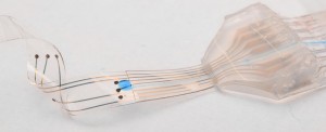
Here’s what the implant looks like,
Courtesy: EPFL
The press release describes how the implant is placed (Note: A link has been removed),
Flexible and stretchy, the implant developed at EPFL is placed beneath the dura mater, directly onto the spinal cord. Its elasticity and its potential for deformation are almost identical to the living tissue surrounding it. This reduces friction and inflammation to a minimum. When implanted into rats, the e-Dura prototype caused neither damage nor rejection, even after two months. More rigid traditional implants would have caused significant nerve tissue damage during this period of time.
The researchers tested the device prototype by applying their rehabilitation protocol — which combines electrical and chemical stimulation – to paralyzed rats. Not only did the implant prove its biocompatibility, but it also did its job perfectly, allowing the rats to regain the ability to walk on their own again after a few weeks of training.
“Our e-Dura implant can remain for a long period of time on the spinal cord or the cortex, precisely because it has the same mechanical properties as the dura mater itself. This opens up new therapeutic possibilities for patients suffering from neurological trauma or disorders, particularly individuals who have become paralyzed following spinal cord injury,” explains Lacour, co-author of the paper, and holder of EPFL’s Bertarelli Chair in Neuroprosthetic Technology.
The press release goes on to describe the engineering achievements,
Developing the e-Dura implant was quite a feat of engineering. As flexible and stretchable as living tissue, it nonetheless includes electronic elements that stimulate the spinal cord at the point of injury. The silicon substrate is covered with cracked gold electric conducting tracks that can be pulled and stretched. The electrodes are made of an innovative composite of silicon and platinum microbeads. They can be deformed in any direction, while still ensuring optimal electrical conductivity. Finally, a fluidic microchannel enables the delivery of pharmacological substances – neurotransmitters in this case – that will reanimate the nerve cells beneath the injured tissue.
The implant can also be used to monitor electrical impulses from the brain in real time. When they did this, the scientists were able to extract with precision the animal’s motor intention before it was translated into movement.
“It’s the first neuronal surface implant designed from the start for long-term application. In order to build it, we had to combine expertise from a considerable number of areas,” explains Courtine, co-author and holder of EPFL’s IRP Chair in Spinal Cord Repair. “These include materials science, electronics, neuroscience, medicine, and algorithm programming. I don’t think there are many places in the world where one finds the level of interdisciplinary cooperation that exists in our Center for Neuroprosthetics.”
For the time being, the e-Dura implant has been primarily tested in cases of spinal cord injury in paralyzed rats. But the potential for applying these surface implants is huge – for example in epilepsy, Parkinson’s disease and pain management. The scientists are planning to move towards clinical trials in humans, and to develop their prototype in preparation for commercialization.
EPFL has provided a video of researcher Stéphanie Lacour describing e-Dura and expressing hopes for its commercialization,
Here’s a link to and a citation for the paper,
Electronic dura mater for long-term multimodal neural interfaces by Ivan R. Minev, Pavel Musienko, Arthur Hirsch, Quentin Barraud, Nikolaus Wenger, Eduardo Martin Moraud, Jérôme Gandar, Marco Capogrosso, Tomislav Milekovic, Léonie Asboth, Rafael Fajardo Torres, Nicolas Vachicouras, Qihan Liu, Natalia Pavlova, Simone Duis, Alexandre Larmagnac, Janos Vörös, Silvestro Micera, Zhigang Suo, Grégoire Courtine, Stéphanie P. Lacour. Science 9 January 2015: Vol. 347 no. 6218 pp. 159-163 DOI: 10.1126/science.1260318
This paper is behind a paywall.
Carbon nanotube fibres could connect to the brain
Researchers at Rice University (Texas, US) are excited about the possibilities that carbon nanotube fibres offer in the field of implantable electronics for the brain. From a March 25, 2015 news item on Nanowerk,
Carbon nanotube fibers invented at Rice University may provide the best way to communicate directly with the brain.
The fibers have proven superior to metal electrodes for deep brain stimulation and to read signals from a neuronal network. Because they provide a two-way connection, they show promise for treating patients with neurological disorders while monitoring the real-time response of neural circuits in areas that control movement, mood and bodily functions.
New experiments at Rice demonstrated the biocompatible fibers are ideal candidates for small, safe electrodes that interact with the brain’s neuronal system, according to the researchers. They could replace much larger electrodes currently used in devices for deep brain stimulation therapies in Parkinson’s disease patients.
They may also advance technologies to restore sensory or motor functions and brain-machine interfaces as well as deep brain stimulation therapies for other neurological disorders, including dystonia and depression, the researchers wrote.
The fibers created by the Rice lab of chemist and chemical engineer Matteo Pasquali consist of bundles of long nanotubes originally intended for aerospace applications where strength, weight and conductivity are paramount.
The individual nanotubes measure only a few nanometers across, but when millions are bundled in a process called wet spinning, they become thread-like fibers about a quarter the width of a human hair.
“We developed these fibers as high-strength, high-conductivity materials,” Pasquali said. “Yet, once we had them in our hand, we realized that they had an unexpected property: They are really soft, much like a thread of silk. Their unique combination of strength, conductivity and softness makes them ideal for interfacing with the electrical function of the human body.”
The simultaneous arrival in 2012 of Caleb Kemere, a Rice assistant professor who brought expertise in animal models of Parkinson’s disease, and lead author Flavia Vitale, a research scientist in Pasquali’s lab with degrees in chemical and biomedical engineering, prompted the investigation.
“The brain is basically the consistency of pudding and doesn’t interact well with stiff metal electrodes,” Kemere said. “The dream is to have electrodes with the same consistency, and that’s why we’re really excited about these flexible carbon nanotube fibers and their long-term biocompatibility.”
Weeks-long tests on cells and then in rats with Parkinson’s symptoms proved the fibers are stable and as efficient as commercial platinum electrodes at only a fraction of the size. The soft fibers caused little inflammation, which helped maintain strong electrical connections to neurons by preventing the body’s defenses from scarring and encapsulating the site of the injury.
The highly conductive carbon nanotube fibers also show much more favorable impedance – the quality of the electrical connection — than state-of-the-art metal electrodes, making for better contact at lower voltages over long periods, Kemere said.
The working end of the fiber is the exposed tip, which is about the width of a neuron. The rest is encased with a three-micron layer of a flexible, biocompatible polymer with excellent insulating properties.
The challenge is in placing the tips. “That’s really just a matter of having a brain atlas, and during the experiment adjusting the electrodes very delicately and putting them into the right place,” said Kemere, whose lab studies ways to connect signal-processing systems and the brain’s memory and cognitive centers.
Doctors who implant deep brain stimulation devices start with a recording probe able to “listen” to neurons that emit characteristic signals depending on their functions, Kemere said. Once a surgeon finds the right spot, the probe is removed and the stimulating electrode gently inserted. Rice carbon nanotube fibers that send and receive signals would simplify implantation, Vitale said.
The fibers could lead to self-regulating therapeutic devices for Parkinson’s and other patients. Current devices include an implant that sends electrical signals to the brain to calm the tremors that afflict Parkinson’s patients.
“But our technology enables the ability to record while stimulating,” Vitale said. “Current electrodes can only stimulate tissue. They’re too big to detect any spiking activity, so basically the clinical devices send continuous pulses regardless of the response of the brain.”
Kemere foresees a closed-loop system that can read neuronal signals and adapt stimulation therapy in real time. He anticipates building a device with many electrodes that can be addressed individually to gain fine control over stimulation and monitoring from a small, implantable device.
“Interestingly, conductivity is not the most important electrical property of the nanotube fibers,” Pasquali said. “These fibers are intrinsically porous and extremely stable, which are both great advantages over metal electrodes for sensing electrochemical signals and maintaining performance over long periods of time.”
The paper is open access provided you register on the website.
Remote control for stimulation of the brain
Mo Costandi, neuroscientist and freelance science writer, has written a March 24, 2015 post for the Guardian science blog network focusing on neuronal remote control,
Two teams of scientists have developed new ways of stimulating neurons with nanoparticles, allowing them to activate brain cells remotely using light or magnetic fields. The new methods are quicker and far less invasive than other hi-tech methods available, so could be more suitable for potential new treatments for human diseases.
Researchers have various methods for manipulating brain cell activity, arguably the most powerful being optogenetics, which enables them to switch specific brain cells on or off with unprecedented precision, and simultaneously record their behaviour, using pulses of light.
This is very useful for probing neural circuits and behaviour, but involves first creating genetically engineered mice with light-sensitive neurons, and then inserting the optical fibres that deliver light into the brain, so there are major technical and ethical barriers to its use in humans.
Nanomedicine could get around this. Francisco Bezanilla of the University of Chicago and his colleagues knew that gold nanoparticles can absorb light and convert it into heat, and several years ago they discovered that infrared light can make neurons fire nervous impulses by heating up their cell membranes.
…
Polina Anikeeva’s team at the Massachusetts Institute of Technology adopted a slightly different approach, using spherical iron oxide particles that give off heat when exposed to an alternating magnetic field.
…
Although still in the experimental stages, research like this may eventually allow for wireless and minimally invasive deep brain stimulation of the human brain. Bezanilla’s group aim to apply their method to develop treatments for macular degeneration and other conditions that kill off light-sensitive cells in the retina. This would involve injecting nanoparticles into the eye so that they bind to other retinal cells, allowing natural light to excite them into firing impulses to the optic nerve.
Costandi’s article is intended for an audience that either understands the science or can deal with the uncertainty of not understanding absolutely everything. Provided you fall into either of those categories, the article is well written and it provides links and citations to the papers for both research teams being featured.
Taken together, the research at EPFL, Rice University, University of Chicago, and Massachusetts Institute of Technology provides a clue as to how much money and intellectual power is being directed at the brain.