Tag Archives: brain-machine interfaces

A biohybrid artificial synapse that can communicate with living cells

As I noted in my June 16, 2020 posting, we may have more than one kind of artificial brain in our future. This latest work features a biohybrid. From a June 15, 2020 news item on ScienceDaily,

In 2017, Stanford University researchers presented a new device that mimics the brain’s efficient and low-energy neural learning process [see my March 8, 2017 posting for more]. It was an artificial version of a synapse — the gap across which neurotransmitters travel to communicate between neurons — made from organic materials. In 2019, the researchers assembled nine of their artificial synapses together in an array, showing that they could be simultaneously programmed to mimic the parallel operation of the brain [see my Sept. 17, 2019 posting].

Now, in a paper published June 15 [2020] in Nature Materials, they have tested the first biohybrid version of their artificial synapse and demonstrated that it can communicate with living cells. Future technologies stemming from this device could function by responding directly to chemical signals from the brain. The research was conducted in collaboration with researchers at Istituto Italiano di Tecnologia (Italian Institute of Technology — IIT) in Italy and at Eindhoven University of Technology (Netherlands).

“This paper really highlights the unique strength of the materials that we use in being able to interact with living matter,” said Alberto Salleo, professor of materials science and engineering at Stanford and co-senior author of the paper. “The cells are happy sitting on the soft polymer. But the compatibility goes deeper: These materials work with the same molecules neurons use naturally.”

While other brain-integrated devices require an electrical signal to detect and process the brain’s messages, the communications between this device and living cells occur through electrochemistry — as though the material were just another neuron receiving messages from its neighbor.

A June 15, 2020 Stanford University news release (also on EurekAlert) by Taylor Kubota, which originated the news item, delves further into this recent work,

How neurons learn

The biohybrid artificial synapse consists of two soft polymer electrodes, separated by a trench filled with electrolyte solution – which plays the part of the synaptic cleft that separates communicating neurons in the brain. When living cells are placed on top of one electrode, neurotransmitters that those cells release can react with that electrode to produce ions. Those ions travel across the trench to the second electrode and modulate the conductive state of this electrode. Some of that change is preserved, simulating the learning process occurring in nature.

“In a biological synapse, essentially everything is controlled by chemical interactions at the synaptic junction. Whenever the cells communicate with one another, they’re using chemistry,” said Scott Keene, a graduate student at Stanford and co-lead author of the paper. “Being able to interact with the brain’s natural chemistry gives the device added utility.”

This process mimics the same kind of learning seen in biological synapses, which is highly efficient in terms of energy because computing and memory storage happen in one action. In more traditional computer systems, the data is processed first and then later moved to storage.

To test their device, the researchers used rat neuroendocrine cells that release the neurotransmitter dopamine. Before they ran their experiment, they were unsure how the dopamine would interact with their material – but they saw a permanent change in the state of their device upon the first reaction.

“We knew the reaction is irreversible, so it makes sense that it would cause a permanent change in the device’s conductive state,” said Keene. “But, it was hard to know whether we’d achieve the outcome we predicted on paper until we saw it happen in the lab. That was when we realized the potential this has for emulating the long-term learning process of a synapse.”

A first step

This biohybrid design is in such early stages that the main focus of the current research was simply to make it work.

“It’s a demonstration that this communication melding chemistry and electricity is possible,” said Salleo. “You could say it’s a first step toward a brain-machine interface, but it’s a tiny, tiny very first step.”

Now that the researchers have successfully tested their design, they are figuring out the best paths for future research, which could include work on brain-inspired computers, brain-machine interfaces, medical devices or new research tools for neuroscience. Already, they are working on how to make the device function better in more complex biological settings that contain different kinds of cells and neurotransmitters.

Here’s a link to and a citation for the paper,

A biohybrid synapse with neurotransmitter-mediated plasticity by Scott T. Keene, Claudia Lubrano, Setareh Kazemzadeh, Armantas Melianas, Yaakov Tuchman, Giuseppina Polino, Paola Scognamiglio, Lucio Cinà, Alberto Salleo, Yoeri van de Burgt & Francesca Santoro. Nature Materials (2020) DOI: https://doi.org/10.1038/s41563-020-0703-y Published: 15 June 2020

This paper is behind a paywall.
Soft things for your brain

A March 5, 2018 news item on Nanowerk describes the latest stretchable electrode (Note: A link has been removed),

Klas Tybrandt, principal investigator at the Laboratory of Organic Electronics at Linköping University [Sweden], has developed new technology for long-term stable neural recording. It is based on a novel elastic material composite, which is biocompatible and retains high electrical conductivity even when stretched to double its original length.

The result has been achieved in collaboration with colleagues in Zürich and New York. The breakthrough, which is crucial for many applications in biomedical engineering, is described in an article published in the prestigious scientific journal Advanced Materials (“High-Density Stretchable Electrode Grids for Chronic Neural Recording”).

A March 5, 2018 Linköping University press release, which originated the news item, gives more detail but does not mention that the nanowires are composed of titanium dioxide (you can find additional details in the abstract for the paper; link and citation will be provided later in this posting)),

The coupling between electronic components and nerve cells is crucial not only to collect information about cell signalling, but also to diagnose and treat neurological disorders and diseases, such as epilepsy.

It is very challenging to achieve long-term stable connections that do not damage neurons or tissue, since the two systems, the soft and elastic tissue of the body and the hard and rigid electronic components, have completely different mechanical properties.

Stretchable soft electrodeThe soft electrode stretched to twice its length Photo credit: Thor Balkhed

“As human tissue is elastic and mobile, damage and inflammation arise at the interface with rigid electronic components. It not only causes damage to tissue; it also attenuates neural signals,” says Klas Tybrandt, leader of the Soft Electronics group at the Laboratory of Organic Electronics, Linköping University, Campus Norrköping.

New conductive material

Klas Tybrandt has developed a new conductive material that is as soft as human tissue and can be stretched to twice its length. The material consists of gold coated titanium dioxide nanowires, embedded into silicone rubber. The material is biocompatible – which means it can be in contact with the body without adverse effects – and its conductivity remains stable over time.

“The microfabrication of soft electrically conductive composites involves several challenges. We have developed a process to manufacture small electrodes that also preserves the biocompatibility of the materials. The process uses very little material, and this means that we can work with a relatively expensive material such as gold, without the cost becoming prohibitive,” says Klas Tybrandt.

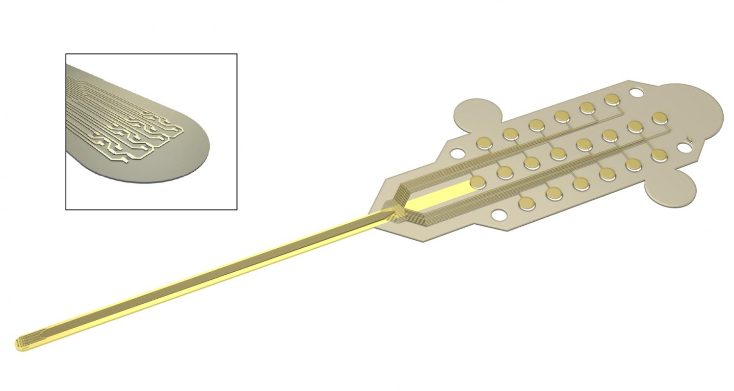
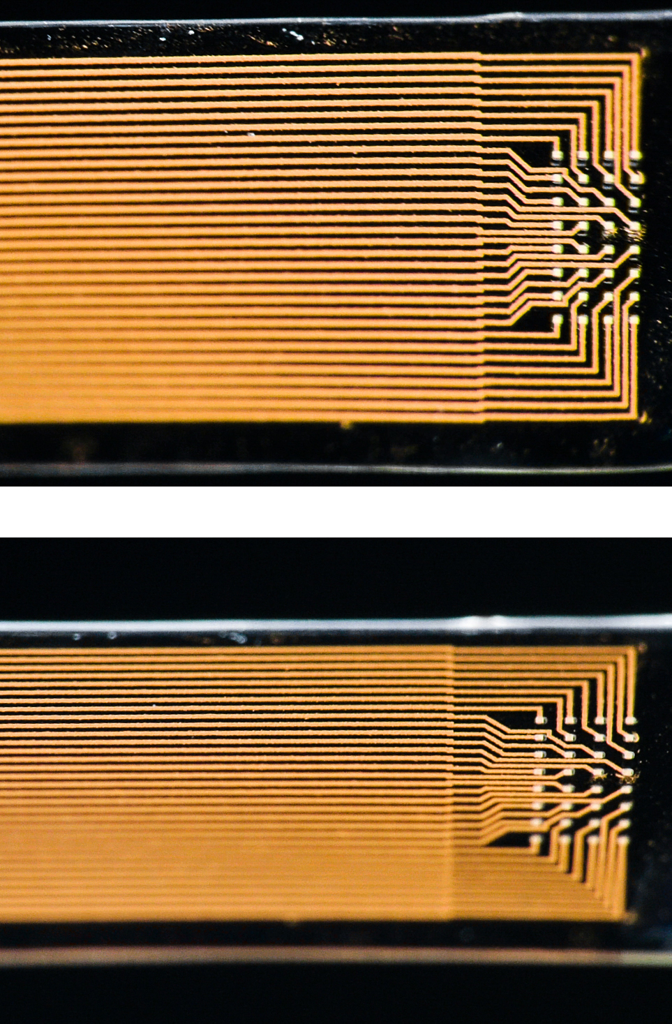
The electrodes are 50 µm [microns or micrometres] in size and are located at a distance of 200 µm from each other. The fabrication procedure allows 32 electrodes to be placed onto a very small surface. The final probe, shown in the photograph, has a width of 3.2 mm and a thickness of 80 µm.

The soft microelectrodes have been developed at Linköping University and ETH Zürich, and researchers at New York University and Columbia University have subsequently implanted them in the brain of rats. The researchers were able to collect high-quality neural signals from the freely moving rats for 3 months. The experiments have been subject to ethical review, and have followed the strict regulations that govern animal experiments.

Important future applications

Klas Tybrandt, researcher at Laboratory for Organic ElectronicsKlas Tybrandt, researcher at Laboratory for Organic Electronics Photo credit: Thor Balkhed

“When the neurons in the brain transmit signals, a voltage is formed that the electrodes detect and transmit onwards through a tiny amplifier. We can also see which electrodes the signals came from, which means that we can estimate the location in the brain where the signals originated. This type of spatiotemporal information is important for future applications. We hope to be able to see, for example, where the signal that causes an epileptic seizure starts, a prerequisite for treating it. Another area of application is brain-machine interfaces, by which future technology and prostheses can be controlled with the aid of neural signals. There are also many interesting applications involving the peripheral nervous system in the body and the way it regulates various organs,” says Klas Tybrandt.

The breakthrough is the foundation of the research area Soft Electronics, currently being established at Linköping University, with Klas Tybrandt as principal investigator.
liu.se/soft-electronics

A video has been made available (Note: For those who find any notion of animal testing disturbing; don’t watch the video even though it is an animation and does not feature live animals),

Here’s a link to and a citation for the paper,

High-Density Stretchable Electrode Grids for Chronic Neural Recording by Klas Tybrandt, Dion Khodagholy, Bernd Dielacher, Flurin Stauffer, Aline F. Renz, György Buzsáki, and János Vörös. Advanced Materials 2018. DOI: 10.1002/adma.201706520
 First published 28 February 2018

This paper is open access.

Graphene-based neural probes

I have two news bits (dated almost one month apart) about the use of graphene in neural probes, one from the European Union and the other from Korea.

European Union (EU)

This work is being announced by the European Commission’s (a subset of the EU) Graphene Flagship (one of two mega-funding projects announced in 2013; 1B Euros each over ten years for the Graphene Flagship and the Human Brain Project).

According to a March 27, 2017 news item on ScienceDaily, researchers have developed a graphene-based neural probe that has been tested on rats,

Measuring brain activity with precision is essential to developing further understanding of diseases such as epilepsy and disorders that affect brain function and motor control. Neural probes with high spatial resolution are needed for both recording and stimulating specific functional areas of the brain. Now, researchers from the Graphene Flagship have developed a new device for recording brain activity in high resolution while maintaining excellent signal to noise ratio (SNR). Based on graphene field-effect transistors, the flexible devices open up new possibilities for the development of functional implants and interfaces.

The research, published in 2D Materials, was a collaborative effort involving Flagship partners Technical University of Munich (TU Munich; Germany), Institut d’Investigacions Biomèdiques August Pi i Sunyer (IDIBAPS; Spain), Spanish National Research Council (CSIC; Spain), The Biomedical Research Networking Center in Bioengineering, Biomaterials and Nanomedicine (CIBER-BBN; Spain) and the Catalan Institute of Nanoscience and Nanotechnology (ICN2; Spain).

Caption: Graphene transistors integrated in a flexible neural probe enables electrical signals from neurons to be measured with high accuracy and density. Inset: The tip of the probe contains 16 flexible graphene transistors. Credit: ICN2

A March 27, 2017 Graphene Flagship press release on EurekAlert, which originated the news item, describes the work,  in more detail,

The devices were used to record the large signals generated by pre-epileptic activity in rats, as well as the smaller levels of brain activity during sleep and in response to visual light stimulation. These types of activities lead to much smaller electrical signals, and are at the level of typical brain activity. Neural activity is detected through the highly localised electric fields generated when neurons fire, so densely packed, ultra-small measuring devices is important for accurate brain readings.

The neural probes are placed directly on the surface of the brain, so safety is of paramount importance for the development of graphene-based neural implant devices. Importantly, the researchers determined that the graphene-based probes are non-toxic, and did not induce any significant inflammation.

Devices implanted in the brain as neural prosthesis for therapeutic brain stimulation technologies and interfaces for sensory and motor devices, such as artificial limbs, are an important goal for improving quality of life for patients. This work represents a first step towards the use of graphene in research as well as clinical neural devices, showing that graphene-based technologies can deliver the high resolution and high SNR needed for these applications.

First author Benno Blaschke (TU Munich) said “Graphene is one of the few materials that allows recording in a transistor configuration and simultaneously complies with all other requirements for neural probes such as flexibility, biocompability and chemical stability. Although graphene is ideally suited for flexible electronics, it was a great challenge to transfer our fabrication process from rigid substrates to flexible ones. The next step is to optimize the wafer-scale fabrication process and improve device flexibility and stability.”

Jose Antonio Garrido (ICN2), led the research. He said “Mechanical compliance is an important requirement for safe neural probes and interfaces. Currently, the focus is on ultra-soft materials that can adapt conformally to the brain surface. Graphene neural interfaces have shown already great potential, but we have to improve on the yield and homogeneity of the device production in order to advance towards a real technology. Once we have demonstrated the proof of concept in animal studies, the next goal will be to work towards the first human clinical trial with graphene devices during intraoperative mapping of the brain. This means addressing all regulatory issues associated to medical devices such as safety, biocompatibility, etc.”

Caption: The graphene-based neural probes were used to detect rats’ responses to visual stimulation, as well as neural signals during sleep. Both types of signals are small, and typically difficult to measure. Credit: ICN2

Here’s a link to and a citation for the paper,

Mapping brain activity with flexible graphene micro-transistors by Benno M Blaschke, Núria Tort-Colet, Anton Guimerà-Brunet, Julia Weinert, Lionel Rousseau, Axel Heimann, Simon Drieschner, Oliver Kempski, Rosa Villa, Maria V Sanchez-Vives. 2D Materials, Volume 4, Number 2 DOI https://doi.org/10.1088/2053-1583/aa5eff Published 24 February 2017

© 2017 IOP Publishing Ltd

This paper is behind a paywall.

Korea

While this research from Korea was published more recently, the probe itself has not been subjected to in vivo (animal testing). From an April 19, 2017 news item on ScienceDaily,

Electrodes placed in the brain record neural activity, and can help treat neural diseases like Parkinson’s and epilepsy. Interest is also growing in developing better brain-machine interfaces, in which electrodes can help control prosthetic limbs. Progress in these fields is hindered by limitations in electrodes, which are relatively stiff and can damage soft brain tissue.

Designing smaller, gentler electrodes that still pick up brain signals is a challenge because brain signals are so weak. Typically, the smaller the electrode, the harder it is to detect a signal. However, a team from the Daegu Gyeongbuk Institute of Science & Technology [DGIST} in Korea developed new probes that are small, flexible and read brain signals clearly.

This is a pretty interesting way to illustrate the research,

Caption: Graphene and gold make a better brain probe. Credit: DGIST

An April 19, 2017 DGIST press release (also on EurekAlert), which originated the news item, expands on the theme (Note: A link has been removed),

The probe consists of an electrode, which records the brain signal. The signal travels down an interconnection line to a connector, which transfers the signal to machines measuring and analysing the signals.

The electrode starts with a thin gold base. Attached to the base are tiny zinc oxide nanowires, which are coated in a thin layer of gold, and then a layer of conducting polymer called PEDOT. These combined materials increase the probe’s effective surface area, conducting properties, and strength of the electrode, while still maintaining flexibility and compatibility with soft tissue.

Packing several long, thin nanowires together onto one probe enables the scientists to make a smaller electrode that retains the same effective surface area of a larger, flat electrode. This means the electrode can shrink, but not reduce signal detection. The interconnection line is made of a mix of graphene and gold. Graphene is flexible and gold is an excellent conductor. The researchers tested the probe and found it read rat brain signals very clearly, much better than a standard flat, gold electrode.

“Our graphene and nanowires-based flexible electrode array can be useful for monitoring and recording the functions of the nervous system, or to deliver electrical signals to the brain,” the researchers conclude in their paper recently published in the journal ACS Applied Materials and Interfaces.

The probe requires further clinical tests before widespread commercialization. The researchers are also interested in developing a wireless version to make it more convenient for a variety of applications.

Here’s a link to and a citation for the paper,

Enhancement of Interface Characteristics of Neural Probe Based on Graphene, ZnO Nanowires, and Conducting Polymer PEDOT by Mingyu Ryu, Jae Hoon Yang, Yumi Ahn, Minkyung Sim, Kyung Hwa Lee, Kyungsoo Kim, Taeju Lee, Seung-Jun Yoo, So Yeun Kim, Cheil Moon, Minkyu Je, Ji-Woong Choi, Youngu Lee, and Jae Eun Jang. ACS Appl. Mater. Interfaces, 2017, 9 (12), pp 10577–10586 DOI: 10.1021/acsami.7b02975 Publication Date (Web): March 7, 2017

Copyright © 2017 American Chemical Society

This paper is behind a paywall.

High-performance, low-energy artificial synapse for neural network computing

This artificial synapse is apparently an improvement on the standard memristor-based artificial synapse but that doesn’t become clear until reading the abstract for the paper. First, there’s a Feb. 20, 2017 Stanford University news release by Taylor Kubota (dated Feb. 21, 2017 on EurekAlert), Note: Links have been removed,

For all the improvements in computer technology over the years, we still struggle to recreate the low-energy, elegant processing of the human brain. Now, researchers at Stanford University and Sandia National Laboratories have made an advance that could help computers mimic one piece of the brain’s efficient design – an artificial version of the space over which neurons communicate, called a synapse.

“It works like a real synapse but it’s an organic electronic device that can be engineered,” said Alberto Salleo, associate professor of materials science and engineering at Stanford and senior author of the paper. “It’s an entirely new family of devices because this type of architecture has not been shown before. For many key metrics, it also performs better than anything that’s been done before with inorganics.”

The new artificial synapse, reported in the Feb. 20 issue of Nature Materials, mimics the way synapses in the brain learn through the signals that cross them. This is a significant energy savings over traditional computing, which involves separately processing information and then storing it into memory. Here, the processing creates the memory.

This synapse may one day be part of a more brain-like computer, which could be especially beneficial for computing that works with visual and auditory signals. Examples of this are seen in voice-controlled interfaces and driverless cars. Past efforts in this field have produced high-performance neural networks supported by artificially intelligent algorithms but these are still distant imitators of the brain that depend on energy-consuming traditional computer hardware.

Building a brain

When we learn, electrical signals are sent between neurons in our brain. The most energy is needed the first time a synapse is traversed. Every time afterward, the connection requires less energy. This is how synapses efficiently facilitate both learning something new and remembering what we’ve learned. The artificial synapse, unlike most other versions of brain-like computing, also fulfills these two tasks simultaneously, and does so with substantial energy savings.

“Deep learning algorithms are very powerful but they rely on processors to calculate and simulate the electrical states and store them somewhere else, which is inefficient in terms of energy and time,” said Yoeri van de Burgt, former postdoctoral scholar in the Salleo lab and lead author of the paper. “Instead of simulating a neural network, our work is trying to make a neural network.”

The artificial synapse is based off a battery design. It consists of two thin, flexible films with three terminals, connected by an electrolyte of salty water. The device works as a transistor, with one of the terminals controlling the flow of electricity between the other two.

Like a neural path in a brain being reinforced through learning, the researchers program the artificial synapse by discharging and recharging it repeatedly. Through this training, they have been able to predict within 1 percent of uncertainly what voltage will be required to get the synapse to a specific electrical state and, once there, it remains at that state. In other words, unlike a common computer, where you save your work to the hard drive before you turn it off, the artificial synapse can recall its programming without any additional actions or parts.

Testing a network of artificial synapses

Only one artificial synapse has been produced but researchers at Sandia used 15,000 measurements from experiments on that synapse to simulate how an array of them would work in a neural network. They tested the simulated network’s ability to recognize handwriting of digits 0 through 9. Tested on three datasets, the simulated array was able to identify the handwritten digits with an accuracy between 93 to 97 percent.

Although this task would be relatively simple for a person, traditional computers have a difficult time interpreting visual and auditory signals.

“More and more, the kinds of tasks that we expect our computing devices to do require computing that mimics the brain because using traditional computing to perform these tasks is becoming really power hungry,” said A. Alec Talin, distinguished member of technical staff at Sandia National Laboratories in Livermore, California, and senior author of the paper. “We’ve demonstrated a device that’s ideal for running these type of algorithms and that consumes a lot less power.”

This device is extremely well suited for the kind of signal identification and classification that traditional computers struggle to perform. Whereas digital transistors can be in only two states, such as 0 and 1, the researchers successfully programmed 500 states in the artificial synapse, which is useful for neuron-type computation models. In switching from one state to another they used about one-tenth as much energy as a state-of-the-art computing system needs in order to move data from the processing unit to the memory.

This, however, means they are still using about 10,000 times as much energy as the minimum a biological synapse needs in order to fire. The researchers are hopeful that they can attain neuron-level energy efficiency once they test the artificial synapse in smaller devices.

Organic potential

Every part of the device is made of inexpensive organic materials. These aren’t found in nature but they are largely composed of hydrogen and carbon and are compatible with the brain’s chemistry. Cells have been grown on these materials and they have even been used to make artificial pumps for neural transmitters. The voltages applied to train the artificial synapse are also the same as those that move through human neurons.

All this means it’s possible that the artificial synapse could communicate with live neurons, leading to improved brain-machine interfaces. The softness and flexibility of the device also lends itself to being used in biological environments. Before any applications to biology, however, the team plans to build an actual array of artificial synapses for further research and testing.

Additional Stanford co-authors of this work include co-lead author Ewout Lubberman, also of the University of Groningen in the Netherlands, Scott T. Keene and Grégorio C. Faria, also of Universidade de São Paulo, in Brazil. Sandia National Laboratories co-authors include Elliot J. Fuller and Sapan Agarwal in Livermore and Matthew J. Marinella in Albuquerque, New Mexico. Salleo is an affiliate of the Stanford Precourt Institute for Energy and the Stanford Neurosciences Institute. Van de Burgt is now an assistant professor in microsystems and an affiliate of the Institute for Complex Molecular Studies (ICMS) at Eindhoven University of Technology in the Netherlands.

This research was funded by the National Science Foundation, the Keck Faculty Scholar Funds, the Neurofab at Stanford, the Stanford Graduate Fellowship, Sandia’s Laboratory-Directed Research and Development Program, the U.S. Department of Energy, the Holland Scholarship, the University of Groningen Scholarship for Excellent Students, the Hendrik Muller National Fund, the Schuurman Schimmel-van Outeren Foundation, the Foundation of Renswoude (The Hague and Delft), the Marco Polo Fund, the Instituto Nacional de Ciência e Tecnologia/Instituto Nacional de Eletrônica Orgânica in Brazil, the Fundação de Amparo à Pesquisa do Estado de São Paulo and the Brazilian National Council.

Here’s an abstract for the researchers’ paper (link to paper provided after abstract) and it’s where you’ll find the memristor connection explained,

The brain is capable of massively parallel information processing while consuming only ~1–100fJ per synaptic event1, 2. Inspired by the efficiency of the brain, CMOS-based neural architectures3 and memristors4, 5 are being developed for pattern recognition and machine learning. However, the volatility, design complexity and high supply voltages for CMOS architectures, and the stochastic and energy-costly switching of memristors complicate the path to achieve the interconnectivity, information density, and energy efficiency of the brain using either approach. Here we describe an electrochemical neuromorphic organic device (ENODe) operating with a fundamentally different mechanism from existing memristors. ENODe switches at low voltage and energy (<10pJ for 103μm2 devices), displays >500 distinct, non-volatile conductance states within a ~1V range, and achieves high classification accuracy when implemented in neural network simulations. Plastic ENODes are also fabricated on flexible substrates enabling the integration of neuromorphic functionality in stretchable electronic systems6, 7. Mechanical flexibility makes ENODes compatible with three-dimensional architectures, opening a path towards extreme interconnectivity comparable to the human brain.

Here’s a link to and a citation for the paper,

A non-volatile organic electrochemical device as a low-voltage artificial synapse for neuromorphic computing by Yoeri van de Burgt, Ewout Lubberman, Elliot J. Fuller, Scott T. Keene, Grégorio C. Faria, Sapan Agarwal, Matthew J. Marinella, A. Alec Talin, & Alberto Salleo. Nature Materials (2017) doi:10.1038/nmat4856 Published online 20 February 2017

This paper is behind a paywall.

ETA March 8, 2017 10:28 PST: You may find this this piece on ferroelectricity and neuromorphic engineering of interest (March 7, 2017 posting titled: Ferroelectric roadmap to neuromorphic computing).

Long-term brain mapping with injectable electronics

Charles Lieber and his team at Harvard University announced a success with their work on injectable electronics last year (see my June 11, 2015 posting for more) and now they are reporting on their work with more extensive animal studies according to an Aug. 29, 2016 news item on psypost.org,

Scientists in recent years have made great strides in the quest to understand the brain by using implanted probes to explore how specific neural circuits work.

Though effective, those probes also come with their share of problems as a result of rigidity. The inflammation they produce induces chronic recording instability and means probes must be relocated every few days, leaving some of the central questions of neuroscience – like how the neural circuits are reorganized during development, learning and aging- beyond scientists’ reach.

But now, it seems, things are about to change.

Led by Charles Lieber, The Mark Hyman Jr. Professor of Chemistry and chair of the Department of Chemistry and Chemical Biology, a team of researchers that included graduate student Tian-Ming Fu, postdoctoral fellow Guosong Hong, graduate student Tao Zhou and others, has demonstrated that syringe-injectable mesh electronics can stably record neural activity in mice for eight months or more, with none of the inflammation

An Aug. 29, 2016 Harvard University press release, which originated the news item, provides more detail,

“With the ability to follow the same individual neurons in a circuit chronically…there’s a whole suite of things this opens up,” Lieber said. “The eight months we demonstrate in this paper is not a limit, but what this does show is that mesh electronics could be used…to investigate neuro-degenerative diseases like Alzheimer’s, or processes that occur over long time, like aging or learning.”

Lieber and colleagues also demonstrated that the syringe-injectable mesh electronics could be used to deliver electrical stimulation to the brain over three months or more.

“Ultimately, our aim is to create these with the goal of finding clinical applications,” Lieber said. “What we found is that, because of the lack of immune response (to the mesh electronics), which basically insulates neurons, we can deliver stimulation in a much more subtle way, using lower voltages that don’t damage tissue.”

The possibilities, however, don’t end there.

The seamless integration of the electronics and biology, Lieber said, could open the door to an entirely new class of brain-machine interfaces and vast improvements in prosthetics, among other fields.

“Today, brain-machine interfaces are based on traditional implanted probes, and there has been some impressive work that’s been done in that field,” Lieber said. “But all the interfaces rely on the same technique to decode neural signals.”

Because traditional rigid implanted probes are invariably unstable, he explained, researchers and clinicians rely on decoding what they call the “population average” – essentially taking a host of neural signals and applying complex computational tools to determine what they mean.

Using tissue-like mesh electronics, by comparison, researchers may be able to read signals from specific neurons over time, potentially allowing for the development of improved brain-machine interfaces for prosthetics.

“We think this is going to be very powerful, because we can identify circuits and both record and stimulate in a way that just hasn’t been possible before,” Lieber said. “So what I like to say is: I think therefore it happens.”

Lieber even held out the possibility that the syringe-injectable mesh electronics could one day be used to treat catastrophic injuries to the brain and spinal cord.

“I don’t think that’s science-fiction,” he said. “Other people may say that will be possible through, for example, regenerative medicine, but we are pursuing this from a different angle.

“My feeling is that this is about a seamless integration between the biological and the electronic systems, so they’re not distinct entities,” he continued. “If we can make the electronics look like the neural network, they will work together…and that’s where you want to be if you want to exploit the strengths of both.”

In the 2015 posting, Lieber was discussing cyborgs, here he broaches the concept without using the word, “… seamless integration between the biological and the electronic systems, so they’re not distinct entities.”

Here’s a link to and a citation for the paper,

Stable long-term chronic brain mapping at the single-neuron level by Tian-Ming Fu, Guosong Hong, Tao Zhou, Thomas G Schuhmann, Robert D Viveros, & Charles M Lieber. Nature Methods (2016) doi:10.1038/nmeth.3969 Published online 29 August 2016

This paper is behind a paywall.