Tag Archives: neuron

Two approaches to memristors

Within one day of each other in October 2018, two different teams working on memristors with applications to neuroprosthetics and neuromorphic computing (brainlike computing) announced their results.

Russian team

An October 15, 2018 (?) Lobachevsky University press release (also published on October 15, 2018 on EurekAlert) describes a new approach to memristors,

Biological neurons are coupled unidirectionally through a special junction called a synapse. An electrical signal is transmitted along a neuron after some biochemical reactions initiate a chemical release to activate an adjacent neuron. These junctions are crucial for cognitive functions, such as perception, learning and memory.

A group of researchers from Lobachevsky University in Nizhny Novgorod investigates the dynamics of an individual memristive device when it receives a neuron-like signal as well as the dynamics of a network of analog electronic neurons connected by means of a memristive device. According to Svetlana Gerasimova, junior researcher at the Physics and Technology Research Institute and at the Neurotechnology Department of Lobachevsky University, this system simulates the interaction between synaptically coupled brain neurons while the memristive device imitates a neuron axon.

A memristive device is a physical model of Chua’s [Dr. Leon Chua, University of California at Berkeley; see my May 9, 2008 posting for a brief description Dr. Chua’s theory] memristor, which is an electric circuit element capable of changing its resistance depending on the electric signal received at the input. The device based on a Au/ZrO2(Y)/TiN/Ti structure demonstrates reproducible bipolar switching between the low and high resistance states. Resistive switching is determined by the oxidation and reduction of segments of conducting channels (filaments) in the oxide film when voltage with different polarity is applied to it. In the context of the present work, the ability of a memristive device to change conductivity under the action of pulsed signals makes it an almost ideal electronic analog of a synapse.

Lobachevsky University scientists and engineers supported by the Russian Science Foundation (project No.16-19-00144) have experimentally implemented and theoretically described the synaptic connection of neuron-like generators using the memristive interface and investigated the characteristics of this connection.

“Each neuron is implemented in the form of a pulse signal generator based on the FitzHugh-Nagumo model. This model provides a qualitative description of the main neurons’ characteristics: the presence of the excitation threshold, the presence of excitable and self-oscillatory regimes with the possibility of a changeover. At the initial time moment, the master generator is in the self-oscillatory mode, the slave generator is in the excitable mode, and the memristive device is used as a synapse. The signal from the master generator is conveyed to the input of the memristive device, the signal from the output of the memristive device is transmitted to the input of the slave generator via the loading resistance. When the memristive device switches from a high resistance to a low resistance state, the connection between the two neuron-like generators is established. The master generator goes into the oscillatory mode and the signals of the generators are synchronized. Different signal modulation mode synchronizations were demonstrated for the Au/ZrO2(Y)/TiN/Ti memristive device,” – says Svetlana Gerasimova.

UNN researchers believe that the next important stage in the development of neuromorphic systems based on memristive devices is to apply such systems in neuroprosthetics. Memristive systems will provide a highly efficient imitation of synaptic connection due to the stochastic nature of the memristive phenomenon and can be used to increase the flexibility of the connections for neuroprosthetic purposes. Lobachevsky University scientists have vast experience in the development of neurohybrid systems. In particular, a series of experiments was performed with the aim of connecting the FitzHugh-Nagumo oscillator with a biological object, a rat brain hippocampal slice. The signal from the electronic neuron generator was transmitted through the optic fiber communication channel to the bipolar electrode which stimulated Schaffer collaterals (axons of pyramidal neurons in the CA3 field) in the hippocampal slices. “We are going to combine our efforts in the design of artificial neuromorphic systems and our experience of working with living cells to improve flexibility of prosthetics,” concludes S. Gerasimova.

The results of this research were presented at the 38th International Conference on Nonlinear Dynamics (Dynamics Days Europe) at Loughborough University (Great Britain).

This diagram illustrates an aspect of the work,

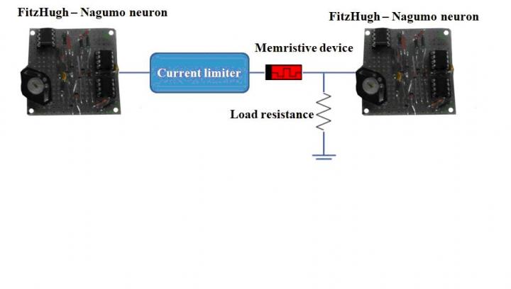
Caption: Schematic of electronic neurons coupling via a memristive device. Credit: Lobachevsky University

US team

The American Institute of Physics (AIP) announced the publication of a ‘memristor paper’ by a team from the University of Southern California (USC) in an October 16, 2018 news item on phys.org,

Just like their biological counterparts, hardware that mimics the neural circuitry of the brain requires building blocks that can adjust how they synapse, with some connections strengthening at the expense of others. One such approach, called memristors, uses current resistance to store this information. New work looks to overcome reliability issues in these devices by scaling memristors to the atomic level.

An October 16, 2018 AIP news release (also on EurekAlert), which originated the news item, delves further into the particulars of this particular piece of memristor research,

A group of researchers demonstrated a new type of compound synapse that can achieve synaptic weight programming and conduct vector-matrix multiplication with significant advances over the current state of the art. Publishing its work in the Journal of Applied Physics, from AIP Publishing, the group’s compound synapse is constructed with atomically thin boron nitride memristors running in parallel to ensure efficiency and accuracy.

The article appears in a special topic section of the journal devoted to “New Physics and Materials for Neuromorphic Computation,” which highlights new developments in physical and materials science research that hold promise for developing the very large-scale, integrated “neuromorphic” systems of tomorrow that will carry computation beyond the limitations of current semiconductors today.

“There’s a lot of interest in using new types of materials for memristors,” said Ivan Sanchez Esqueda, an author on the paper. “What we’re showing is that filamentary devices can work well for neuromorphic computing applications, when constructed in new clever ways.”

Current memristor technology suffers from a wide variation in how signals are stored and read across devices, both for different types of memristors as well as different runs of the same memristor. To overcome this, the researchers ran several memristors in parallel. The combined output can achieve accuracies up to five times those of conventional devices, an advantage that compounds as devices become more complex.

The choice to go to the subnanometer level, Sanchez said, was born out of an interest to keep all of these parallel memristors energy-efficient. An array of the group’s memristors were found to be 10,000 times more energy-efficient than memristors currently available.

“It turns out if you start to increase the number of devices in parallel, you can see large benefits in accuracy while still conserving power,” Sanchez said. Sanchez said the team next looks to further showcase the potential of the compound synapses by demonstrating their use completing increasingly complex tasks, such as image and pattern recognition.

Here’s an image illustrating the parallel artificial synapses,

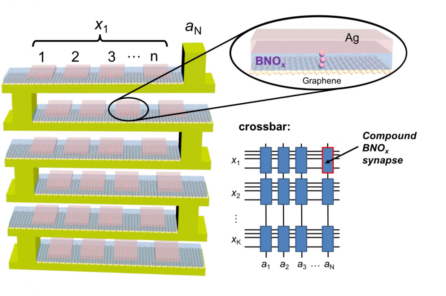
Caption: Hardware that mimics the neural circuitry of the brain requires building blocks that can adjust how they synapse. One such approach, called memristors, uses current resistance to store this information. New work looks to overcome reliability issues in these devices by scaling memristors to the atomic level. Researchers demonstrated a new type of compound synapse that can achieve synaptic weight programming and conduct vector-matrix multiplication with significant advances over the current state of the art. They discuss their work in this week’s Journal of Applied Physics. This image shows a conceptual schematic of the 3D implementation of compound synapses constructed with boron nitride oxide (BNOx) binary memristors, and the crossbar array with compound BNOx synapses for neuromorphic computing applications. Credit: Ivan Sanchez Esqueda

Here’s a link to and a citation for the paper,

Efficient learning and crossbar operations with atomically-thin 2-D material compound synapses by Ivan Sanchez Esqueda, Huan Zhao and Han Wang. The article will appear in the Journal of Applied Physics Oct. 16, 2018 (DOI: 10.1063/1.5042468).

This paper is behind a paywall.

*Title corrected from ‘Two approaches to memristors featuring’ to ‘Two approaches to memristors’ on May 31, 2019 at 1455 hours PDT.

Crowdsourcing brain research at Princeton University to discover 6 new neuron types

Spritely music!

There were already 1/4M registered players as of May 17, 2018 but I’m sure there’s room for more should you be inspired. A May 17, 2018 Princeton University news release (also on EurekAlert) reveals more about the game and about the neurons,

With the help of a quarter-million video game players, Princeton researchers have created and shared detailed maps of more than 1,000 neurons — and they’re just getting started.

“Working with Eyewirers around the world, we’ve made a digital museum that shows off the intricate beauty of the retina’s neural circuits,” said Sebastian Seung, the Evnin Professor in Neuroscience and a professor of computer science and the Princeton Neuroscience Institute (PNI). The related paper is publishing May 17 [2018] in the journal Cell.

Seung is unveiling the Eyewire Museum, an interactive archive of neurons available to the general public and neuroscientists around the world, including the hundreds of researchers involved in the federal Brain Research through Advancing Innovative Neurotechnologies (BRAIN) Initiative.

“This interactive viewer is a huge asset for these larger collaborations, especially among people who are not physically in the same lab,” said Amy Robinson Sterling, a crowdsourcing specialist with PNI and the executive director of Eyewire, the online gaming platform for the citizen scientists who have created this data set.

“This museum is something like a brain atlas,” said Alexander Bae, a graduate student in electrical engineering and one of four co-first authors on the paper. “Previous brain atlases didn’t have a function where you could visualize by individual cell, or a subset of cells, and interact with them. Another novelty: Not only do we have the morphology of each cell, but we also have the functional data, too.”

The neural maps were developed by Eyewirers, members of an online community of video game players who have devoted hundreds of thousands of hours to painstakingly piecing together these neural cells, using data from a mouse retina gathered in 2009.

Eyewire pairs machine learning with gamers who trace the twisting and branching paths of each neuron. Humans are better at visually identifying the patterns of neurons, so every player’s moves are recorded and checked against each other by advanced players and Eyewire staffers, as well as by software that is improving its own pattern recognition skills.

Since Eyewire’s launch in 2012, more than 265,000 people have signed onto the game, and they’ve collectively colored in more than 10 million 3-D “cubes,” resulting in the mapping of more than 3,000 neural cells, of which about a thousand are displayed in the museum.

Each cube is a tiny subset of a single cell, about 4.5 microns across, so a 10-by-10 block of cubes would be the width of a human hair. Every cell is reviewed by between 5 and 25 gamers before it is accepted into the system as complete.

“Back in the early years it took weeks to finish a single cell,” said Sterling. “Now players complete multiple neurons per day.” The Eyewire user experience stays focused on the larger mission — “For science!” is a common refrain — but it also replicates a typical gaming environment, with achievement badges, a chat feature to connect with other players and technical support, and the ability to unlock privileges with increasing skill. “Our top players are online all the time — easily 30 hours a week,” Sterling said.

Dedicated Eyewirers have also contributed in other ways, including donating the swag that gamers win during competitions and writing program extensions “to make game play more efficient and more fun,” said Sterling, including profile histories, maps of player activity, a top 100 leaderboard and ever-increasing levels of customizability.

“The community has really been the driving force behind why Eyewire has been successful,” Sterling said. “You come in, and you’re not alone. Right now, there are 43 people online. Some of them will be admins from Boston or Princeton, but most are just playing — now it’s 46.”

For science!

With 100 billion neurons linked together via trillions of connections, the brain is immeasurably complex, and neuroscientists are still assembling its “parts list,” said Nicholas Turner, a graduate student in computer science and another of the co-first authors. “If you know what parts make up the machine you’re trying to break apart, you’re set to figure out how it all works,” he said.

The researchers have started by tackling Eyewire-mapped ganglion cells from the retina of a mouse. “The retina doesn’t just sense light,” Seung said. “Neural circuits in the retina perform the first steps of visual perception.”

The retina grows from the same embryonic tissue as the brain, and while much simpler than the brain, it is still surprisingly complex, Turner said. “Hammering out these details is a really valuable effort,” he said, “showing the depth and complexity that exists in circuits that we naively believe are simple.”

The researchers’ fundamental question is identifying exactly how the retina works, said Bae. “In our case, we focus on the structural morphology of the retinal ganglion cells.”

“Why the ganglion cells of the eye?” asked Shang Mu, an associate research scholar in PNI and fellow first author. “Because they’re the connection between the retina and the brain. They’re the only cell class that go back into the brain.” Different types of ganglion cells are known to compute different types of visual features, which is one reason the museum has linked shape to functional data.

Using Eyewire-produced maps of 396 ganglion cells, the researchers in Seung’s lab successfully classified these cells more thoroughly than has ever been done before.

“The number of different cell types was a surprise,” said Mu. “Just a few years ago, people thought there were only 15 to 20 ganglion cell types, but we found more than 35 — we estimate between 35 and 50 types.”

Of those, six appear to be novel, in that the researchers could not find any matching descriptions in a literature search.

A brief scroll through the digital museum reveals just how remarkably flat the neurons are — nearly all of the branching takes place along a two-dimensional plane. Seung’s team discovered that different cells grow along different planes, with some reaching high above the nucleus before branching out, while others spread out close to the nucleus. Their resulting diagrams resemble a rainforest, with ground cover, an understory, a canopy and an emergent layer overtopping the rest.

All of these are subdivisions of the inner plexiform layer, one of the five previously recognized layers of the retina. The researchers also identified a “density conservation principle” that they used to distinguish types of neurons.

One of the biggest surprises of the research project has been the extraordinary richness of the original sample, said Seung. “There’s a little sliver of a mouse retina, and almost 10 years later, we’re still learning things from it.”

Of course, it’s a mouse’s brain that you’ll be examining and while there are differences between a mouse brain and a human brain, mouse brains still provide valuable data as they did in the case of some groundbreaking research published in October 2017. James Hamblin wrote about it in an Oct. 7, 2017 article for The Atlantic (Note: Links have been removed),


Scientists Somehow Just Discovered a New System of Vessels in Our Brains

It is unclear what they do—but they likely play a central role in aging and disease.

A transparent model of the brain with a network of vessels filled in
Daniel Reich / National Institute of Neurological Disorders and Stroke

You are now among the first people to see the brain’s lymphatic system. The vessels in the photo above transport fluid that is likely crucial to metabolic and inflammatory processes. Until now, no one knew for sure that they existed.

Doctors practicing today have been taught that there are no lymphatic vessels inside the skull. Those deep-purple vessels were seen for the first time in images published this week by researchers at the U.S. National Institute of Neurological Disorders and Stroke.

In the rest of the body, the lymphatic system collects and drains the fluid that bathes our cells, in the process exporting their waste. It also serves as a conduit for immune cells, which go out into the body looking for adversaries and learning how to distinguish self from other, and then travel back to lymph nodes and organs through lymphatic vessels.

So how was it even conceivable that this process wasn’t happening in our brains?

Reich (Daniel Reich, senior investigator) started his search in 2015, after a major study in Nature reported a similar conduit for lymph in mice. The University of Virginia team wrote at the time, “The discovery of the central-nervous-system lymphatic system may call for a reassessment of basic assumptions in neuroimmunology.” The study was regarded as a potential breakthrough in understanding how neurodegenerative disease is associated with the immune system.

Around the same time, researchers discovered fluid in the brains of mice and humans that would become known as the “glymphatic system.” [emphasis mine] It was described by a team at the University of Rochester in 2015 as not just the brain’s “waste-clearance system,” but as potentially helping fuel the brain by transporting glucose, lipids, amino acids, and neurotransmitters. Although since “the central nervous system completely lacks conventional lymphatic vessels,” the researchers wrote at the time, it remained unclear how this fluid communicated with the rest of the body.

There are occasional references to the idea of a lymphatic system in the brain in historic literature. Two centuries ago, the anatomist Paolo Mascagni made full-body models of the lymphatic system that included the brain, though this was dismissed as an error. [emphases mine]  A historical account in The Lancet in 2003 read: “Mascagni was probably so impressed with the lymphatic system that he saw lymph vessels even where they did not exist—in the brain.”

I couldn’t resist the reference to someone whose work had been dismissed summarily being proved right, eventually, and with the help of mouse brains. Do read Hamblin’s article in its entirety if you have time as these excerpts don’t do it justice.

Getting back to Princeton’s research, here’s their research paper,

Digital museum of retinal ganglion cells with dense anatomy and physiology,” by Alexander Bae, Shang Mu, Jinseop Kim, Nicholas Turner, Ignacio Tartavull, Nico Kemnitz, Chris Jordan, Alex Norton, William Silversmith, Rachel Prentki, Marissa Sorek, Celia David, Devon Jones, Doug Bland, Amy Sterling, Jungman Park, Kevin Briggman, Sebastian Seung and the Eyewirers, was published May 17 in the journal Cell with DOI 10.1016/j.cell.2018.04.040.

The research was supported by the Gatsby Charitable Foundation, National Institute of Health-National Institute of Neurological Disorders and Stroke (U01NS090562 and 5R01NS076467), Defense Advanced Research Projects Agency (HR0011-14-2- 0004), Army Research Office (W911NF-12-1-0594), Intelligence Advanced Research Projects Activity (D16PC00005), KT Corporation, Amazon Web Services Research Grants, Korea Brain Research Institute (2231-415) and Korea National Research Foundation Brain Research Program (2017M3C7A1048086).

This paper is behind a paywall. For the players amongst us, here’s the Eyewire website. Go forth,  play, and, maybe, discover new neurons!

Announcing the ‘memtransistor’

Yet another advance toward ‘brainlike’ computing (how many times have I written this or a variation thereof in the last 10 years? See: Dexter Johnson’s take on the situation at the end of this post): Northwestern University announced their latest memristor research in a February 21, 2018 news item on Nanowerk,

Computer algorithms might be performing brain-like functions, such as facial recognition and language translation, but the computers themselves have yet to operate like brains.

“Computers have separate processing and memory storage units, whereas the brain uses neurons to perform both functions,” said Northwestern University’s Mark C. Hersam. “Neural networks can achieve complicated computation with significantly lower energy consumption compared to a digital computer.”

A February 21, 2018 Northwestern University news release (also on EurekAlert), which originated the news item, provides more information about the latest work from this team,

In recent years, researchers have searched for ways to make computers more neuromorphic, or brain-like, in order to perform increasingly complicated tasks with high efficiency. Now Hersam, a Walter P. Murphy Professor of Materials Science and Engineering in Northwestern’s McCormick School of Engineering, and his team are bringing the world closer to realizing this goal.

The research team has developed a novel device called a “memtransistor,” which operates much like a neuron by performing both memory and information processing. With combined characteristics of a memristor and transistor, the memtransistor also encompasses multiple terminals that operate more similarly to a neural network.

Supported by the National Institute of Standards and Technology and the National Science Foundation, the research was published online today, February 22 [2018], in Nature. Vinod K. Sangwan and Hong-Sub Lee, postdoctoral fellows advised by Hersam, served as the paper’s co-first authors.

The memtransistor builds upon work published in 2015, in which Hersam, Sangwan, and their collaborators used single-layer molybdenum disulfide (MoS2) to create a three-terminal, gate-tunable memristor for fast, reliable digital memory storage. Memristor, which is short for “memory resistors,” are resistors in a current that “remember” the voltage previously applied to them. Typical memristors are two-terminal electronic devices, which can only control one voltage channel. By transforming it into a three-terminal device, Hersam paved the way for memristors to be used in more complex electronic circuits and systems, such as neuromorphic computing.

To develop the memtransistor, Hersam’s team again used atomically thin MoS2 with well-defined grain boundaries, which influence the flow of current. Similar to the way fibers are arranged in wood, atoms are arranged into ordered domains – called “grains” – within a material. When a large voltage is applied, the grain boundaries facilitate atomic motion, causing a change in resistance.

“Because molybdenum disulfide is atomically thin, it is easily influenced by applied electric fields,” Hersam explained. “This property allows us to make a transistor. The memristor characteristics come from the fact that the defects in the material are relatively mobile, especially in the presence of grain boundaries.”

But unlike his previous memristor, which used individual, small flakes of MoS2, Hersam’s memtransistor makes use of a continuous film of polycrystalline MoS2 that comprises a large number of smaller flakes. This enabled the research team to scale up the device from one flake to many devices across an entire wafer.

“When length of the device is larger than the individual grain size, you are guaranteed to have grain boundaries in every device across the wafer,” Hersam said. “Thus, we see reproducible, gate-tunable memristive responses across large arrays of devices.”

After fabricating memtransistors uniformly across an entire wafer, Hersam’s team added additional electrical contacts. Typical transistors and Hersam’s previously developed memristor each have three terminals. In their new paper, however, the team realized a seven-terminal device, in which one terminal controls the current among the other six terminals.

“This is even more similar to neurons in the brain,” Hersam said, “because in the brain, we don’t usually have one neuron connected to only one other neuron. Instead, one neuron is connected to multiple other neurons to form a network. Our device structure allows multiple contacts, which is similar to the multiple synapses in neurons.”

Next, Hersam and his team are working to make the memtransistor faster and smaller. Hersam also plans to continue scaling up the device for manufacturing purposes.

“We believe that the memtransistor can be a foundational circuit element for new forms of neuromorphic computing,” he said. “However, making dozens of devices, as we have done in our paper, is different than making a billion, which is done with conventional transistor technology today. Thus far, we do not see any fundamental barriers that will prevent further scale up of our approach.”

The researchers have made this illustration available,

Caption: This is the memtransistor symbol overlaid on an artistic rendering of a hypothetical circuit layout in the shape of a brain. Credit; Hersam Research Group

Here’s a link to and a citation for the paper,

Multi-terminal memtransistors from polycrystalline monolayer molybdenum disulfide by Vinod K. Sangwan, Hong-Sub Lee, Hadallia Bergeron, Itamar Balla, Megan E. Beck, Kan-Sheng Chen, & Mark C. Hersam. Nature volume 554, pages 500–504 (22 February 2018 doi:10.1038/nature25747 Published online: 21 February 2018

This paper is behind a paywall.

The team’s earlier work referenced in the news release was featured here in an April 10, 2015 posting.

Dexter Johnson

From a Feb. 23, 2018 posting by Dexter Johnson on the Nanoclast blog (on the IEEE [Institute of Electrical and Electronics Engineers] website),

While this all seems promising, one of the big shortcomings in neuromorphic computing has been that it doesn’t mimic the brain in a very important way. In the brain, for every neuron there are a thousand synapses—the electrical signal sent between the neurons of the brain. This poses a problem because a transistor only has a single terminal, hardly an accommodating architecture for multiplying signals.

Now researchers at Northwestern University, led by Mark Hersam, have developed a new device that combines memristors—two-terminal non-volatile memory devices based on resistance switching—with transistors to create what Hersam and his colleagues have dubbed a “memtransistor” that performs both memory storage and information processing.

This most recent research builds on work that Hersam and his team conducted back in 2015 in which the researchers developed a three-terminal, gate-tunable memristor that operated like a kind of synapse.

While this work was recognized as mimicking the low-power computing of the human brain, critics didn’t really believe that it was acting like a neuron since it could only transmit a signal from one artificial neuron to another. This was far short of a human brain that is capable of making tens of thousands of such connections.

“Traditional memristors are two-terminal devices, whereas our memtransistors combine the non-volatility of a two-terminal memristor with the gate-tunability of a three-terminal transistor,” said Hersam to IEEE Spectrum. “Our device design accommodates additional terminals, which mimic the multiple synapses in neurons.”

Hersam believes that these unique attributes of these multi-terminal memtransistors are likely to present a range of new opportunities for non-volatile memory and neuromorphic computing.

If you have the time and the interest, Dexter’s post provides more context,

Narrating neuroscience in Toronto (Canada) on Oct. 20, 2017 and knitting a neuron

What is it with the Canadian neuroscience community? First, there’s The Beautiful Brain an exhibition of the extraordinary drawings of Santiago Ramón y Cajal (1852–1934) at the Belkin Gallery on the University of British Columbia (UBC) campus in Vancouver and a series of events marking the exhibition (for more see my Sept. 11, 2017 posting ; scroll down about 30% for information about the drawings and the events still to come).

I guess there must be some money floating around for raising public awareness because now there’s a neuroscience and ‘storytelling’ event (Narrating Neuroscience) in Toronto, Canada. From a Sept. 25, 2017 ArtSci Salon announcement (received via email),

With NARRATING NEUROSCIENCE we plan to initiate a discussion on the  role and the use of storytelling and art (both in verbal and visual  forms) to communicate abstract and complex concepts in neuroscience to  very different audiences, ranging from fellow scientists, clinicians and patients, to social scientists and the general public. We invited four guests to share their research through case studies and experiences stemming directly from their research or from other practices they have adopted and incorporated into their research, where storytelling and the arts have played a crucial role not only in communicating cutting edge research in neuroscience, but also in developing and advancing it.

OUR GUESTS

MATTEO FARINELLA, PhD, Presidential Scholar in Society and Neuroscience – Columbia University

SHELLEY WALL , AOCAD, MSc, PhD – Assistant professor, Biomedical Communications Graduate Program and Department of Biology, UTM

ALFONSO FASANO, MD, PhD, Associate Professor – University of Toronto Clinician Investigator – Krembil Research Institute Movement Disorders Centre – Toronto Western Hospital

TAHANI BAAKDHAH, MD, MSc, PhD candidate – University of Toronto

DATE: October 20, 2017
TIME: 6:00-8:00 pm
LOCATION: The Fields Institute for Research in Mathematical Sciences
222 College Street, Toronto, ON

Events Facilitators: Roberta Buiani and Stephen Morris (ArtSci Salon) and Nina Czegledy (Leonardo Network)

TAHANI BAAKDHAH is a PhD student at the University of Toronto studying how the stem cells built our retina during development, the mechanism by which the light sensing cells inside the eye enable us to see this beautiful world and how we can regenerate these cells in case of disease or injury.

MATTEO FARINELLA combines a background in neuroscience with a lifelong passion for drawing, making comics and illustrations about the brain. He is the author of _Neurocomic_ (Nobrow 2013) published with the support of the Wellcome Trust, _Cervellopoli_ (Editoriale Scienza 2017) and he has collaborated with universities and educational institutions around
the world to make science more clear and accessible. In 2016 Matteo joined Columbia University as a Presidential Scholar in Society and Neuroscience, where he investigates the role of visual narratives in science communication. Working with science journalists, educators and cognitive neuroscientists he aims to understand how these tools may
affect the public perception of science and increase scientific literacy (cartoonscience.org [2]).

ALFONSO FASANO graduated from the Catholic University of Rome, Italy, in 2002 and became a neurologist in 2007. After a 2-year fellowship at the University of Kiel, Germany, he completed a PhD in neuroscience at the Catholic University of Rome. In 2013 he joined the Movement Disorder Centre at Toronto Western Hospital, where he is the co-director of the
surgical program for movement disorders. He is also an associate professor of medicine in the Division of Neurology at the University of Toronto and clinician investigator at the Krembil Research Institute. Dr. Fasano’s main areas of interest are the treatment of movement  disorders with advanced technology (infusion pumps and neuromodulation), pathophysiology and treatment of tremor and gait disorders. He is author of more than 170 papers and book chapters. He is principal investigator of several clinical trials.

SHELLEY WALL is an assistant professor in the University of Toronto’s Biomedical Communications graduate program, a certified medical illustrator, and inaugural Illustrator-in-Residence in the Faculty of Medicine, University of Toronto. One of her primary areas of research, teaching, and creation is graphic medicine—the intersection of comics with illness, medicine, and caregiving—and one of her ongoing projects is a series of comics about caregiving and young onset Parkinson’s disease.

You can register for this free Toronto event here.

One brief observation, there aren’t any writers (other than academics) or storytellers included in this ‘storytelling’ event. The ‘storytelling’ being featured is visual. To be blunt I’m not of the ‘one picture is worth a thousand words’ school of thinking (see my Feb. 22, 2011 posting). Yes, sometimes pictures are all you need but that tiresome aphorism which suggests  communication can be reduced to one means of communication really needs to be retired. As for academic writing, it’s not noted for its storytelling qualities or experimentation. Academics are not judged on their writing or storytelling skills although there are some who are very good.

Getting back to the Toronto event, they seem to have the visual part of their focus  ” … discussion on the  role and the use of storytelling and art (both in verbal and visual  forms) … ” covered. Having recently attended a somewhat similar event in Vancouver, which was announced n my Sept. 11, 2017 posting, there were some exciting images and ideas presented.

The ArtSci Salon folks also announced this (from the Sept. 25, 2017 ArtSci Salon announcement; received via email),

ATTENTION ARTSCI SALONISTAS AND FANS OF ART AND SCIENCE!!
CALL FOR KNITTING AND CROCHET LOVERS!

In addition to being a PhD student at the University of Toronto, Tahani Baakdhah is a prolific knitter and crocheter and has been the motor behind two successful Knit-a-Neuron Toronto initiatives. We invite all Knitters and Crocheters among our ArtSci Salonistas to pick a pattern
(link below) and knit a neuron (or 2! Or as many as you want!!)

http://bit.ly/2y05hRR

BRING THEM TO OUR OCTOBER 20 ARTSCI SALON!
Come to the ArtSci Salon and knit there!
You can’t come?
Share a picture with @ArtSci_Salon @SciCommTO #KnitANeuronTO [3] on
social media
Or…Drop us a line at artscisalon@gmail.com !

I think it’s been a few years since my last science knitting post. No, it was Oct. 18, 2016. Moving on, I found more neuron knitting while researching this piece. Here’s the Neural Knitworks group, which is part of Australia’s National Science Week (11-19 August 2018) initiative (from the Neural Knitworks webpage),

Neural Knitworks is a collaborative project about mind and brain health.

Whether you’re a whiz with yarn, or just discovering the joy of craft, now you can crochet wrap, knit or knot—and find out about neuroscience.

During 2014 an enormous number of handmade neurons were donated (1665 in total!) and used to build a giant walk-in brain, as seen here at Hazelhurst Gallery [scroll to end of this post]. Since then Neural Knitworks have been held in dozens of communities across Australia, with installations created in Queensland, the ACT, Singapore, as part of the Cambridge Science Festival in the UK and in Philadelphia, USA.

In 2017, the Neural Knitworks team again invites you to host your own home-grown Neural Knitwork for National Science Week*. Together we’ll create a giant ‘virtual’ neural network by linking your displays visually online.

* If you wish to host a Neural Knitwork event outside of National Science Week or internationally we ask that you contact us to seek permission to use the material, particularly if you intend to create derivative works or would like to exhibit the giant brain. Please outline your plans in an email.

Your creation can be big or small, part of a formal display, or simply consist of neighbourhood neuron ‘yarn-bombings’. Knitworks can be created at home, at work or at school. No knitting experience is required and all ages can participate.

See below for how to register your event and download our scientifically informed patterns.

What is a neuron?

Neurons are electrically excitable cells of the brain, spinal cord and peripheral nerves. The billions of neurons in your body connect to each other in neural networks. They receive signals from every sense, control movement, create memories, and form the neural basis of every thought.

Check out the neuron microscopy gallery for some real-world inspiration.

What happens at a Neural Knitwork?

Neural Knitworks are based on the principle that yarn craft, with its mental challenges, social connection and mindfulness, helps keep our brains and minds sharp, engaged and healthy.

Have fun as you

  • design your own woolly neurons, or get inspired by our scientifically-informed knitting, crochet or knot patterns;
  • natter with neuroscientists and teach them a few of your crafty tricks;
  • contribute to a travelling textile brain exhibition;
  • increase your attention span and test your memory.

Calm your mind and craft your own brain health as you

  • forge friendships;
  • solve creative and mental challenges;
  • practice mindfulness and relaxation;
  • teach and learn;
  • develop eye-hand coordination and fine motor dexterity.

Interested in hosting a Neural Knitwork?

  1. Log your event on the National Science Week calendar to take advantage of multi-channel promotion.
  2. Share the link^ for this Neural Knitwork page on your own website or online newsletter and add information your own event details.
  3. Use this flyer template (2.5 MB .docx) to promote your event in local shop windows and on noticeboards.
  4. Read our event organisers toolbox for tips on hosting a successful event.
  5. You’ll need plenty of yarn, needles, copies of our scientifically-based neuron crafting pattern books (3.4 MB PDF) and a comfy spot in which to create.
  6. Gather together a group of friends who knit, crochet, design, spin, weave and anyone keen to give it a go. Those who know how to knit can teach others how to do it, and there’s even an easy no knit pattern that you can knot.
  7. Download a neuroscience podcast to listen to, and you’ve got a Neural Knitwork!
  8. Join the Neural Knitworks community on Facebook  to share and find information about events including public talks featuring neuroscientists.
  9. Tweet #neuralknitworks to show us your creations.
  10. Find display ideas in the pattern book and on our Facebook page.

Finally,, the knitted neurons from Australia’s 2014 National Science Week brain exhibit,

[downloaded from https://www.scienceweek.net.au/neural-knitworks/]

ETA Oct. 24, 2017: If you’re interested on how the talk was received, there’s an Oct. 24, 2017 posting by Magosia Pakulska for the Research2Reality blog.

Neuristors and brainlike computing

As you might suspect, a neuristor is based on a memristor .(For a description of a memristor there’s this Wikipedia entry and you can search this blog with the tags ‘memristor’ and neuromorphic engineering’ for more here.)

Being new to neuristors ,I needed a little more information before reading the latest and found this Dec. 24, 2012 article by John Timmer for Ars Technica (Note: Links have been removed),

Computing hardware is composed of a series of binary switches; they’re either on or off. The other piece of computational hardware we’re familiar with, the brain, doesn’t work anything like that. Rather than being on or off, individual neurons exhibit brief spikes of activity, and encode information in the pattern and timing of these spikes. The differences between the two have made it difficult to model neurons using computer hardware. In fact, the recent, successful generation of a flexible neural system required that each neuron be modeled separately in software in order to get the sort of spiking behavior real neurons display.

But researchers may have figured out a way to create a chip that spikes. The people at HP labs who have been working on memristors have figured out a combination of memristors and capacitors that can create a spiking output pattern. Although these spikes appear to be more regular than the ones produced by actual neurons, it might be possible to create versions that are a bit more variable than this one. And, more significantly, it should be possible to fabricate them in large numbers, possibly right on a silicon chip.

The key to making the devices is something called a Mott insulator. These are materials that would normally be able to conduct electricity, but are unable to because of interactions among their electrons. Critically, these interactions weaken with elevated temperatures. So, by heating a Mott insulator, it’s possible to turn it into a conductor. In the case of the material used here, NbO2, the heat is supplied by resistance itself. By applying a voltage to the NbO2 in the device, it becomes a resistor, heats up, and, when it reaches a critical temperature, turns into a conductor, allowing current to flow through. But, given the chance to cool off, the device will return to its resistive state. Formally, this behavior is described as a memristor.

To get the sort of spiking behavior seen in a neuron, the authors turned to a simplified model of neurons based on the proteins that allow them to transmit electrical signals. When a neuron fires, sodium channels open, allowing ions to rush into a nerve cell, and changing the relative charges inside and outside its membrane. In response to these changes, potassium channels then open, allowing different ions out, and restoring the charge balance. That shuts the whole thing down, and allows various pumps to start restoring the initial ion balance.

Here’s a link to and a citation for the research paper described in Timmer’s article,

A scalable neuristor built with Mott memristors by Matthew D. Pickett, Gilberto Medeiros-Ribeiro, & R. Stanley Williams. Nature Materials 12, 114–117 (2013) doi:10.1038/nmat3510 Published online 16 December 2012

This paper is behind a paywall.

A July 28, 2017 news item on Nanowerk provides an update on neuristors,

A future android brain like that of Star Trek’s Commander Data might contain neuristors, multi-circuit components that emulate the firings of human neurons.

Neuristors already exist today in labs, in small quantities, and to fuel the quest to boost neuristors’ power and numbers for practical use in brain-like computing, the U.S. Department of Defense has awarded a $7.1 million grant to a research team led by the Georgia Institute of Technology. The researchers will mainly work on new metal oxide materials that buzz electronically at the nanoscale to emulate the way human neural networks buzz with electric potential on a cellular level.

A July 28, 2017 Georgia Tech news release, which originated the news item, delves further into neuristors and the proposed work leading to an artificial retina that can learn (!). This was not where I was expecting things to go,

But let’s walk expectations back from the distant sci-fi future into the scientific present: The research team is developing its neuristor materials to build an intelligent light sensor, and not some artificial version of the human brain, which would require hundreds of trillions of circuits.

“We’re not going to reach circuit complexities of that magnitude, not even a tenth,” said Alan Doolittle, a professor at Georgia Tech’s School of Electrical and Computer Engineering. “Also, currently science doesn’t really know yet very well how the human brain works, so we can’t duplicate it.”

Intelligent retina

But an artificial retina that can learn autonomously appears well within reach of the research team from Georgia Tech and Binghamton University. Despite the term “retina,” the development is not intended as a medical implant, but it could be used in advanced image recognition cameras for national defense and police work.

At the same time, it would significantly advance brain-mimicking, or neuromorphic, computing. The research field that takes its cues from what science already does know about how the brain computes to develop exponentially more powerful computing.

The retina would be comprised of an array of ultra-compact circuits called neuristors (a word combining “neuron” and “transistor”) that sense light, compute an image out of it and store the image. All three of the functions would occur simultaneously and nearly instantaneously.

“The same device senses, computes and stores the image,” Doolittle said. “The device is the sensor, and it’s the processor, and it’s the memory all at the same time.” A neuristor itself is comprised in part of devices called memristors inspired by the way human neurons work.

Brain vs. PC

That cuts out loads of processing and memory lag time that are inherent in traditional computing.

Take the device you’re reading this article on: Its microprocessor has to tap a separate memory component to get data, then do some processing, tap memory again for more data, process some more, etc. “That back-and-forth from memory to microprocessor has created a bottleneck,” Doolittle said.

A neuristor array breaks the bottleneck by emulating the extreme flexibility of biological nervous systems: When a brain computes, it uses a broad set of neural pathways that flash with enormous data. Then, later, to compute the same thing again, it will use quite different neural paths.

Traditional computer pathways, by contrast, are hardwired. For example, look at a present-day processor and you’ll see lines etched into it. Those are pathways that computational signals are limited to.

The new memristor materials at the heart of the neuristor are not etched, and signals flow through the surface very freely, more like they do through the brain, exponentially increasing the number of possible pathways computation can take. That helps the new intelligent retina compute powerfully and swiftly.

Terrorists, missing children

The retina’s memory could also store thousands of photos, allowing it to immediately match up what it sees with the saved images. The retina could pinpoint known terror suspects in a crowd, find missing children, or identify enemy aircraft virtually instantaneously, without having to trawl databases to correctly identify what is in the images.

Even if you take away the optics, the new neuristor arrays still advance artificial intelligence. Instead of light, a surface of neuristors could absorb massive data streams at once, compute them, store them, and compare them to patterns of other data, immediately. It could even autonomously learn to extrapolate further information, like calculating the third dimension out of data from two dimensions.

“It will work with anything that has a repetitive pattern like radar signatures, for example,” Doolittle said. “Right now, that’s too challenging to compute, because radar information is flying out at such a high data rate that no computer can even think about keeping up.”

Smart materials

The research project’s title acronym CEREBRAL may hint at distant dreams of an artificial brain, but what it stands for spells out the present goal in neuromorphic computing: Cross-disciplinary Electronic-ionic Research Enabling Biologically Realistic Autonomous Learning.

The intelligent retina’s neuristors are based on novel metal oxide nanotechnology materials, unique to Georgia Tech. They allow computing signals to flow flexibly across pathways that are electronic, which is customary in computing, and at the same time make use of ion motion, which is more commonly know from the way batteries and biological systems work.

The new materials have already been created, and they work, but the researchers don’t yet fully understand why.

Much of the project is dedicated to examining quantum states in the materials and how those states help create useful electronic-ionic properties. Researchers will view them by bombarding the metal oxides with extremely bright x-ray photons at the recently constructed National Synchrotron Light Source II.

Grant sub-awardee Binghamton University is located close by, and Binghamton physicists will run experiments and hone them via theoretical modeling.

‘Sea of lithium’

The neuristors are created mainly by the way the metal oxide materials are grown in the lab, which has advantages over building neuristors in a more wired way.

This materials-growing approach is conducive to mass production. Also, though neuristors in general free signals to take multiple pathways, Georgia Tech’s neuristors do it much more flexibly thanks to chemical properties.

“We also have a sea of lithium, and it’s like an infinite reservoir of computational ionic fluid,” Doolittle said. The lithium niobite imitates the way ionic fluid bathes biological neurons and allows them to flash with electric potential while signaling. In a neuristor array, the lithium niobite helps computational signaling move in myriad directions.

“It’s not like the typical semiconductor material, where you etch a line, and only that line has the computational material,” Doolittle said.

Commander Data’s brain?

“Unlike any other previous neuristors, our neuristors will adapt themselves in their computational-electronic pulsing on the fly, which makes them more like a neurological system,” Doolittle said. “They mimic biology in that we have ion drift across the material to create the memristors (the memory part of neuristors).”

Brains are far superior to computers at most things, but not all. Brains recognize objects and do motor tasks much better. But computers are much better at arithmetic and data processing.

Neuristor arrays can meld both types of computing, making them biological and algorithmic at once, a bit like Commander Data’s brain.

The research is being funded through the U.S. Department of Defense’s Multidisciplinary University Research Initiatives (MURI) Program under grant number FOA: N00014-16-R-FO05. Any opinions, findings, and conclusions or recommendations expressed in this material are those of the authors and do not necessarily reflect the views of those agencies.

Fascinating, non?