Tag Archives: peripheral nerves

Prosthetic pain

“Feeling no pain” can be a euphemism for being drunk. However, there are some people for whom it’s not a euphemism and they literally feel no pain for one reason or another. One group of people who feel no pain are amputees and a researcher at Johns Hopkins University (Maryland, US) has found a way so they can feel pain again.

A June 20, 2018 news item on ScienceDaily provides an introduction to the research and to the reason for it,

Amputees often experience the sensation of a “phantom limb” — a feeling that a missing body part is still there.

That sensory illusion is closer to becoming a reality thanks to a team of engineers at the Johns Hopkins University that has created an electronic skin. When layered on top of prosthetic hands, this e-dermis brings back a real sense of touch through the fingertips.

“After many years, I felt my hand, as if a hollow shell got filled with life again,” says the anonymous amputee who served as the team’s principal volunteer tester.

Made of fabric and rubber laced with sensors to mimic nerve endings, e-dermis recreates a sense of touch as well as pain by sensing stimuli and relaying the impulses back to the peripheral nerves.

A June 20, 2018 Johns Hopkins University news release (also on EurekAlert), which originated the news item, explores the research in more depth,

“We’ve made a sensor that goes over the fingertips of a prosthetic hand and acts like your own skin would,” says Luke Osborn, a graduate student in biomedical engineering. “It’s inspired by what is happening in human biology, with receptors for both touch and pain.

“This is interesting and new,” Osborn said, “because now we can have a prosthetic hand that is already on the market and fit it with an e-dermis that can tell the wearer whether he or she is picking up something that is round or whether it has sharp points.”

The work – published June 20 in the journal Science Robotics – shows it is possible to restore a range of natural, touch-based feelings to amputees who use prosthetic limbs. The ability to detect pain could be useful, for instance, not only in prosthetic hands but also in lower limb prostheses, alerting the user to potential damage to the device.

Human skin contains a complex network of receptors that relay a variety of sensations to the brain. This network provided a biological template for the research team, which includes members from the Johns Hopkins departments of Biomedical Engineering, Electrical and Computer Engineering, and Neurology, and from the Singapore Institute of Neurotechnology.

Bringing a more human touch to modern prosthetic designs is critical, especially when it comes to incorporating the ability to feel pain, Osborn says.

“Pain is, of course, unpleasant, but it’s also an essential, protective sense of touch that is lacking in the prostheses that are currently available to amputees,” he says. “Advances in prosthesis designs and control mechanisms can aid an amputee’s ability to regain lost function, but they often lack meaningful, tactile feedback or perception.”

That is where the e-dermis comes in, conveying information to the amputee by stimulating peripheral nerves in the arm, making the so-called phantom limb come to life. The e-dermis device does this by electrically stimulating the amputee’s nerves in a non-invasive way, through the skin, says the paper’s senior author, Nitish Thakor, a professor of biomedical engineering and director of the Biomedical Instrumentation and Neuroengineering Laboratory at Johns Hopkins.

“For the first time, a prosthesis can provide a range of perceptions, from fine touch to noxious to an amputee, making it more like a human hand,” says Thakor, co-founder of Infinite Biomedical Technologies, the Baltimore-based company that provided the prosthetic hardware used in the study.

Inspired by human biology, the e-dermis enables its user to sense a continuous spectrum of tactile perceptions, from light touch to noxious or painful stimulus. The team created a “neuromorphic model” mimicking the touch and pain receptors of the human nervous system, allowing the e-dermis to electronically encode sensations just as the receptors in the skin would. Tracking brain activity via electroencephalography, or EEG, the team determined that the test subject was able to perceive these sensations in his phantom hand.

The researchers then connected the e-dermis output to the volunteer by using a noninvasive method known as transcutaneous electrical nerve stimulation, or TENS. In a pain-detection task, the team determined that the test subject and the prosthesis were able to experience a natural, reflexive reaction to both pain while touching a pointed object and non-pain when touching a round object.

The e-dermis is not sensitive to temperature–for this study, the team focused on detecting object curvature (for touch and shape perception) and sharpness (for pain perception). The e-dermis technology could be used to make robotic systems more human, and it could also be used to expand or extend to astronaut gloves and space suits, Osborn says.

The researchers plan to further develop the technology and better understand how to provide meaningful sensory information to amputees in the hopes of making the system ready for widespread patient use.

Johns Hopkins is a pioneer in the field of upper limb dexterous prostheses. More than a decade ago, the university’s Applied Physics Laboratory led the development of the advanced Modular Prosthetic Limb, which an amputee patient controls with the muscles and nerves that once controlled his or her real arm or hand.

In addition to the funding from Space@Hopkins, which fosters space-related collaboration across the university’s divisions, the team also received grants from the Applied Physics Laboratory Graduate Fellowship Program and the Neuroengineering Training Initiative through the National Institute of Biomedical Imaging and Bioengineering through the National Institutes of Health under grant T32EB003383.

The e-dermis was tested over the course of one year on an amputee who volunteered in the Neuroengineering Laboratory at Johns Hopkins. The subject frequently repeated the testing to demonstrate consistent sensory perceptions via the e-dermis. The team has worked with four other amputee volunteers in other experiments to provide sensory feedback.

Here’s a video about this work,

Sarah Zhang’s June 20, 2018 article for The Atlantic reveals a few more details while covering some of the material in the news release,

Osborn and his team added one more feature to make the prosthetic hand, as he puts it, “more lifelike, more self-aware”: When it grasps something too sharp, it’ll open its fingers and immediately drop it—no human control necessary. The fingers react in just 100 milliseconds, the speed of a human reflex. Existing prosthetic hands have a similar degree of theoretically helpful autonomy: If an object starts slipping, the hand will grasp more tightly. Ideally, users would have a way to override a prosthesis’s reflex, like how you can hold your hand on a stove if you really, really want to. After all, the whole point of having a hand is being able to tell it what to do.

Here’s a link to and a citation for the paper,

Prosthesis with neuromorphic multilayered e-dermis perceives touch and pain by Luke E. Osborn, Andrei Dragomir, Joseph L. Betthauser, Christopher L. Hunt, Harrison H. Nguyen, Rahul R. Kaliki, and Nitish V. Thakor. Science Robotics 20 Jun 2018: Vol. 3, Issue 19, eaat3818 DOI: 10.1126/scirobotics.aat3818

This paper is behind a paywall.
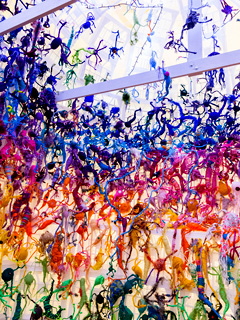
Narrating neuroscience in Toronto (Canada) on Oct. 20, 2017 and knitting a neuron

What is it with the Canadian neuroscience community? First, there’s The Beautiful Brain an exhibition of the extraordinary drawings of Santiago Ramón y Cajal (1852–1934) at the Belkin Gallery on the University of British Columbia (UBC) campus in Vancouver and a series of events marking the exhibition (for more see my Sept. 11, 2017 posting ; scroll down about 30% for information about the drawings and the events still to come).

I guess there must be some money floating around for raising public awareness because now there’s a neuroscience and ‘storytelling’ event (Narrating Neuroscience) in Toronto, Canada. From a Sept. 25, 2017 ArtSci Salon announcement (received via email),

With NARRATING NEUROSCIENCE we plan to initiate a discussion on the  role and the use of storytelling and art (both in verbal and visual  forms) to communicate abstract and complex concepts in neuroscience to  very different audiences, ranging from fellow scientists, clinicians and patients, to social scientists and the general public. We invited four guests to share their research through case studies and experiences stemming directly from their research or from other practices they have adopted and incorporated into their research, where storytelling and the arts have played a crucial role not only in communicating cutting edge research in neuroscience, but also in developing and advancing it.

OUR GUESTS

MATTEO FARINELLA, PhD, Presidential Scholar in Society and Neuroscience – Columbia University

SHELLEY WALL , AOCAD, MSc, PhD – Assistant professor, Biomedical Communications Graduate Program and Department of Biology, UTM

ALFONSO FASANO, MD, PhD, Associate Professor – University of Toronto Clinician Investigator – Krembil Research Institute Movement Disorders Centre – Toronto Western Hospital

TAHANI BAAKDHAH, MD, MSc, PhD candidate – University of Toronto

DATE: October 20, 2017
TIME: 6:00-8:00 pm
LOCATION: The Fields Institute for Research in Mathematical Sciences
222 College Street, Toronto, ON

Events Facilitators: Roberta Buiani and Stephen Morris (ArtSci Salon) and Nina Czegledy (Leonardo Network)

TAHANI BAAKDHAH is a PhD student at the University of Toronto studying how the stem cells built our retina during development, the mechanism by which the light sensing cells inside the eye enable us to see this beautiful world and how we can regenerate these cells in case of disease or injury.

MATTEO FARINELLA combines a background in neuroscience with a lifelong passion for drawing, making comics and illustrations about the brain. He is the author of _Neurocomic_ (Nobrow 2013) published with the support of the Wellcome Trust, _Cervellopoli_ (Editoriale Scienza 2017) and he has collaborated with universities and educational institutions around
the world to make science more clear and accessible. In 2016 Matteo joined Columbia University as a Presidential Scholar in Society and Neuroscience, where he investigates the role of visual narratives in science communication. Working with science journalists, educators and cognitive neuroscientists he aims to understand how these tools may
affect the public perception of science and increase scientific literacy (cartoonscience.org [2]).

ALFONSO FASANO graduated from the Catholic University of Rome, Italy, in 2002 and became a neurologist in 2007. After a 2-year fellowship at the University of Kiel, Germany, he completed a PhD in neuroscience at the Catholic University of Rome. In 2013 he joined the Movement Disorder Centre at Toronto Western Hospital, where he is the co-director of the
surgical program for movement disorders. He is also an associate professor of medicine in the Division of Neurology at the University of Toronto and clinician investigator at the Krembil Research Institute. Dr. Fasano’s main areas of interest are the treatment of movement  disorders with advanced technology (infusion pumps and neuromodulation), pathophysiology and treatment of tremor and gait disorders. He is author of more than 170 papers and book chapters. He is principal investigator of several clinical trials.

SHELLEY WALL is an assistant professor in the University of Toronto’s Biomedical Communications graduate program, a certified medical illustrator, and inaugural Illustrator-in-Residence in the Faculty of Medicine, University of Toronto. One of her primary areas of research, teaching, and creation is graphic medicine—the intersection of comics with illness, medicine, and caregiving—and one of her ongoing projects is a series of comics about caregiving and young onset Parkinson’s disease.

You can register for this free Toronto event here.

One brief observation, there aren’t any writers (other than academics) or storytellers included in this ‘storytelling’ event. The ‘storytelling’ being featured is visual. To be blunt I’m not of the ‘one picture is worth a thousand words’ school of thinking (see my Feb. 22, 2011 posting). Yes, sometimes pictures are all you need but that tiresome aphorism which suggests  communication can be reduced to one means of communication really needs to be retired. As for academic writing, it’s not noted for its storytelling qualities or experimentation. Academics are not judged on their writing or storytelling skills although there are some who are very good.

Getting back to the Toronto event, they seem to have the visual part of their focus  ” … discussion on the  role and the use of storytelling and art (both in verbal and visual  forms) … ” covered. Having recently attended a somewhat similar event in Vancouver, which was announced n my Sept. 11, 2017 posting, there were some exciting images and ideas presented.

The ArtSci Salon folks also announced this (from the Sept. 25, 2017 ArtSci Salon announcement; received via email),

ATTENTION ARTSCI SALONISTAS AND FANS OF ART AND SCIENCE!!
CALL FOR KNITTING AND CROCHET LOVERS!

In addition to being a PhD student at the University of Toronto, Tahani Baakdhah is a prolific knitter and crocheter and has been the motor behind two successful Knit-a-Neuron Toronto initiatives. We invite all Knitters and Crocheters among our ArtSci Salonistas to pick a pattern
(link below) and knit a neuron (or 2! Or as many as you want!!)

http://bit.ly/2y05hRR

BRING THEM TO OUR OCTOBER 20 ARTSCI SALON!
Come to the ArtSci Salon and knit there!
You can’t come?
Share a picture with @ArtSci_Salon @SciCommTO #KnitANeuronTO [3] on
social media
Or…Drop us a line at artscisalon@gmail.com !

I think it’s been a few years since my last science knitting post. No, it was Oct. 18, 2016. Moving on, I found more neuron knitting while researching this piece. Here’s the Neural Knitworks group, which is part of Australia’s National Science Week (11-19 August 2018) initiative (from the Neural Knitworks webpage),

Neural Knitworks is a collaborative project about mind and brain health.

Whether you’re a whiz with yarn, or just discovering the joy of craft, now you can crochet wrap, knit or knot—and find out about neuroscience.

During 2014 an enormous number of handmade neurons were donated (1665 in total!) and used to build a giant walk-in brain, as seen here at Hazelhurst Gallery [scroll to end of this post]. Since then Neural Knitworks have been held in dozens of communities across Australia, with installations created in Queensland, the ACT, Singapore, as part of the Cambridge Science Festival in the UK and in Philadelphia, USA.

In 2017, the Neural Knitworks team again invites you to host your own home-grown Neural Knitwork for National Science Week*. Together we’ll create a giant ‘virtual’ neural network by linking your displays visually online.

* If you wish to host a Neural Knitwork event outside of National Science Week or internationally we ask that you contact us to seek permission to use the material, particularly if you intend to create derivative works or would like to exhibit the giant brain. Please outline your plans in an email.

Your creation can be big or small, part of a formal display, or simply consist of neighbourhood neuron ‘yarn-bombings’. Knitworks can be created at home, at work or at school. No knitting experience is required and all ages can participate.

See below for how to register your event and download our scientifically informed patterns.

What is a neuron?

Neurons are electrically excitable cells of the brain, spinal cord and peripheral nerves. The billions of neurons in your body connect to each other in neural networks. They receive signals from every sense, control movement, create memories, and form the neural basis of every thought.

Check out the neuron microscopy gallery for some real-world inspiration.

What happens at a Neural Knitwork?

Neural Knitworks are based on the principle that yarn craft, with its mental challenges, social connection and mindfulness, helps keep our brains and minds sharp, engaged and healthy.

Have fun as you

  • design your own woolly neurons, or get inspired by our scientifically-informed knitting, crochet or knot patterns;
  • natter with neuroscientists and teach them a few of your crafty tricks;
  • contribute to a travelling textile brain exhibition;
  • increase your attention span and test your memory.

Calm your mind and craft your own brain health as you

  • forge friendships;
  • solve creative and mental challenges;
  • practice mindfulness and relaxation;
  • teach and learn;
  • develop eye-hand coordination and fine motor dexterity.

Interested in hosting a Neural Knitwork?

  1. Log your event on the National Science Week calendar to take advantage of multi-channel promotion.
  2. Share the link^ for this Neural Knitwork page on your own website or online newsletter and add information your own event details.
  3. Use this flyer template (2.5 MB .docx) to promote your event in local shop windows and on noticeboards.
  4. Read our event organisers toolbox for tips on hosting a successful event.
  5. You’ll need plenty of yarn, needles, copies of our scientifically-based neuron crafting pattern books (3.4 MB PDF) and a comfy spot in which to create.
  6. Gather together a group of friends who knit, crochet, design, spin, weave and anyone keen to give it a go. Those who know how to knit can teach others how to do it, and there’s even an easy no knit pattern that you can knot.
  7. Download a neuroscience podcast to listen to, and you’ve got a Neural Knitwork!
  8. Join the Neural Knitworks community on Facebook  to share and find information about events including public talks featuring neuroscientists.
  9. Tweet #neuralknitworks to show us your creations.
  10. Find display ideas in the pattern book and on our Facebook page.

Finally,, the knitted neurons from Australia’s 2014 National Science Week brain exhibit,

[downloaded from https://www.scienceweek.net.au/neural-knitworks/]

ETA Oct. 24, 2017: If you’re interested on how the talk was received, there’s an Oct. 24, 2017 posting by Magosia Pakulska for the Research2Reality blog.