Tag Archives: retina

New nanotubes discovered in the eye

I was half-expecting to read about some sort of fancy carbon nanotubes—I was wrong. From an August 12, 2020 news item on ScienceDaily where the researchers keep the mystery going for a while,

A new mechanism of blood redistribution that is essential for the proper functioning of the adult retina has just been discovered in vivo by researchers at the University of Montreal Hospital Research Centre (CRCHUM).

“For the first time, we have identified a communication structure between cells that is required to coordinate blood supply in the living retina,” said Dr. Adriana Di Polo, a neuroscience professor at Université de Montréal and holder of a Canada Research Chair in glaucoma and age-related neurodegeneration, who supervised the study.

“We already knew that activated retinal areas receive more blood than non-activated ones,” she said, “but until now no one understood how this essential blood delivery was finely regulated.”

The study was conducted on mice by two members of Di Polo’s lab: Dr. Luis Alarcon-Martinez, a postdoctoral fellow, and Deborah Villafranca-Baughman, a PhD student. Both are the first co-authors of this study.

In living animals, as in humans, the retina uses the oxygen and nutrients contained in the blood to fully function. This vital exchange takes place through capillaries, the thinnest blood vessels in all organs of the body. When the blood supply is dramatically reduced or cut off — such as in ischemia or stroke — the retina does not receive the oxygen it needs. In this condition, the cells begin to die and the retina stops working as it should.

An August 12, 2020 University of Montreal Hospital Research Centre (CRCHUM) news release on EurekAlert clears up the mystery,

Tunnelling between cells

Wrapped around the capillaries are pericytes, cells that have the ability to control the amount of blood passing through a single capillary simply by squeezing and releasing it.

“Using a microscopy technique to visualize vascular changes in living mice, we showed that pericytes project very thin tubes, called inter-pericyte tunnelling nanotubes, [emphasis mine] to communicate with other pericytes located in distant capillaries,” said Alarcon-Martinez. “Through these nanotubes, the pericytes can talk to each other to deliver blood where it is most needed.”

Another important feature, added Villafranca-Baughman, is that “the capillaries lose their ability to shuttle blood where it is required when the tunnelling nanotubes are damaged–after an ischemic stroke, for example. The lack of blood supply that follows has a detrimental effect on neurons and the overall tissue function.”

The team’s findings suggest that microvascular deficits observed in neurodegenerative diseases like strokes, glaucoma, and Alzheimer’s disease might result from the loss of tunnelling nanotubes and impaired blood distribution. Strategies that protect these nanostructures should then be beneficial, but remain to be demonstrated.

Here’s a link to and a citation for the paper

Interpericyte tunnelling nanotubes regulate neurovascular coupling by Luis Alarcon-Martinez, Deborah Villafranca-Baughman, Heberto Quintero, J. Benjamin Kacerovsky, Florence Dotigny, Keith K. Murai, Alexandre Prat, Pierre Drapeau & Adriana Di Polo. Nature (2020) Published: 12 August 2020 DOI: https://doi.org/10.1038/s41586-020-2589-x

This paper is behind a paywall.

World’s first liquid retina prosthesis

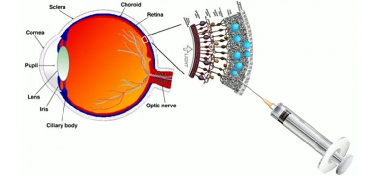
The new artificial liquid retina is biomimetic and consists of an aqueous component in which photoactive polymeric nanoparticles (whose size is of 350 nanometres, thus about 1/100 of the diameter of a hair) are suspended, going to replace the damaged photoreceptors. Credit: IIT-Istituto Italiano di Tecnologia [image downloaded from https://www.medgadget.com/2020/06/injectable-liquid-prosthesis-to-treat-retinal-diseases-developed.html]

A June 29, 2020 news item on Nanowerk announces the world’s first liquid retina prosthesis,

Researchers at IIT-Istituto Italiano di Tecnologia (Italian Institute of Technology) has led to the revolutionary development of an artificial liquid retinal prosthesis to counteract the effects of diseases such as retinitis pigmentosa and age-related macular degeneration that cause the progressive degeneration of photoreceptors of the retina, resulting in blindness.

The multidisciplinary team is composed by researchers from the IIT’s Center for Synaptic Neuroscience and Technology in Genoa coordinated by Fabio Benfenati and a team from the IIT’s Center for Nano Science and Technology in Milan coordinated by Guglielmo Lanzani, and it also involves the IRCCS Ospedale Sacrocuore Don Calabria in Negrar (Verona) with the team lead by Grazia Pertile, the IRCCS Ospedale Policlinico San Martino in Genoa and the CNR in Bologna. The research has been supported by Fondazione 13 Marzo Onlus, Fondazione Ra.Mo., Rare Partners srl and Fondazione Cariplo.

The study represents the state of the art in retinal prosthetics and is an evolution of the planar artificial retinal model developed by the same team in 2017 and based on organic semiconductor materials (Nature Materials 2017, 16: 681-689).

A June 30, 2020 IIT-Istituto Italiano di Tecnologia (Italian Institute of Technology) press release (also on EurekAlert but published June 29, 2020) provides more detail,

The “second generation” artificial retina is biomimetic, offers high spatial resolution and consists of an aqueous component in which photoactive polymeric nanoparticles (whose size is of 350 nanometres, thus about 1/100 of the diameter of a hair) are suspended, going to replace the damaged photoreceptors.

The experimental results show that the natural light stimulation of nanoparticles, in fact, causes the activation of retinal neurons spared from degeneration, thus mimicking the functioning of photoreceptors in healthy subjects.

Compared to other existing approaches, the new liquid nature of the prosthesis ensures fast and less traumatic surgery that consist of microinjections of nanoparticles directly under the retina, where they remain trapped and replace the degenerated photoreceptors; this method also ensures an increased effectiveness.

The data collected show also that the innovative experimental technique represents a valid alternative to the methods used to date to restore the photoreceptive capacity of retinal neurons while preserving their spatial resolution, laying a solid foundation for future clinical trials in humans. Moreover, the development of these photosensitive nanomaterials opens the way to new future applications in neuroscience and medicine.

“Our experimental results highlight the potential relevance of nanomaterials in the development of second-generation retinal prostheses to treat degenerative retinal blindness, and represents a major step forward” Fabio Benfenati commented. “The creation of a liquid artificial retinal implant has great potential to ensure a wide-field vision and high-resolution vision. Enclosing the photoactive polymers in particles that are smaller than the photoreceptors, increases the active surface of interaction with the retinal neurons, allows to easily cover the entire retinal surface and to scale the photoactivation at the level of a single photoreceptor.”

“In this research we have applied nanotechnology to medicine” concludes Guglielmo Lanzani. “In particular in our labs we have realized polymer nanoparticles that behave like tiny photovoltaic cells, based on carbon and hydrogen, fundamental components of the biochemistry of life. Once injected into the retina, these nanoparticles form small aggregates the size of which is comparable to that of neurons, that effectively behave like photoreceptors.”

“The surgical procedure for the subretinal injection of photoactive nanoparticles is minimally invasive and potentially replicable over time, unlike planar retinal prostheses” adds Grazia Pertile, Director at Operating Unit of Ophthalmology at IRCCS Ospedale Sacro Cuore Don Calabria. “At the same time maintaining the advantages of polymeric prosthesis, which is naturally sensitive to the light entering the eye and does not require glasses, cameras or external energy sources.”

The research study is based on preclinical models and further experimentations will be fundamental to make the technique a clinical treatment for diseases such as retinitis pigmentosa and age-related macular degeneration.

Here’s a link to and a citation for the paper,

Subretinally injected semiconducting polymer nanoparticles rescue vision in a rat model of retinal dystrophy by José Fernando Maya-Vetencourt, Giovanni Manfredi, Maurizio Mete, Elisabetta Colombo, Mattia Bramini, Stefano Di Marco, Dmytro Shmal, Giulia Mantero, Michele Dipalo, Anna Rocchi, Mattia L. DiFrancesco, Ermanno D. Papaleo, Angela Russo, Jonathan Barsotti, Cyril Eleftheriou, Francesca Di Maria, Vanessa Cossu, Fabio Piazza, Laura Emionite, Flavia Ticconi, Cecilia Marini, Gianmario Sambuceti, Grazia Pertile, Guglielmo Lanzani & Fabio Benfenati. Nature Nanotechnology (2020) DOI: https://doi.org/10.1038/s41565-020-0696-3 Published: 29 June 2020

This paper is behind a paywall.

Crowdsourcing brain research at Princeton University to discover 6 new neuron types

Spritely music!

There were already 1/4M registered players as of May 17, 2018 but I’m sure there’s room for more should you be inspired. A May 17, 2018 Princeton University news release (also on EurekAlert) reveals more about the game and about the neurons,

With the help of a quarter-million video game players, Princeton researchers have created and shared detailed maps of more than 1,000 neurons — and they’re just getting started.

“Working with Eyewirers around the world, we’ve made a digital museum that shows off the intricate beauty of the retina’s neural circuits,” said Sebastian Seung, the Evnin Professor in Neuroscience and a professor of computer science and the Princeton Neuroscience Institute (PNI). The related paper is publishing May 17 [2018] in the journal Cell.

Seung is unveiling the Eyewire Museum, an interactive archive of neurons available to the general public and neuroscientists around the world, including the hundreds of researchers involved in the federal Brain Research through Advancing Innovative Neurotechnologies (BRAIN) Initiative.

“This interactive viewer is a huge asset for these larger collaborations, especially among people who are not physically in the same lab,” said Amy Robinson Sterling, a crowdsourcing specialist with PNI and the executive director of Eyewire, the online gaming platform for the citizen scientists who have created this data set.

“This museum is something like a brain atlas,” said Alexander Bae, a graduate student in electrical engineering and one of four co-first authors on the paper. “Previous brain atlases didn’t have a function where you could visualize by individual cell, or a subset of cells, and interact with them. Another novelty: Not only do we have the morphology of each cell, but we also have the functional data, too.”

The neural maps were developed by Eyewirers, members of an online community of video game players who have devoted hundreds of thousands of hours to painstakingly piecing together these neural cells, using data from a mouse retina gathered in 2009.

Eyewire pairs machine learning with gamers who trace the twisting and branching paths of each neuron. Humans are better at visually identifying the patterns of neurons, so every player’s moves are recorded and checked against each other by advanced players and Eyewire staffers, as well as by software that is improving its own pattern recognition skills.

Since Eyewire’s launch in 2012, more than 265,000 people have signed onto the game, and they’ve collectively colored in more than 10 million 3-D “cubes,” resulting in the mapping of more than 3,000 neural cells, of which about a thousand are displayed in the museum.

Each cube is a tiny subset of a single cell, about 4.5 microns across, so a 10-by-10 block of cubes would be the width of a human hair. Every cell is reviewed by between 5 and 25 gamers before it is accepted into the system as complete.

“Back in the early years it took weeks to finish a single cell,” said Sterling. “Now players complete multiple neurons per day.” The Eyewire user experience stays focused on the larger mission — “For science!” is a common refrain — but it also replicates a typical gaming environment, with achievement badges, a chat feature to connect with other players and technical support, and the ability to unlock privileges with increasing skill. “Our top players are online all the time — easily 30 hours a week,” Sterling said.

Dedicated Eyewirers have also contributed in other ways, including donating the swag that gamers win during competitions and writing program extensions “to make game play more efficient and more fun,” said Sterling, including profile histories, maps of player activity, a top 100 leaderboard and ever-increasing levels of customizability.

“The community has really been the driving force behind why Eyewire has been successful,” Sterling said. “You come in, and you’re not alone. Right now, there are 43 people online. Some of them will be admins from Boston or Princeton, but most are just playing — now it’s 46.”

For science!

With 100 billion neurons linked together via trillions of connections, the brain is immeasurably complex, and neuroscientists are still assembling its “parts list,” said Nicholas Turner, a graduate student in computer science and another of the co-first authors. “If you know what parts make up the machine you’re trying to break apart, you’re set to figure out how it all works,” he said.

The researchers have started by tackling Eyewire-mapped ganglion cells from the retina of a mouse. “The retina doesn’t just sense light,” Seung said. “Neural circuits in the retina perform the first steps of visual perception.”

The retina grows from the same embryonic tissue as the brain, and while much simpler than the brain, it is still surprisingly complex, Turner said. “Hammering out these details is a really valuable effort,” he said, “showing the depth and complexity that exists in circuits that we naively believe are simple.”

The researchers’ fundamental question is identifying exactly how the retina works, said Bae. “In our case, we focus on the structural morphology of the retinal ganglion cells.”

“Why the ganglion cells of the eye?” asked Shang Mu, an associate research scholar in PNI and fellow first author. “Because they’re the connection between the retina and the brain. They’re the only cell class that go back into the brain.” Different types of ganglion cells are known to compute different types of visual features, which is one reason the museum has linked shape to functional data.

Using Eyewire-produced maps of 396 ganglion cells, the researchers in Seung’s lab successfully classified these cells more thoroughly than has ever been done before.

“The number of different cell types was a surprise,” said Mu. “Just a few years ago, people thought there were only 15 to 20 ganglion cell types, but we found more than 35 — we estimate between 35 and 50 types.”

Of those, six appear to be novel, in that the researchers could not find any matching descriptions in a literature search.

A brief scroll through the digital museum reveals just how remarkably flat the neurons are — nearly all of the branching takes place along a two-dimensional plane. Seung’s team discovered that different cells grow along different planes, with some reaching high above the nucleus before branching out, while others spread out close to the nucleus. Their resulting diagrams resemble a rainforest, with ground cover, an understory, a canopy and an emergent layer overtopping the rest.

All of these are subdivisions of the inner plexiform layer, one of the five previously recognized layers of the retina. The researchers also identified a “density conservation principle” that they used to distinguish types of neurons.

One of the biggest surprises of the research project has been the extraordinary richness of the original sample, said Seung. “There’s a little sliver of a mouse retina, and almost 10 years later, we’re still learning things from it.”

Of course, it’s a mouse’s brain that you’ll be examining and while there are differences between a mouse brain and a human brain, mouse brains still provide valuable data as they did in the case of some groundbreaking research published in October 2017. James Hamblin wrote about it in an Oct. 7, 2017 article for The Atlantic (Note: Links have been removed),


Scientists Somehow Just Discovered a New System of Vessels in Our Brains

It is unclear what they do—but they likely play a central role in aging and disease.

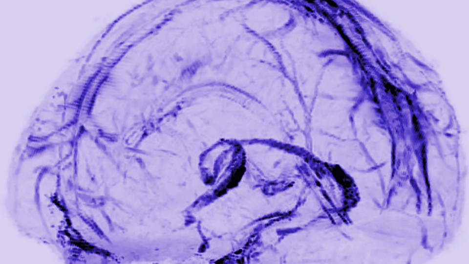
A transparent model of the brain with a network of vessels filled in
Daniel Reich / National Institute of Neurological Disorders and Stroke

You are now among the first people to see the brain’s lymphatic system. The vessels in the photo above transport fluid that is likely crucial to metabolic and inflammatory processes. Until now, no one knew for sure that they existed.

Doctors practicing today have been taught that there are no lymphatic vessels inside the skull. Those deep-purple vessels were seen for the first time in images published this week by researchers at the U.S. National Institute of Neurological Disorders and Stroke.

In the rest of the body, the lymphatic system collects and drains the fluid that bathes our cells, in the process exporting their waste. It also serves as a conduit for immune cells, which go out into the body looking for adversaries and learning how to distinguish self from other, and then travel back to lymph nodes and organs through lymphatic vessels.

So how was it even conceivable that this process wasn’t happening in our brains?

Reich (Daniel Reich, senior investigator) started his search in 2015, after a major study in Nature reported a similar conduit for lymph in mice. The University of Virginia team wrote at the time, “The discovery of the central-nervous-system lymphatic system may call for a reassessment of basic assumptions in neuroimmunology.” The study was regarded as a potential breakthrough in understanding how neurodegenerative disease is associated with the immune system.

Around the same time, researchers discovered fluid in the brains of mice and humans that would become known as the “glymphatic system.” [emphasis mine] It was described by a team at the University of Rochester in 2015 as not just the brain’s “waste-clearance system,” but as potentially helping fuel the brain by transporting glucose, lipids, amino acids, and neurotransmitters. Although since “the central nervous system completely lacks conventional lymphatic vessels,” the researchers wrote at the time, it remained unclear how this fluid communicated with the rest of the body.

There are occasional references to the idea of a lymphatic system in the brain in historic literature. Two centuries ago, the anatomist Paolo Mascagni made full-body models of the lymphatic system that included the brain, though this was dismissed as an error. [emphases mine]  A historical account in The Lancet in 2003 read: “Mascagni was probably so impressed with the lymphatic system that he saw lymph vessels even where they did not exist—in the brain.”

I couldn’t resist the reference to someone whose work had been dismissed summarily being proved right, eventually, and with the help of mouse brains. Do read Hamblin’s article in its entirety if you have time as these excerpts don’t do it justice.
Getting back to Princeton’s research, here’s their research paper,

Digital museum of retinal ganglion cells with dense anatomy and physiology,” by Alexander Bae, Shang Mu, Jinseop Kim, Nicholas Turner, Ignacio Tartavull, Nico Kemnitz, Chris Jordan, Alex Norton, William Silversmith, Rachel Prentki, Marissa Sorek, Celia David, Devon Jones, Doug Bland, Amy Sterling, Jungman Park, Kevin Briggman, Sebastian Seung and the Eyewirers, was published May 17 in the journal Cell with DOI 10.1016/j.cell.2018.04.040.

The research was supported by the Gatsby Charitable Foundation, National Institute of Health-National Institute of Neurological Disorders and Stroke (U01NS090562 and 5R01NS076467), Defense Advanced Research Projects Agency (HR0011-14-2- 0004), Army Research Office (W911NF-12-1-0594), Intelligence Advanced Research Projects Activity (D16PC00005), KT Corporation, Amazon Web Services Research Grants, Korea Brain Research Institute (2231-415) and Korea National Research Foundation Brain Research Program (2017M3C7A1048086).

This paper is behind a paywall. For the players amongst us, here’s the Eyewire website. Go forth,  play, and, maybe, discover new neurons!

Using CRISPR to reverse retinosa pigmentosa (eye disease)

Years ago I worked as a publicist for the BC (British Columbia) Motorcycle Federation’s Ride for Sight; they were raising funds for research into retinitis pigmentosa (RP). I hadn’t thought about that in years but it all came back when I saw this April 21, 2017 news item on ScienceDaily,

Using the gene-editing tool CRISPR/Cas9, researchers at University of California San Diego [UCSD] School of Medicine and Shiley Eye Institute at UC San Diego Health, with colleagues in China, have reprogrammed mutated rod photoreceptors to become functioning cone photoreceptors, reversing cellular degeneration and restoring visual function in two mouse models of retinitis pigmentosa.

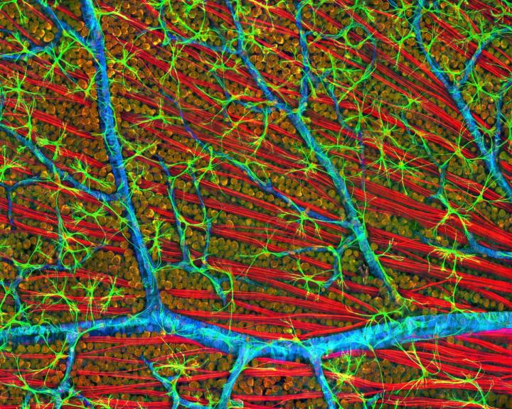
Caption: This is a confocal micrograph of mouse retina depicting optic fiber layer. Credit: Image courtesy of National Center for Microscopy and Imaging Research, UC San Diego.

An April 21, 2017 UCSD news release by Scott LaFee (also on EurekAlert), which originated the news item, delves further into retinitis pigmentosa and this CRISPR research,

Retinitis pigmentosa (RP) is a group of inherited vision disorders caused by numerous mutations in more than 60 genes. The mutations affect the eyes’ photoreceptors, specialized cells in the retina that sense and convert light images into electrical signals sent to the brain. There are two types: rod cells that function for night vision and peripheral vision, and cone cells that provide central vision (visual acuity) and discern color. The human retina typically contains 120 million rod cells and 6 million cone cells.

In RP, which affects approximately 100,000 Americans and 1 in 4,000 persons worldwide, rod-specific genetic mutations cause rod photoreceptor cells to dysfunction and degenerate over time. Initial symptoms are loss of peripheral and night vision, followed by diminished visual acuity and color perception as cone cells also begin to fail and die. There is no treatment for RP. The eventual result may be legal blindness.

In their published research, a team led by senior author Kang Zhang, MD, PhD, chief of ophthalmic genetics, founding director of the Institute for Genomic Medicine and co-director of biomaterials and tissue engineering at the Institute of Engineering in Medicine, both at UC San Diego School of Medicine, used CRISPR/Cas9 to deactivate a master switch gene called Nrl and a downstream transcription factor called Nr2e3.

CRISPR, which stands for Clustered Regularly Interspaced Short Palindromic Repeats, allows researchers to target specific stretches of genetic code and edit DNA at precise locations, modifying select gene functions. Deactivating either Nrl or Nr2e3 reprogrammed rod cells to become cone cells.

“Cone cells are less vulnerable to the genetic mutations that cause RP,” said Zhang. “Our strategy was to use gene therapy to make the underlying mutations irrelevant, resulting in the preservation of tissue and vision.”

The scientists tested their approach in two different mouse models of RP. In both cases, they found an abundance of reprogrammed cone cells and preserved cellular architecture in the retinas. Electroretinography testing of rod and cone receptors in live mice show improved function.

Zhang said a recent independent study led by Zhijian Wu, PhD, at National Eye Institute, part of the National Institutes of Health, also reached similar conclusions.

The researchers used adeno-associated virus (AAV) to perform the gene therapy, which they said should help advance their work to human clinical trials quicker. “AAV is a common cold virus and has been used in many successful gene therapy treatments with a relatively good safely profile,” said Zhang. “Human clinical trials could be planned soon after completion of preclinical study. There is no treatment for RP so the need is great and pressing. In addition, our approach of reprogramming mutation-sensitive cells to mutation-resistant cells may have broader application to other human diseases, including cancer.”

Here’s a link to and a citation for the paper,

Gene and mutation independent therapy via CRISPR-Cas9 mediated cellular reprogramming in rod photoreceptors by Jie Zhu, Chang Ming, Xin Fu, Yaou Duan, Duc Anh Hoang, Jeffrey Rutgard, Runze Zhang, Wenqiu Wang, Rui Hou, Daniel Zhang, Edward Zhang, Charlotte Zhang, Xiaoke Hao, Wenjun Xiong, and Kang Zhang. Cell Research advance online publication 21 April 2017; doi: 10.1038/cr.2017.57

This paper (it’s in the form of a letter to the editor) is open access.

Fractal imagery (from nature or from art or from mathematics) soothes

Jackson Pollock’s work is often cited when fractal art is discussed. I think it’s largely because he likely produced the art without knowing about the concept.

No. 5, 1948 (Jackson Pollock, downloaded from Wikipedia essay about No. 5, 1948)

Richard Taylor, a professor of physics at the University of Oregon, provides more information about how fractals affect us and how this is relevant to his work with retinal implants in a March 30, 2017 essay for The Conversation (h/t Mar. 31, 2017 news item on phys.org), Note: Links have been removed),

Humans are visual creatures. Objects we call “beautiful” or “aesthetic” are a crucial part of our humanity. Even the oldest known examples of rock and cave art served aesthetic rather than utilitarian roles. Although aesthetics is often regarded as an ill-defined vague quality, research groups like mine are using sophisticated techniques to quantify it – and its impact on the observer.

We’re finding that aesthetic images can induce staggering changes to the body, including radical reductions in the observer’s stress levels. Job stress alone is estimated to cost American businesses many billions of dollars annually, so studying aesthetics holds a huge potential benefit to society.

Researchers are untangling just what makes particular works of art or natural scenes visually appealing and stress-relieving – and one crucial factor is the presence of the repetitive patterns called fractals.

When it comes to aesthetics, who better to study than famous artists? They are, after all, the visual experts. My research group took this approach with Jackson Pollock, who rose to the peak of modern art in the late 1940s by pouring paint directly from a can onto horizontal canvases laid across his studio floor. Although battles raged among Pollock scholars regarding the meaning of his splattered patterns, many agreed they had an organic, natural feel to them.

My scientific curiosity was stirred when I learned that many of nature’s objects are fractal, featuring patterns that repeat at increasingly fine magnifications. For example, think of a tree. First you see the big branches growing out of the trunk. Then you see smaller versions growing out of each big branch. As you keep zooming in, finer and finer branches appear, all the way down to the smallest twigs. Other examples of nature’s fractals include clouds, rivers, coastlines and mountains.

In 1999, my group used computer pattern analysis techniques to show that Pollock’s paintings are as fractal as patterns found in natural scenery. Since then, more than 10 different groups have performed various forms of fractal analysis on his paintings. Pollock’s ability to express nature’s fractal aesthetics helps explain the enduring popularity of his work.

The impact of nature’s aesthetics is surprisingly powerful. In the 1980s, architects found that patients recovered more quickly from surgery when given hospital rooms with windows looking out on nature. Other studies since then have demonstrated that just looking at pictures of natural scenes can change the way a person’s autonomic nervous system responds to stress.

Are fractals the secret to some soothing natural scenes? Ronan, CC BY-NC-ND

For me, this raises the same question I’d asked of Pollock: Are fractals responsible? Collaborating with psychologists and neuroscientists, we measured people’s responses to fractals found in nature (using photos of natural scenes), art (Pollock’s paintings) and mathematics (computer generated images) and discovered a universal effect we labeled “fractal fluency.”

Through exposure to nature’s fractal scenery, people’s visual systems have adapted to efficiently process fractals with ease. We found that this adaptation occurs at many stages of the visual system, from the way our eyes move to which regions of the brain get activated. This fluency puts us in a comfort zone and so we enjoy looking at fractals. Crucially, we used EEG to record the brain’s electrical activity and skin conductance techniques to show that this aesthetic experience is accompanied by stress reduction of 60 percent – a surprisingly large effect for a nonmedicinal treatment. This physiological change even accelerates post-surgical recovery rates.

Pollock’s motivation for continually increasing the complexity of his fractal patterns became apparent recently when I studied the fractal properties of Rorschach inkblots. These abstract blots are famous because people see imaginary forms (figures and animals) in them. I explained this process in terms of the fractal fluency effect, which enhances people’s pattern recognition processes. The low complexity fractal inkblots made this process trigger-happy, fooling observers into seeing images that aren’t there.

Pollock disliked the idea that viewers of his paintings were distracted by such imaginary figures, which he called “extra cargo.” He intuitively increased the complexity of his works to prevent this phenomenon.

Pollock’s abstract expressionist colleague, Willem De Kooning, also painted fractals. When he was diagnosed with dementia, some art scholars called for his retirement amid concerns that that it would reduce the nurture component of his work. Yet, although they predicted a deterioration in his paintings, his later works conveyed a peacefulness missing from his earlier pieces. Recently, the fractal complexity of his paintings was shown to drop steadily as he slipped into dementia. The study focused on seven artists with different neurological conditions and highlighted the potential of using art works as a new tool for studying these diseases. To me, the most inspiring message is that, when fighting these diseases, artists can still create beautiful artworks.

Recognizing how looking at fractals reduces stress means it’s possible to create retinal implants that mimic the mechanism. Nautilus image via www.shutterstock.com.

My main research focuses on developing retinal implants to restore vision to victims of retinal diseases. At first glance, this goal seems a long way from Pollock’s art. Yet, it was his work that gave me the first clue to fractal fluency and the role nature’s fractals can play in keeping people’s stress levels in check. To make sure my bio-inspired implants induce the same stress reduction when looking at nature’s fractals as normal eyes do, they closely mimic the retina’s design.

When I started my Pollock research, I never imagined it would inform artificial eye designs. This, though, is the power of interdisciplinary endeavors – thinking “out of the box” leads to unexpected but potentially revolutionary ideas.

Fabulous essay, eh?

I have previously featured Jackson Pollock in a June 30, 2011 posting titled: Jackson Pollock’s physics and and briefly mentioned him in a May 11, 2010 visual arts commentary titled: Rennie Collection’s latest: Richard Jackson, Georges Seurat & Jackson Pollock, guns, the act of painting, and women (scroll down about 45% of the way).