Tag Archives: eyes

Nanobody could lead to treatment for retinitis pigmentosa (a condition that leads to blindness)

This is an image illustrating the work but you’ll probably need to read the news release to understand the explanation offered,

Caption: This image depicts the crystal structure of two nanobodies binding to a rhodopsin dimer. The rhodopsin molecules are shown in green and blue, with 11-cis-retinal displayed in red. The figure emphasizes the significant interactions between the nanobodies (represented in a semi-transparent surface cartoon) and the extracellular surface of rhodopsin, including its N-terminal glycans highlighted in orange.. Credit: UCI [University of California at Irvine] School of Medicine

The research from the University of California at Irvine (UCI) has been featured twice, in an August 31, 2023 news item on phys.org and again in a September 7, 2023 news item on ScienceDaily.

An August 29, 2023 UCI news release (also on EurekAlert but published Sept. 6, 2023), which originated the news items, provides information about RP and the nanobodies,

A team of scientists from the University of California, Irvine, believe they have discovered a special antibody which may lead to a treatment for Retinitis Pigmentosa, a condition that causes loss of central vision, as well as night and color vision. 

The study, Structural basis for the allosteric modulation of rhodopsin by nanobody binding to its extracellular domain, was published in Nature Communications. Authors of the study were Arum Wu, PhD, David Salom, PhD, John D. Hong, Aleksander Tworak, PhD, Philip D. Kiser, PharmD, PhD, and Krzysztof Palczewski, PhD, in the Department of Ophthalmology, Gavin Herbert Eye Institute, at the University of California, Irvine. Research was conducted  in collaboration with Jan Steyaert, PhD, at the Vrije Universiteit Brussel (VUB).

Retinitis Pigmentosa (RP) is a group of inherited eye diseases that affect the retina in the back of the eye. It is caused by the death of cells that detect light signals, known as photoreceptor cells. There is no known cure for RP, and the development of new treatments for this condition relies on cell and gene therapies. 

UCI researchers have targeted their study on a specific molecule which they believe will provide a treatment for Rhodopsin-associated autosomal dominant RP (adRP). The molecule, Rhodopsin, is a key light-sensing molecule in the human retina. It is found in rod photoreceptor cells, and mutations in the Rhodopsin gene are a primary cause of adRP. 

“More than 150 mutations in rhodopsin can cause Retinitis Pigmentosa, making it challenging to develop targeted gene therapies,” said Krzysztof Palczewski, PhD, Donald Bren Professor, UCI School of Medicine. “However due to the high prevalence of RP, there has been significant investment in research and development efforts to find novel treatments.”

Although Rhodopsin has been studied for over a century, key details of its mechanism for converting light into a cellular signal have been difficult to experimentally address.

For this study, researchers used a special type of llama-derived antibody, known as a nanobody, that can halt the process of Rhodopsin photoactivation, allowing it to be investigated at high resolution. 

“Our team has developed nanobodies that work through a novel mechanism of action. These nanobodies have high specificity and can recognize the target rhodopsin extracellularly,” said David Salom , PhD, researcher and project scientist, UCI School of Medicine. “This enables us to lock this GPCR in a non-signaling state.” 

Scientists discovered that these nanobodies target an unexpected site on the Rhodopsin molecule, near the location where retinaldehyde binds. They also found that the stabilizing effect of these nanobodies can also be applied to Rhodopsin mutants that are associated with retinal disease, suggesting their use as therapeutics. 

“In the future, we hope to involve the in vitro evolution of these initial set of nanobodies,” said Arum Wu, PhD, researcher and project scientist, UCI School of Medicine. “We will also evaluate the safety and effectiveness of a future nanobody gene therapy for RP.”

Researchers hope to improve nanobodies’ ability to recognize Rhodopsin from other species including mice, for which several pre-clinical models of adRP are available. They also have plans to use these nanobodies to address a long-term goal in the field of structurally resolving the key intermediate states of Rhodopsin from the inactive state to the fully ligand-activated state.

Here’s a link to and a citation for the paper,

Structural basis for the allosteric modulation of rhodopsin by nanobody binding to its extracellular domain by Arum Wu, David Salom, John D. Hong, Aleksander Tworak, Kohei Watanabe, Els Pardon, Jan Steyaert, Hideki Kandori, Kota Katayama, Philip D. Kiser & Krzysztof Palczewski. Nature Communications volume 14, Article number: 5209 (2023) DOI: https://doi.org/10.1038/s41467-023-40911-9 Published: 25 August 2023

This paper is open access.

New nanotubes discovered in the eye

I was half-expecting to read about some sort of fancy carbon nanotubes—I was wrong. From an August 12, 2020 news item on ScienceDaily where the researchers keep the mystery going for a while,

A new mechanism of blood redistribution that is essential for the proper functioning of the adult retina has just been discovered in vivo by researchers at the University of Montreal Hospital Research Centre (CRCHUM).

“For the first time, we have identified a communication structure between cells that is required to coordinate blood supply in the living retina,” said Dr. Adriana Di Polo, a neuroscience professor at Université de Montréal and holder of a Canada Research Chair in glaucoma and age-related neurodegeneration, who supervised the study.

“We already knew that activated retinal areas receive more blood than non-activated ones,” she said, “but until now no one understood how this essential blood delivery was finely regulated.”

The study was conducted on mice by two members of Di Polo’s lab: Dr. Luis Alarcon-Martinez, a postdoctoral fellow, and Deborah Villafranca-Baughman, a PhD student. Both are the first co-authors of this study.

In living animals, as in humans, the retina uses the oxygen and nutrients contained in the blood to fully function. This vital exchange takes place through capillaries, the thinnest blood vessels in all organs of the body. When the blood supply is dramatically reduced or cut off — such as in ischemia or stroke — the retina does not receive the oxygen it needs. In this condition, the cells begin to die and the retina stops working as it should.

An August 12, 2020 University of Montreal Hospital Research Centre (CRCHUM) news release on EurekAlert clears up the mystery,

Tunnelling between cells

Wrapped around the capillaries are pericytes, cells that have the ability to control the amount of blood passing through a single capillary simply by squeezing and releasing it.

“Using a microscopy technique to visualize vascular changes in living mice, we showed that pericytes project very thin tubes, called inter-pericyte tunnelling nanotubes, [emphasis mine] to communicate with other pericytes located in distant capillaries,” said Alarcon-Martinez. “Through these nanotubes, the pericytes can talk to each other to deliver blood where it is most needed.”

Another important feature, added Villafranca-Baughman, is that “the capillaries lose their ability to shuttle blood where it is required when the tunnelling nanotubes are damaged–after an ischemic stroke, for example. The lack of blood supply that follows has a detrimental effect on neurons and the overall tissue function.”

The team’s findings suggest that microvascular deficits observed in neurodegenerative diseases like strokes, glaucoma, and Alzheimer’s disease might result from the loss of tunnelling nanotubes and impaired blood distribution. Strategies that protect these nanostructures should then be beneficial, but remain to be demonstrated.

Here’s a link to and a citation for the paper

Interpericyte tunnelling nanotubes regulate neurovascular coupling by Luis Alarcon-Martinez, Deborah Villafranca-Baughman, Heberto Quintero, J. Benjamin Kacerovsky, Florence Dotigny, Keith K. Murai, Alexandre Prat, Pierre Drapeau & Adriana Di Polo. Nature (2020) Published: 12 August 2020 DOI: https://doi.org/10.1038/s41586-020-2589-x

This paper is behind a paywall.

World’s first liquid retina prosthesis

The new artificial liquid retina is biomimetic and consists of an aqueous component in which photoactive polymeric nanoparticles (whose size is of 350 nanometres, thus about 1/100 of the diameter of a hair) are suspended, going to replace the damaged photoreceptors. Credit: IIT-Istituto Italiano di Tecnologia [image downloaded from https://www.medgadget.com/2020/06/injectable-liquid-prosthesis-to-treat-retinal-diseases-developed.html]

A June 29, 2020 news item on Nanowerk announces the world’s first liquid retina prosthesis,

Researchers at IIT-Istituto Italiano di Tecnologia (Italian Institute of Technology) has led to the revolutionary development of an artificial liquid retinal prosthesis to counteract the effects of diseases such as retinitis pigmentosa and age-related macular degeneration that cause the progressive degeneration of photoreceptors of the retina, resulting in blindness.

The multidisciplinary team is composed by researchers from the IIT’s Center for Synaptic Neuroscience and Technology in Genoa coordinated by Fabio Benfenati and a team from the IIT’s Center for Nano Science and Technology in Milan coordinated by Guglielmo Lanzani, and it also involves the IRCCS Ospedale Sacrocuore Don Calabria in Negrar (Verona) with the team lead by Grazia Pertile, the IRCCS Ospedale Policlinico San Martino in Genoa and the CNR in Bologna. The research has been supported by Fondazione 13 Marzo Onlus, Fondazione Ra.Mo., Rare Partners srl and Fondazione Cariplo.

The study represents the state of the art in retinal prosthetics and is an evolution of the planar artificial retinal model developed by the same team in 2017 and based on organic semiconductor materials (Nature Materials 2017, 16: 681-689).

A June 30, 2020 IIT-Istituto Italiano di Tecnologia (Italian Institute of Technology) press release (also on EurekAlert but published June 29, 2020) provides more detail,

The “second generation” artificial retina is biomimetic, offers high spatial resolution and consists of an aqueous component in which photoactive polymeric nanoparticles (whose size is of 350 nanometres, thus about 1/100 of the diameter of a hair) are suspended, going to replace the damaged photoreceptors.

The experimental results show that the natural light stimulation of nanoparticles, in fact, causes the activation of retinal neurons spared from degeneration, thus mimicking the functioning of photoreceptors in healthy subjects.

Compared to other existing approaches, the new liquid nature of the prosthesis ensures fast and less traumatic surgery that consist of microinjections of nanoparticles directly under the retina, where they remain trapped and replace the degenerated photoreceptors; this method also ensures an increased effectiveness.

The data collected show also that the innovative experimental technique represents a valid alternative to the methods used to date to restore the photoreceptive capacity of retinal neurons while preserving their spatial resolution, laying a solid foundation for future clinical trials in humans. Moreover, the development of these photosensitive nanomaterials opens the way to new future applications in neuroscience and medicine.

“Our experimental results highlight the potential relevance of nanomaterials in the development of second-generation retinal prostheses to treat degenerative retinal blindness, and represents a major step forward” Fabio Benfenati commented. “The creation of a liquid artificial retinal implant has great potential to ensure a wide-field vision and high-resolution vision. Enclosing the photoactive polymers in particles that are smaller than the photoreceptors, increases the active surface of interaction with the retinal neurons, allows to easily cover the entire retinal surface and to scale the photoactivation at the level of a single photoreceptor.”

“In this research we have applied nanotechnology to medicine” concludes Guglielmo Lanzani. “In particular in our labs we have realized polymer nanoparticles that behave like tiny photovoltaic cells, based on carbon and hydrogen, fundamental components of the biochemistry of life. Once injected into the retina, these nanoparticles form small aggregates the size of which is comparable to that of neurons, that effectively behave like photoreceptors.”

“The surgical procedure for the subretinal injection of photoactive nanoparticles is minimally invasive and potentially replicable over time, unlike planar retinal prostheses” adds Grazia Pertile, Director at Operating Unit of Ophthalmology at IRCCS Ospedale Sacro Cuore Don Calabria. “At the same time maintaining the advantages of polymeric prosthesis, which is naturally sensitive to the light entering the eye and does not require glasses, cameras or external energy sources.”

The research study is based on preclinical models and further experimentations will be fundamental to make the technique a clinical treatment for diseases such as retinitis pigmentosa and age-related macular degeneration.

Here’s a link to and a citation for the paper,

Subretinally injected semiconducting polymer nanoparticles rescue vision in a rat model of retinal dystrophy by José Fernando Maya-Vetencourt, Giovanni Manfredi, Maurizio Mete, Elisabetta Colombo, Mattia Bramini, Stefano Di Marco, Dmytro Shmal, Giulia Mantero, Michele Dipalo, Anna Rocchi, Mattia L. DiFrancesco, Ermanno D. Papaleo, Angela Russo, Jonathan Barsotti, Cyril Eleftheriou, Francesca Di Maria, Vanessa Cossu, Fabio Piazza, Laura Emionite, Flavia Ticconi, Cecilia Marini, Gianmario Sambuceti, Grazia Pertile, Guglielmo Lanzani & Fabio Benfenati. Nature Nanotechnology (2020) DOI: https://doi.org/10.1038/s41565-020-0696-3 Published: 29 June 2020

This paper is behind a paywall.

Non-viral ocular gene therapy with gold nanoparticles and femtosecond lasers

I love the stylistic choice the writer made (pay special attention to the second paragraph) when producing this November 19, 2018 Polytechnique Montréal news release (also on EurekAlert),

A scientific breakthrough by Professor Michel Meunier of Polytechnique Montréal and his collaborators offers hope for people with glaucoma, retinitis or macular degeneration.

In January 2009, the life of engineer Michel Meunier, a professor at Polytechnique Montréal, changed dramatically. Like others, he had observed that the extremely short pulse of a femtosecond laser (0.000000000000001 second) could make nanometre-sized holes appear in silicon when it was covered by gold nanoparticles. But this researcher, recognized internationally for his skills in laser and nanotechnology, decided to go a step further with what was then just a laboratory curiosity. He wondered if it was possible to go from silicon to living matter, from inorganic to organic. Could the gold nanoparticles and the femtosecond laser, this “light scalpel,” reproduce the same phenomenon with living cells?

A very pretty image illustrating the work,

Caption: Gold nanoparticles, which act like “nanolenses,” concentrate the energy produced by the extremely short pulse of a femtosecond laser to create a nanoscale incision on the surface of the eye’s retina cells. This technology, which preserves cell integrity, can be used to effectively inject drugs or genes into specific areas of the eye, offering new hope to people with glaucoma, retinitis or macular degeneration. Credit and Copyright: Polytechnique Montréal

The news release goes on to describe the technology in more detail,

Professor Meunier started working on cells in vitro in his Polytechnique laboratory. The challenge was to make a nanometric incision in the cells’ extracellular membrane without damaging it. Using gold nanoparticles that acted as “nanolenses,” Professor Meunier realized that it was possible to concentrate the light energy coming from the laser at a wavelength of 800 nanometres. Since there is very little energy absorption by the cells at this wavelength, their integrity is preserved. Mission accomplished!

Based on this finding, Professor Meunier decided to work on cells in vivo, cells that are part of a complex living cell structure, such as the eye for example.

The eye and the light scalpel

In April 2012, Professor Meunier met Przemyslaw Sapieha, an internationally renowned eye specialist, particularly recognized for his work on the retina. “Mike”, as he goes by, is a professor in the Department of Ophthalmology at Université de Montréal and a researcher at Centre intégré universitaire de santé et de services sociaux (CIUSSS) de l’Est-de-l’Île-de-Montréal. He immediately saw the potential of this new technology and everything that could be done in the eye if you could block the ripple effect that occurs following a trigger that leads to glaucoma or macular degeneration, for example, by injecting drugs, proteins or even genes.

Using a femtosecond laser to treat the eye–a highly specialized and fragile organ–is very complex, however. The eye is part of the central nervous system, and therefore many of the cells or families of cells that compose it are neurons. And when a neuron dies, it does not regenerate like other cells do. Mike Sapieha’s first task was therefore to ensure that a femtosecond laser could be used on one or several neurons without affecting them. This is what is referred to as “proof of concept.”

Proof of concept

Mike and Michel called on biochemistry researcher Ariel Wilson, an expert in eye structures and vision mechanisms, as well as Professor Santiago Costantino and his team from the Department of Ophthalmology at Université de Montréal and the CIUSSS de l’Est-de-l’Île-de-Montréal for their expertise in biophotonics. The team first decided to work on healthy cells, because they are better understood than sick cells. They injected gold nanoparticles combined with antibodies to target specific neuronal cells in the eye, and then waited for the nanoparticles to settle around the various neurons or families of neurons, such as the retina. Following the bright flash generated by the femtosecond laser, the expected phenomenon occurred: small holes appeared in the cells of the eye’s retina, making it possible to effectively inject drugs or genes in specific areas of the eye. It was another victory for Michel Meunier and his collaborators, with these conclusive results now opening the path to new treatments.

The key feature of the technology developed by the researchers from Polytechnique and CIUSSS de l’Est-de-l’Île-de-Montréal is its extreme precision. With the use of functionalized gold nanoparticles, the light scalpel makes it possible to precisely locate the family of cells where the doctor will have to intervene.

Having successfully demonstrated proof of concept, Professor Meunier and his team filed a patent application in the United States. This tremendous work was also the subject of a paper reviewed by an impressive reading committee and published in the renowned journal Nano Letters in October 2018.

While there is still a lot of research to be done–at least 10 years’ worth, first on animals and then on humans–this technology could make all the difference in an aging population suffering from eye deterioration for which there are still no effective long-term treatments. It also has the advantage of avoiding the use of viruses commonly employed in gene therapy. These researchers are looking at applications of this technology in all eye diseases, but more particularly in glaucoma, retinitis and macular degeneration.

This light scalpel is unprecedented.

Here’s a link to and a citation for the paper,

In Vivo Laser-Mediated Retinal Ganglion Cell Optoporation Using KV1.1 Conjugated Gold Nanoparticles by Ariel M. Wilson, Javier Mazzaferri, Éric Bergeron, Sergiy Patskovsky, Paule Marcoux-Valiquette, Santiago Costantino, Przemyslaw Sapieha, Michel Meunier. Nano Lett.201818116981-6988 DOI: https://doi.org/10.1021/acs.nanolett.8b02896 Publication Date: October 4, 2018  Copyright © 2018 American Chemical Society

This paper is behind a paywall.

Fractal imagery (from nature or from art or from mathematics) soothes

Jackson Pollock’s work is often cited when fractal art is discussed. I think it’s largely because he likely produced the art without knowing about the concept.

No. 5, 1948 (Jackson Pollock, downloaded from Wikipedia essay about No. 5, 1948)

Richard Taylor, a professor of physics at the University of Oregon, provides more information about how fractals affect us and how this is relevant to his work with retinal implants in a March 30, 2017 essay for The Conversation (h/t Mar. 31, 2017 news item on phys.org), Note: Links have been removed),

Humans are visual creatures. Objects we call “beautiful” or “aesthetic” are a crucial part of our humanity. Even the oldest known examples of rock and cave art served aesthetic rather than utilitarian roles. Although aesthetics is often regarded as an ill-defined vague quality, research groups like mine are using sophisticated techniques to quantify it – and its impact on the observer.

We’re finding that aesthetic images can induce staggering changes to the body, including radical reductions in the observer’s stress levels. Job stress alone is estimated to cost American businesses many billions of dollars annually, so studying aesthetics holds a huge potential benefit to society.

Researchers are untangling just what makes particular works of art or natural scenes visually appealing and stress-relieving – and one crucial factor is the presence of the repetitive patterns called fractals.

When it comes to aesthetics, who better to study than famous artists? They are, after all, the visual experts. My research group took this approach with Jackson Pollock, who rose to the peak of modern art in the late 1940s by pouring paint directly from a can onto horizontal canvases laid across his studio floor. Although battles raged among Pollock scholars regarding the meaning of his splattered patterns, many agreed they had an organic, natural feel to them.

My scientific curiosity was stirred when I learned that many of nature’s objects are fractal, featuring patterns that repeat at increasingly fine magnifications. For example, think of a tree. First you see the big branches growing out of the trunk. Then you see smaller versions growing out of each big branch. As you keep zooming in, finer and finer branches appear, all the way down to the smallest twigs. Other examples of nature’s fractals include clouds, rivers, coastlines and mountains.

In 1999, my group used computer pattern analysis techniques to show that Pollock’s paintings are as fractal as patterns found in natural scenery. Since then, more than 10 different groups have performed various forms of fractal analysis on his paintings. Pollock’s ability to express nature’s fractal aesthetics helps explain the enduring popularity of his work.

The impact of nature’s aesthetics is surprisingly powerful. In the 1980s, architects found that patients recovered more quickly from surgery when given hospital rooms with windows looking out on nature. Other studies since then have demonstrated that just looking at pictures of natural scenes can change the way a person’s autonomic nervous system responds to stress.

Are fractals the secret to some soothing natural scenes? Ronan, CC BY-NC-ND

For me, this raises the same question I’d asked of Pollock: Are fractals responsible? Collaborating with psychologists and neuroscientists, we measured people’s responses to fractals found in nature (using photos of natural scenes), art (Pollock’s paintings) and mathematics (computer generated images) and discovered a universal effect we labeled “fractal fluency.”

Through exposure to nature’s fractal scenery, people’s visual systems have adapted to efficiently process fractals with ease. We found that this adaptation occurs at many stages of the visual system, from the way our eyes move to which regions of the brain get activated. This fluency puts us in a comfort zone and so we enjoy looking at fractals. Crucially, we used EEG to record the brain’s electrical activity and skin conductance techniques to show that this aesthetic experience is accompanied by stress reduction of 60 percent – a surprisingly large effect for a nonmedicinal treatment. This physiological change even accelerates post-surgical recovery rates.

Pollock’s motivation for continually increasing the complexity of his fractal patterns became apparent recently when I studied the fractal properties of Rorschach inkblots. These abstract blots are famous because people see imaginary forms (figures and animals) in them. I explained this process in terms of the fractal fluency effect, which enhances people’s pattern recognition processes. The low complexity fractal inkblots made this process trigger-happy, fooling observers into seeing images that aren’t there.

Pollock disliked the idea that viewers of his paintings were distracted by such imaginary figures, which he called “extra cargo.” He intuitively increased the complexity of his works to prevent this phenomenon.

Pollock’s abstract expressionist colleague, Willem De Kooning, also painted fractals. When he was diagnosed with dementia, some art scholars called for his retirement amid concerns that that it would reduce the nurture component of his work. Yet, although they predicted a deterioration in his paintings, his later works conveyed a peacefulness missing from his earlier pieces. Recently, the fractal complexity of his paintings was shown to drop steadily as he slipped into dementia. The study focused on seven artists with different neurological conditions and highlighted the potential of using art works as a new tool for studying these diseases. To me, the most inspiring message is that, when fighting these diseases, artists can still create beautiful artworks.

Recognizing how looking at fractals reduces stress means it’s possible to create retinal implants that mimic the mechanism. Nautilus image via www.shutterstock.com.

My main research focuses on developing retinal implants to restore vision to victims of retinal diseases. At first glance, this goal seems a long way from Pollock’s art. Yet, it was his work that gave me the first clue to fractal fluency and the role nature’s fractals can play in keeping people’s stress levels in check. To make sure my bio-inspired implants induce the same stress reduction when looking at nature’s fractals as normal eyes do, they closely mimic the retina’s design.

When I started my Pollock research, I never imagined it would inform artificial eye designs. This, though, is the power of interdisciplinary endeavors – thinking “out of the box” leads to unexpected but potentially revolutionary ideas.

Fabulous essay, eh?

I have previously featured Jackson Pollock in a June 30, 2011 posting titled: Jackson Pollock’s physics and and briefly mentioned him in a May 11, 2010 visual arts commentary titled: Rennie Collection’s latest: Richard Jackson, Georges Seurat & Jackson Pollock, guns, the act of painting, and women (scroll down about 45% of the way).

Using melanin in bioelectronic devices

Brazilian researchers are working with melanin to make biosensors and other bioelectronic devices according to a Dec. 20, 2016 news item on phys.org,

Bioelectronics, sometimes called the next medical frontier, is a research field that combines electronics and biology to develop miniaturized implantable devices capable of altering and controlling electrical signals in the human body. Large corporations are increasingly interested: a joint venture in the field has recently been announced by Alphabet, Google’s parent company, and pharmaceutical giant GlaxoSmithKline (GSK).

One of the challenges that scientists face in developing bioelectronic devices is identifying and finding ways to use materials that conduct not only electrons but also ions, as most communication and other processes in the human organism use ionic biosignals (e.g., neurotransmitters). In addition, the materials must be biocompatible.

Resolving this challenge is one of the motivations for researchers at São Paulo State University’s School of Sciences (FC-UNESP) at Bauru in Brazil. They have succeeded in developing a novel route to more rapidly synthesize and to enable the use of melanin, a polymeric compound that pigments the skin, eyes and hair of mammals and is considered one of the most promising materials for use in miniaturized implantable devices such as biosensors.

A Dec. 14, 2016 FAPESP (São Paulo Research Foundation) press release, which originated the news item, further describes both the research and a recent meeting where the research was shared (Note: A link has been removed),

Some of the group’s research findings were presented at FAPESP Week Montevideo during a round-table session on materials science and engineering.

The symposium was organized by the Montevideo Group Association of Universities (AUGM), Uruguay’s University of the Republic (UdelaR) and FAPESP and took place on November 17-18 at UdelaR’s campus in Montevideo. Its purpose was to strengthen existing collaborations and establish new partnerships among South American scientists in a range of knowledge areas. Researchers and leaders of institutions in Uruguay, Brazil, Argentina, Chile and Paraguay attended the meeting.

“All the materials that have been tested to date for applications in bioelectronics are entirely synthetic,” said Carlos Frederico de Oliveira Graeff, a professor at UNESP Bauru and principal investigator for the project, in an interview given to Agência FAPESP.

“One of the great advantages of melanin is that it’s a totally natural compound and biocompatible with the human body: hence its potential use in electronic devices that interface with brain neurons, for example.”

Application challenges

According to Graeff, the challenges of using melanin as a material for the development of bioelectronic devices include the fact that like other carbon-based materials, such as graphene, melanin is not easily dispersible in an aqueous medium, a characteristic that hinders its application in thin-film production.

Furthermore, the conventional process for synthesizing melanin is complex: several steps are hard to control, it can last up to 56 days, and it can result in disorderly structures.

In a series of studies performed in recent years at the Center for Research and Development of Functional Materials (CDFM), where Graeff is a leading researcher and which is one of the Research, Innovation and Dissemination Centers (RIDCs) funded by FAPESP, he and his collaborators managed to obtain biosynthetic melanin with good dispersion in water and a strong resemblance to natural melanin using a novel synthesis route.

The process developed by the group at CDMF takes only a few hours and is based on changes in parameters such as temperature and the application of oxygen pressure to promote oxidation of the material.

By applying oxygen pressure, the researchers were able to increase the density of carboxyl groups, which are organic functional groups consisting of a carbon atom double bonded to an oxygen atom and single bonded to a hydroxyl group (oxygen + hydrogen). This enhances solubility and facilitates the suspension of biosynthetic melanin in water.

“The production of thin films of melanin with high homogeneity and quality is made far easier by these characteristics,” Graeff said.

By increasing the density of carboxyl groups, the researchers were also able to make biosynthetic melanin more similar to the biological compound.

In living organisms, an enzyme that participates in the synthesis of melanin facilitates the production of carboxylic acids. The new melanin synthesis route enabled the researchers to mimic the role of this enzyme chemically while increasing carboxyl group density.

“We’ve succeeded in obtaining a material that’s very close to biological melanin by chemical synthesis and in producing high-quality film for use in bioelectronic devices,” Graeff said.

Through collaboration with colleagues at research institutions in Canada [emphasis mine], the Brazilian researchers have begun using the material in a series of applications, including electrical contacts, pH sensors and photovoltaic cells.

More recently, they have embarked on an attempt to develop a transistor, a semiconductor device used to amplify or switch electronic signals and electrical power.

“Above all, we aim to produce transistors precisely in order to enhance this coupling of electronics with biological systems,” Graeff said.

I’m glad to have gotten some information about the work in South America. It’s one of FrogHeart’s shortcomings that I have so little about the research in that area of the world. I believe this is largely due to my lack of Spanish language skills. Perhaps one day there’ll be a universal translator that works well. In the meantime, it was a surprise to see Canada mentioned in this piece. I wonder which Canadian research institutions are involved with this research in South America.

Nanotechnology for better treatment of eye conditions and a perspective on superhuman sight

There are three ‘eye’-related items in this piece, two of them concerning animal eyes and one concerning a camera-eye or the possibility of superhuman sight.

Earlier this week researchers at the University of Reading (UK) announced they have achieved a better understanding of how nanoparticles might be able to bypass some of the eye’s natural barriers in the hopes of making eye drops more effective in an Oct. 7, 2014 news item on Nanowerk,

Sufferers of eye disorders have new hope after researchers at the University of Reading discovered a potential way of making eye drops more effective.

Typically less than 5% of the medicine dose applied as drops actually penetrates the eye – the majority of the dose will be washed off the cornea by tear fluid and lost.

The team, led by Professor Vitaliy Khutoryanskiy, has developed novel nanoparticles that could attach to the cornea and resist the wash out effect for an extended period of time. If these nanoparticles are loaded with a drug, their longer attachment to the cornea will ensure more medicine penetrates the eye and improves drop treatment.

An Oct. 6, 2014 University of Reading press release, which originated the news item, provides more information about the hoped for impact of this work while providing few details about the research (Note: A link has been removed),

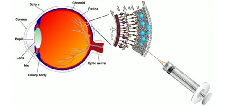
The research could also pave the way for new treatments of currently incurable eye-disorders such as Age-related Macular Degeneration (AMD) – the leading cause of visual impairment with around 500,000 sufferers in the UK.

There is currently no cure for this condition but experts believe the progression of AMD could be slowed considerably using injections of medicines into the eye. However, eye-drops with drug-loaded nanoparticles could be a potentially more effective and desirable course of treatment.

Professor Vitaliy Khutoryanskiy, from the University of Reading’s School of Pharmacy, said: “Treating eye disorders is a challenging task. Our corneas allow us to see and serve as a barrier that protects our eyes from microbial and chemical intervention. Unfortunately this barrier hinders the effectiveness of eye drops. Many medicines administered to the eye are inefficient as they often cannot penetrate the cornea barrier. Only the very small molecules in eye drops can penetrate healthy cornea.

“Many recent breakthroughs to treat eye conditions involve the use of drugs incorporated into nano-containers; their role being to promote drug penetration into the eye.  However the factors affecting this penetration remain poorly understood. Our research also showed that penetration of small drug molecules could be improved by adding enhancers such as cyclodextrins. This means eye drops have the potential to be a more effective, and a more comfortable, future treatment for disorders such as AMD.”

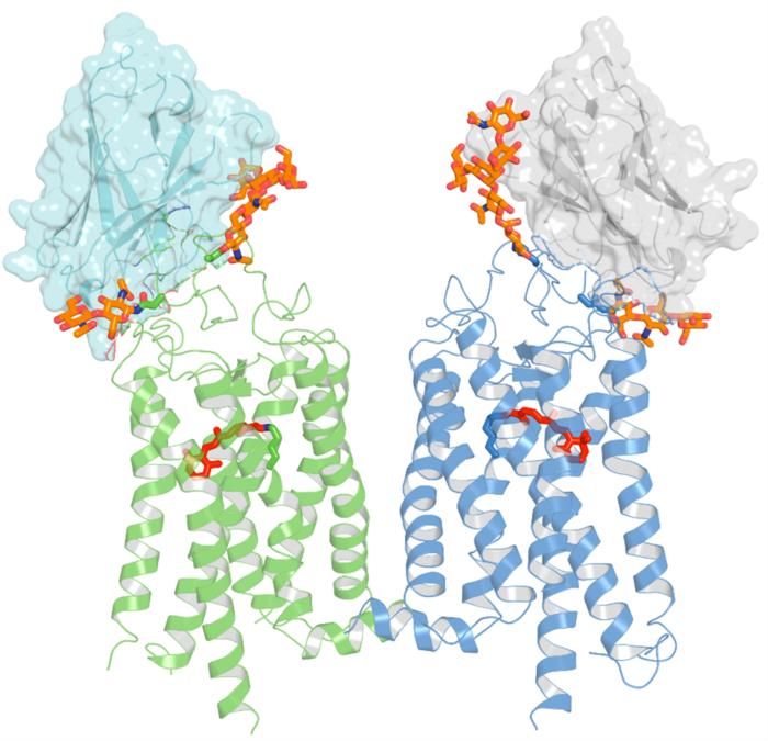
The finding is one of a number of important discoveries highlighted in a paper published today in the journal Molecular Pharmaceutics. The researchers revealed fascinating insights into how the structure of the cornea prevents various small and large molecules, as well as nanoparticles, from entering into the eye. They also examined the effects any damage to the eye would have in allowing these materials to enter the body.

Professor Khutoryanskiy continued: “There is increasing concern about the safety of environmental contaminants, pollutants and nanoparticles and their potential impacts on human health. We tested nanoparticles whose sizes ranged between 21 – 69 nm, similar to the size of viruses such as polio, or similar to airborn particles originating from building industry and found that they could not penetrate healthy and intact cornea irrespective of their chemical nature.

“However if the top layer of the cornea is damaged, either after surgical operation or accidentally, then the eye’s natural defence may be compromised and it becomes susceptible to viral attack which could result in eye infections.

“The results show that our eyes are well-equipped to defend us against potential airborne threats that exist in a fast-developing industrialised world. However we need to be aware of the potential complications that may arise if the cornea is damaged, and not treated quickly and effectively.”

Here’s a link to and a citation for the paper,

On the Barrier Properties of the Cornea: A Microscopy Study of the Penetration of Fluorescently Labeled Nanoparticles, Polymers, and Sodium Fluorescein by Ellina A. Mun, Peter W. J. Morrison, Adrian C. Williams, and Vitaliy V. Khutoryanskiy. Mol. Pharmaceutics, 2014, 11 (10), pp 3556–3564 DOI: 10.1021/mp500332m Publication Date (Web): August 28, 2014

Copyright © 2014 American Chemical Society

There’s a little more information to be had in the paper’s abstract, which is, as these things go, is relatively accessible,

[downloaded from http://pubs.acs.org/doi/abs/10.1021/mp500332m]

[downloaded from http://pubs.acs.org/doi/abs/10.1021/mp500332m]

Overcoming the natural defensive barrier functions of the eye remains one of the greatest challenges of ocular drug delivery. Cornea is a chemical and mechanical barrier preventing the passage of any foreign bodies including drugs into the eye, but the factors limiting penetration of permeants and nanoparticulate drug delivery systems through the cornea are still not fully understood. In this study, we investigate these barrier properties of the cornea using thiolated and PEGylated (750 and 5000 Da) nanoparticles, sodium fluorescein, and two linear polymers (dextran and polyethylene glycol). Experiments used intact bovine cornea in addition to bovine cornea de-epithelialized or tissues pretreated with cyclodextrin. It was shown that corneal epithelium is the major barrier for permeation; pretreatment of the cornea with β-cyclodextrin provides higher permeation of low molecular weight compounds, such as sodium fluorescein, but does not enhance penetration of nanoparticles and larger molecules. Studying penetration of thiolated and PEGylated (750 and 5000 Da) nanoparticles into the de-epithelialized ocular tissue revealed that interactions between corneal surface and thiol groups of nanoparticles were more significant determinants of penetration than particle size (for the sizes used here). PEGylation with polyethylene glycol of a higher molecular weight (5000 Da) allows penetration of nanoparticles into the stroma, which proceeds gradually, after an initial 1 h lag phase.

The paper is behind a paywall. No mention is made in the abstract or in the press release as to how the bovine (ox, cow, or buffalo) eyes were obtained but I gather these body parts are often harvested from animals that have been previously slaughtered for food.

This next item also concerns research about eye drops but this time the work comes from the University of Waterloo (Ontario, Canada). From an Oct. 8, 2014 news item on Azonano,

For the millions of sufferers of dry eye syndrome, their only recourse to easing the painful condition is to use drug-laced eye drops three times a day. Now, researchers from the University of Waterloo have developed a topical solution containing nanoparticles that will combat dry eye syndrome with only one application a week.

An Oct. 8, 2014 University of Waterloo news release (also on EurekAlert), which originated the news item, describes the results of the work without providing much detail about the nanoparticles used to deliver the treatment via eye drops,

The eye drops progressively deliver the right amount of drug-infused nanoparticles to the surface of the eyeball over a period of five days before the body absorbs them.  One weekly dose replaces 15 or more to treat the pain and irritation of dry eyes.

The nanoparticles, about 1/1000th the width of a human hair, stick harmlessly to the eye’s surface and use only five per cent of the drug normally required.

“You can’t tell the difference between these nanoparticle eye drops and water,” said Shengyan (Sandy) Liu, a PhD candidate at Waterloo’s Faculty of Engineering, who led the team of researchers from the Department of Chemical Engineering and the Centre for Contact Lens Research. “There’s no irritation to the eye.”

Dry eye syndrome is a more common ailment for people over the age of 50 and may eventually lead to eye damage. More than six per cent of people in the U.S. have it. Currently, patients must frequently apply the medicine three times a day because of the eye’s ability to self-cleanse—a process that washes away 95 per cent of the drug.

“I knew that if we focused on infusing biocompatible nanoparticles with Cyclosporine A, the drug in the eye drops, and make them stick to the eyeball without irritation for longer periods of time, it would also save patients time and reduce the possibility of toxic exposure due to excessive use of eye drops,” said Liu.

The research team is now focusing on preparing the nanoparticle eye drops for clinical trials with the hope that this nanoparticle therapy could reach the shelves of drugstores within five years.

Here’s a link to and a citation for the paper,

Phenylboronic acid modified mucoadhesive nanoparticle drug carriers facilitate weekly treatment of experimentallyinduced dry eye syndrome by Shengyan Liu, Chu Ning Chang, Mohit S. Verma, Denise Hileeto, Alex Muntz, Ulrike Stahl, Jill Woods, Lyndon W. Jones, and Frank X. Gu. Nano Research (October 2014) DOI: 10.1007/s12274-014-0547-3

This paper is behind a paywall. There is a partial preview available for free. As per the paper’s abstract, research was performed on healthy rabbit eyes.

The last ‘sight’ item I’m featuring here comes from the Massachusetts Institute of Technology (MIT) and does not appear to have been occasioned by the publication of a research paper or some other event. From an Oct. 7, 2014 news item on Azonano,

All through his childhood, Ramesh Raskar wished fervently for eyes in the back of his head. “I had the notion that the world did not exist if I wasn’t looking at it, so I would constantly turn around to see if it was there behind me.” Although this head-spinning habit faded during his teen years, Raskar never lost the desire to possess the widest possible field of vision.

Today, as director of the Camera Culture research group and associate professor of Media Arts and Sciences at the MIT Media Lab, Raskar is realizing his childhood fantasy, and then some. His inventions include a nanocamera that operates at the speed of light and do-it-yourself tools for medical imaging. His scientific mission? “I want to create not just a new kind of vision, but superhuman vision,” Raskar says.

An Oct. 6, 2014 MIT news release, which originated the news item, provides more information about Raskar and his research,

He avoids research projects launched with a goal in mind, “because then you only come up with the same solutions as everyone else.” Discoveries tend to cascade from one area into another. For instance, Raskar’s novel computational methods for reducing motion blur in photography suggested new techniques for analyzing how light propagates. “We do matchmaking; what we do here can be used over there,” says Raskar.

Inspired by the famous microflash photograph of a bullet piercing an apple, created in 1964 by MIT professor and inventor Harold “Doc” Edgerton, Raskar realized, “I can do Edgerton millions of times faster.” This led to one of the Camera Culture group’s breakthrough inventions, femtophotography, a process for recording light in flight.

Manipulating photons into a packet resembling Edgerton’s bullet, Raskar and his team were able to “shoot” ultrashort laser pulses through a Coke bottle. Using a special camera to capture the action of these pulses at half a trillion frames per second with two-trillionths of a second exposure times, they captured moving images of light, complete with wave-like shadows lapping at the exterior of the bottle.

Femtophotography opened up additional avenues of inquiry, as Raskar pondered what other features of the world superfast imaging processes might reveal. He was particularly intrigued by scattered light, the kind in evidence when fog creates the visual equivalent of “noise.”

In one experiment, Raskar’s team concealed an object behind a wall, out of camera view. By firing super-short laser bursts onto a surface nearby, and taking millions of exposures of light bouncing like a pinball around the scene, the group rendered a picture of the hidden object. They had effectively created a camera that peers around corners, an invention that might someday help emergency responders safely investigate a dangerous environment.

Raskar’s objective of “making the invisible visible” extends as well to the human body. The Camera Culture group has developed a technique for taking pictures of the eye using cellphone attachments, spawning inexpensive, patient-managed vision and disease diagnostics. Conventional photography has evolved from time-consuming film development to instantaneous digital snaps, and Raskar believes “the same thing will happen to medical imaging.” His research group intends “to break all the rules and be at the forefront. I think we’ll get there in the next few years,” he says.

Ultimately, Raskar predicts, imaging will serve as a catalyst of transformation in all dimensions of human life — change that can’t come soon enough for him. “I hate ordinary cameras,” he says. “They record only what I see. I want a camera that gives me a superhuman perspective.”

Following the link to the MIT news release will lead you to more information about Raskar and his work. You can also see and hear Raskar talk about his femtophotography in a 2012 TEDGlobal talk here.

Nanodiamond contact lenses in attempt to improve glaucoma treatment

A School of Dentistry, at the University of California at Los Angeles (UCLA) or elsewhere, is not my first thought as a likely source for work on improving glaucoma treatment—it turns out that I’m a bit shortsighted (pun intended).  A Feb. 14, 2014 news item on Azonano describes the issue with glaucoma treatment and a new delivery system for it developed by a research team at UCLA,

By 2020, nearly 80 million people are expected to have glaucoma, a disorder of the eye that, if left untreated, can damage the optic nerve and eventually lead to blindness.

The disease often causes pressure in the eye due to a buildup of fluid and a breakdown of the tissue that is responsible for regulating fluid drainage. Doctors commonly treat glaucoma using eye drops that can help the eye drain or decrease fluid production.

Unfortunately, patients frequently have a hard time sticking to the dosing schedules prescribed by their doctors, and the medication — when administered through drops — can cause side effects in the eye and other parts of the body.

In what could be a significant step toward improving the management of glaucoma, researchers from the UCLA School of Dentistry have created a drug delivery system that may have less severe side effects than traditional glaucoma medication and improve patients’ ability to comply with their prescribed treatments. The scientists bound together glaucoma-fighting drugs with nanodiamonds and embedded them onto contact lenses. The drugs are released into the eye when they interact with the patient’s tears.

The new technology showed great promise for sustained glaucoma treatment and, as a side benefit, the nanodiamond-drug compound even improved the contact lenses’ durability.

The Feb. 13, 2014 UCLA news release by Brianna Deane, which originated the news item, describes the nanodiamonds and how they were employed in this project,

Nanodiamonds, which are byproducts of conventional mining and refining processes, are approximately five nanometers in diameter and are shaped like tiny soccer balls. They can be used to bind a wide spectrum of drug compounds and enable drugs to be released into the body over a long period of time.

To deliver a steady release of medication into the eye, the UCLA researchers combined nanodiamonds with timolol maleate, which is commonly used in eye drops to manage glaucoma. When applied to the nanodiamond-embedded lenses, timolol is released when it comes into contact with lysozyme, an enzyme that is abundant in tears.

“Delivering timolol through exposure to tears may prevent premature drug release when the contact lenses are in storage and may serve as a smarter route toward drug delivery from a contact lens.” said Kangyi Zhang, co-first author of the study and a graduate student in Ho’s lab.

One of the drawbacks of traditional timolol maleate drops is that as little as 5 percent of the drug actually reaches the intended site. Another disadvantage is burst release, where a majority of the drug is delivered too quickly, which can cause significant amounts of the drug to “leak” or spill out of the eye and, in the most serious cases, can cause complications such as an irregular heartbeat. Drops also can be uncomfortable to administer, which leads many patients to stop using their medication.

But the contact lenses developed by the UCLA team successfully avoided the burst release effect. The activity of the released timolol was verified by a primary human-cell study.

“In addition to nanodiamonds’ promise as triggered drug-delivery agents for eye diseases, they can also make the contact lenses more durable during the course of insertion, use and removal, and more comfortable to wear,” said Ho, who is also a professor of bioengineering and a member of the Jonsson Comprehensive Cancer Center and the California NanoSystems Institute.

Even with the nanodiamonds embedded, the lenses still possessed favorable levels of optical clarity. And, although mechanical testing verified that they were stronger than normal lenses, there were no apparent changes to water content, meaning that the contact lenses’ comfort and permeability to oxygen would likely be preserved.

By this time, I was madly curious as to what these contact lenses might look like and so I found this image, accompanying the researchers’ paper,  showing what looks like a standard contact lens with an illustration of how the artist imagines the diamonds and medications are functioning at the nanoscale,

nanodiamonds

[downloaded from http://pubs.acs.org/doi/abs/10.1021/nn5002968]

Here’s a link to and a citation for the paper,

Diamond Nanogel-Embedded Contact Lenses Mediate Lysozyme-Dependent Therapeutic Release by Ho-Joong Kim, Kangyi Zhang, Laura Moore, and Dean Ho. ACS Nano, Article ASAP DOI: 10.1021/nn5002968 Publication Date (Web): February 8, 2014

Copyright © 2014 American Chemical Society

This paper is behind a paywall.

Happy Canada Day to everyone! and news about implantable devices for the prevention diabetes-related vision loss

Although it’s going to be years (I imagine several) before patients at risk for diabetes-related blindness will be able to benefit from this work, it’s certainly exciting news from the University of British Columbia (UBC). One of their research teams has tested a device that could be implanted behind an eye to release medications when an external sensor is activated. ETA July 4, 2011: I took another look at that news release and I’m now not sure that I correctly understood the term “… on-demand release of drugs” which I meant an external locus of control.

Here’s more from the June 29, 2011 UBC  news release,

A team of engineers and scientists at the University of British Columbia has developed a device that can be implanted behind the eye for controlled and on-demand release of drugs to treat retinal damage caused by diabetes.

Diabetic retinopathy is the leading cause of vision loss among patients with diabetes. The disease is caused by the unwanted growth of capillary cells in the retina, which in its advanced stages can result in blindness.

The novel drug delivery mechanism is detailed in the current issue of Lab on a Chip, a multidisciplinary journal on innovative microfluidic and nanofluidic technologies.

The lead authors are recent PhD mechanical engineering graduate Fatemeh Nazly Pirmoradi, who completed the study for her doctoral thesis, and Mechanical Engineering Assoc. Prof. Mu Chiao, who studies nanoscience and microelectromechanical systems for biological applications.

The co-authors are Prof. Helen Burt and research scientist John Jackson at the Faculty of Pharmaceutical Sciences.

“We wanted to come up with a safe and effective way to help diabetic patients safeguard their sight,” says Chiao who has a family member dealing with diabetic retinopathy.

A current treatment for diabetic retinopathy is laser therapy, which has side effects, among them laser burns or the loss of peripheral or night vision. Anti-cancer drugs may also used to treat the disease. However, these compounds clear quickly from the bloodstream so high dosages are required, thus exposing other tissues to toxicity.

Key to UBC’s innovation is the ability to trigger the drug delivery system through an external magnetic field. The team accomplished this by sealing the reservoir of the implantable device – which is no larger than the head of a pin – with an elastic magnetic polydimethylsiloxane (silicone) membrane. A magnetic field causes the membrane to deform and discharge a specific amount of the drug, much like squeezing water out of a flexible bottle.

In a series of lab tests, the UBC researchers loaded the implantable device with the drug docetaxel and triggered the drug release at a dosage suitable for treating diabetic retinopathy. They found that the implantable device kept its integrity with negligible leakage over 35 days.

They also monitored the drug’s biological effectiveness over a given period, testing it against two types of cultured cancer cells, including those found in the prostate. They found that they were able to achieve reliable release rates.

“The docetaxel retained its pharmacological efficacy for more than two months in the device and was able to kill off the cancer cells,” says Pirmoradi.

The UBC device offers improvements upon existing implantable devices for drug delivery, says Chiao.

“Technologies available now are either battery operated and are too large for treating the eye, or they rely on diffusion, which means drug release rates cannot be stopped once the device is implanted – a problem when patients’ conditions change.”

Pirmoradi says it will be several years before the UBC device is ready for patient use. “There’s a lot of work ahead of us in terms of biocompatibility and performance optimization.”

The team is also working to pinpoint all the possible medical applications for their device so that they can tailor the mechanical design to particular diseases

There’s no information as to whether this is work being done at microscale or nanoscale or both for that matter. I do note that the device is described as being “no larger than the head of a pin” so it’s possible to physically see and/or handle it. I wonder what the response would be if the device were invisible to the human eye. I expect that response would be dependent on how unpleasant the effects from the previous technology/ies used have proved to be.