Tag Archives: Samuel I. Stupp

New approach to cartilage regeneration

Not long after announcing their new work on cartilage and ‘dancing molecules’, Samuel I. Stupp and his team at Northwestern University (Chicago, Illinois) have announced work with a new material that does not have dancing molecules in a study using animal models. It’s here in an August 5, 02024 Northwestern University news release (also on EurekAlert and on SciTechDaily and received by email) by Amanda Morris, Note: Links have been removed,

Northwestern University scientists have developed a new bioactive material that successfully regenerated high-quality cartilage in the knee joints of a large-animal model.

Although it looks like a rubbery goo, the material is actually a complex network of molecular components, which work together to mimic cartilage’s natural environment in the body. 

In the new study, the researchers applied the material to damaged cartilage in the animals’ knee joints. Within just six months, the researchers observed evidence of enhanced repair, including the growth of new cartilage containing the natural biopolymers (collagen II and proteoglycans), which enable pain-free mechanical resilience in joints.

With more work, the researchers say the new material someday could potentially be used to prevent full knee replacement surgeries, treat degenerative diseases like osteoarthritis and repair sports-related injuries like ACL [anterior cruciate ligament] tears.

The study will be published during the week of August 5 [2024] in the Proceedings of the National Academy of Sciences.

“Cartilage is a critical component in our joints,” said Northwestern’s Samuel I. Stupp, who led the study. “When cartilage becomes damaged or breaks down over time, it can have a great impact on people’s overall health and mobility. The problem is that, in adult humans, cartilage does not have an inherent ability to heal. Our new therapy can induce repair in a tissue that does not naturally regenerate. We think our treatment could help address a serious, unmet clinical need.”

A pioneer of regenerative nanomedicine, Stupp is Board of Trustees Professor of Materials Science and Engineering, Chemistry, Medicine and Biomedical Engineering at Northwestern, where he is founding director of the Simpson Querrey Institute for BioNanotechnology and its affiliated center, the Center for Regenerative Nanomedicine. Stupp has appointments in the McCormick School of Engineering, Weinberg College of Arts and Sciences and Feinberg School of Medicine. Jacob Lewis, a former Ph.D. student in Stupp’s laboratory, is the paper’s first author.

What’s in the material?

The new study follows recently published work from the Stupp laboratory, in which the team used “dancing molecules” to activate human cartilage cells to boost the production of proteins that build the tissue matrix. Instead of using dancing molecules, the new study evaluates a hybrid biomaterial also developed in Stupp’s lab. The new biomaterial comprises two components: a bioactive peptide that binds to transforming growth factor beta-1 (TGFb-1) — an essential protein for cartilage growth and maintenance — and modified hyaluronic acid, a natural polysaccharide present in cartilage and the lubricating synovial fluid in joints. 

“Many people are familiar with hyaluronic acid because it’s a popular ingredient in skincare products,” Stupp said. “It’s also naturally found in many tissues throughout the human body, including the joints and brain. We chose it because it resembles the natural polymers found in cartilage.”

Stupp’s team integrated the bioactive peptide and chemically modified hyaluronic acid particles to drive the self-organization of nanoscale fibers into bundles that mimic the natural architecture of cartilage. The goal was to create an attractive scaffold for the body’s own cells to regenerate cartilage tissue. Using bioactive signals in the nanoscale fibers, the material encourages cartilage repair by the cells, which populate the scaffold.

Clinically relevant to humans

To evaluate the material’s effectiveness in promoting cartilage growth, the researchers tested it in sheep with cartilage defects in the stifle joint, a complex joint in the hind limbs similar to the human knee. This work was carried out in the laboratory of Mark Markel in the School of Veterinary Medicine at the University of Wisconsin–Madison. 

According to Stupp, testing in a sheep model was vital. Much like humans, sheep cartilage is stubborn and incredibly difficult to regenerate. Sheep stifles and human knees also have similarities in weight bearing, size and mechanical loads.

“A study on a sheep model is more predictive of how the treatment will work in humans,” Stupp said. “In other smaller animals, cartilage regeneration occurs much more readily.”

In the study, researchers injected the thick, paste-like material into cartilage defects, where it transformed into a rubbery matrix. Not only did new cartilage grow to fill the defect as the scaffold degraded, but the repaired tissue was consistently higher quality compared to the control.

A lasting solution

In the future, Stupp imagines the new material could be applied to joints during open-joint or arthroscopic surgeries. The current standard of care is microfracture surgery, during which surgeons create tiny fractures in the underlying bone to induce new cartilage growth.

“The main issue with the microfracture approach is that it often results in the formation of fibrocartilage — the same cartilage in our ears — as opposed to hyaline cartilage, which is the one we need to have functional joints,” Stupp said. “By regenerating hyaline cartilage, our approach should be more resistant to wear and tear, fixing the problem of poor mobility and joint pain for the long term while also avoiding the need for joint reconstruction with large pieces of hardware.”

The study, “A bioactive supramolecular and covalent polymer scaffold for cartilage repair in a sheep model,” was supported by the Mike and Mary Sue Shannon Family Fund for Bio-Inspired and Bioactive Materials Systems for Musculoskeletal Regeneration.

Here’s a link to and a citation for the paper,

A bioactive supramolecular and covalent polymer scaffold for cartilage repair in a sheep model by Jacob A. Lewis, Brett Nemke, Yan Lu, Nicholas A. Sather, Mark T. McClendon, Michael Mullen, Shelby C. Yuan, Sudheer K. Ravuri, Jason A. Bleedorn, Marc J. Philippon, Johnny Huard, Mark D. Markel, and Samuel I. Stupp. Proceedings ot the National Academy of Sciences (PNAS) 121 (33) e2405454121 DOI: https://doi.org/10.1073/pnas.2405454121 August 6, 2024

This paper is behind a paywall.

Healing cartilage damage with ‘dancing molecules’

A July 26, 2024 Northwestern University (Chicago, Illinois) news release (also on EurekAlert) by Amanda Morris describes a new application for ‘dancing molecules’, Note 1: Links have been removed; Note 2: These are ‘in vitro’ (petri dish) experiments ,

In November 2021, Northwestern University researchers introduced an injectable new therapy, which harnessed fast-moving “dancing molecules,” to repair tissues and reverse paralysis after severe spinal cord injuries.

Now, the same research group has applied the therapeutic strategy to damaged human cartilage cells. In the new study, the treatment activated the gene expression necessary to regenerate cartilage within just four hours. And, after only three days, the human cells produced protein components needed for cartilage regeneration.

The researchers also found that, as the molecular motion increased, the treatment’s effectiveness also increased. In other words, the molecules’ “dancing” motions were crucial for triggering the cartilage growth process.

“When we first observed therapeutic effects of dancing molecules, we did not see any reason why it should only apply to the spinal cord,” said Northwestern’s Samuel I. Stupp, who led the study. “Now, we observe the effects in two cell types that are completely disconnected from one another — cartilage cells in our joints and neurons in our brain and spinal cord. This makes me more confident that we might have discovered a universal phenomenon. It could apply to many other tissues.”

An expert in regenerative nanomedicine, Stupp is Board of Trustees Professor of Materials Science and Engineering, Chemistry, Medicine and Biomedical Engineering at Northwestern, where he is founding director of the Simpson Querrey Institute for BioNanotechnology and its affiliated center, the Center for Regenerative Nanomedicine. Stupp has appointments in the McCormick School of Engineering, Weinberg College of Arts and Sciences and Feinberg School of Medicine. Shelby Yuan, a graduate student in the Stupp laboratory, was primary author of the study.

Big problem, few solutions

As of 2019, nearly 530 million people around the globe were living with osteoarthritis, according to the World Health Organization. A degenerative disease in which tissues in joints break down over time, osteoarthritis is a common health problem and leading cause of disability.

In patients with severe osteoarthritis, cartilage can wear so thin that joints essentially transform into bone on bone — without a cushion between. Not only is this incredibly painful, patients’ joints also can no longer properly function. At that point, the only effective treatment is a joint replacement surgery, which is expensive and invasive.

“Current treatments aim to slow disease progression or postpone inevitable joint replacement,” Stupp said. “There are no regenerative options because humans do not have an inherent capacity to regenerate cartilage in adulthood.”

What are ‘dancing molecules’?

Stupp and his team posited that “dancing molecules” might encourage the stubborn tissue to regenerate. Previously invented in Stupp’s laboratory, dancing molecules are assemblies that form synthetic nanofibers comprising tens to hundreds of thousands of molecules with potent signals for cells. By tuning their collective motions through their chemical structure, Stupp discovered the moving molecules could rapidly find and properly engage with cellular receptors, which also are in constant motion and extremely crowded on cell membranes.

“We are beginning to see the tremendous breadth of conditions that this fundamental discovery on ‘dancing molecules’ could apply to.” — Samuel I. Stupp, materials scientist

Once inside the body, the nanofibers mimic the extracellular matrix of the surrounding tissue. By matching the matrix’s structure, mimicking the motion of biological molecules and incorporating bioactive signals for the receptors, the synthetic materials are able to communicate with cells.

“Cellular receptors constantly move around,” Stupp said. “By making our molecules move, ‘dance’ or even leap temporarily out of these structures, known as supramolecular polymers, they are able to connect more effectively with receptors.”

Motion matters

In the new study, Stupp and his team looked to the receptors for a specific protein critical for cartilage formation and maintenance. To target this receptor, the team developed a new circular peptide that mimics the bioactive signal of the protein, which is called transforming growth factor beta-1 (TGFb-1).

Then, the researchers incorporated this peptide into two different molecules that interact to form supramolecular polymers in water, each with the same ability to mimic TGFb-1. The researchers designed one supramolecular polymer with a special structure that enabled its molecules to move more freely within the large assemblies. The other supramolecular polymer, however, restricted molecular movement.

“We wanted to modify the structure in order to compare two systems that differ in the extent of their motion,” Stupp said. “The intensity of supramolecular motion in one is much greater than the motion in the other one.”

Although both polymers mimicked the signal to activate the TGFb-1 receptor, the polymer with rapidly moving molecules was much more effective. In some ways, they were even more effective than the protein that activates the TGFb-1 receptor in nature.

“After three days, the human cells exposed to the long assemblies of more mobile molecules produced greater amounts of the protein components necessary for cartilage regeneration,” Stupp said. “For the production of one of the components in cartilage matrix, known as collagen II, the dancing molecules containing the cyclic peptide that activates the TGF-beta1 receptor were even more effective than the natural protein that has this function in biological systems.”

What’s next?

Stupp’s team is currently testing these systems in animal studies and adding additional signals to create highly bioactive therapies.

“With the success of the study in human cartilage cells, we predict that cartilage regeneration will be greatly enhanced when used in highly translational pre-clinical models,” Stupp said. “It should develop into a novel bioactive material for regeneration of cartilage tissue in joints.”

Stupp’s lab is also testing the ability of dancing molecules to regenerate bone — and already has promising early results, which likely will be published later this year. Simultaneously, he is testing the molecules in human organoids to accelerate the process of discovering and optimizing therapeutic materials.  

Stupp’s team also continues to build its case to the Food and Drug Administration, aiming to gain approval for clinical trials to test the therapy for spinal cord repair.

“We are beginning to see the tremendous breadth of conditions that this fundamental discovery on ‘dancing molecules’ could apply to,” Stupp said. “Controlling supramolecular motion through chemical design appears to be a powerful tool to increase efficacy for a range of regenerative therapies.”

The study, “Supramolecular motion enables chondrogenic bioactivity of a cyclic peptide mimetic of transforming growth factor-β1,” was supported by a gift from Mike and Mary Sue Shannon to Northwestern University for research on musculoskeletal regeneration at the Center for Regenerative Nanomedicine of the Simpson Querrey Institute for BioNanotechnology.

Here’s a link to and a citation for the paper,

Supramolecular Motion Enables Chondrogenic Bioactivity of a Cyclic Peptide Mimetic of Transforming Growth Factor-β1 by Shelby C. Yuan, Zaida Álvarez, Sieun Ruth Lee, Radoslav Z. Pavlović, Chunhua Yuan, Ethan Singer, Steven J. Weigand, Liam C. Palmer, Samuel I. Stupp. Journal of the American Chemical Society Vol 146/Issue 31 (or J. Am. Chem. Soc. 2024, 146, 31, 21555–21567) DOI: https://doi.org/10.1021/jacs.4c05170 Published July 25, 2024 Copyright © 2024 American Chemical Society

This paper is behind a paywall.

Transplanting healthy neurons could be possible with walking molecules and 3D printing

A February 23, 2021 news item on ScienceDaily announces work which may lead to healing brain injuries and diseases,

Imagine if surgeons could transplant healthy neurons into patients living with neurodegenerative diseases or brain and spinal cord injuries. And imagine if they could “grow” these neurons in the laboratory from a patient’s own cells using a synthetic, highly bioactive material that is suitable for 3D printing.

By discovering a new printable biomaterial that can mimic properties of brain tissue, Northwestern University researchers are now closer to developing a platform capable of treating these conditions using regenerative medicine.

A February 22, 2021 Northwestern University news release (also received by email and available on EurekAlert) by Lila Reynolds, which originated the news item, delves further into self-assembling ‘walking’ molecules and the nanofibers resulting in a new material designed to promote the growth of healthy neurons,

A key ingredient to the discovery is the ability to control the self-assembly processes of molecules within the material, enabling the researchers to modify the structure and functions of the systems from the nanoscale to the scale of visible features. The laboratory of Samuel I. Stupp published a 2018 paper in the journal Science which showed that materials can be designed with highly dynamic molecules programmed to migrate over long distances and self-organize to form larger, “superstructured” bundles of nanofibers.

Now, a research group led by Stupp has demonstrated that these superstructures can enhance neuron growth, an important finding that could have implications for cell transplantation strategies for neurodegenerative diseases such as Parkinson’s and Alzheimer’s disease, as well as spinal cord injury.

“This is the first example where we’ve been able to take the phenomenon of molecular reshuffling we reported in 2018 and harness it for an application in regenerative medicine,” said Stupp, the lead author on the study and the director of Northwestern’s Simpson Querrey Institute. “We can also use constructs of the new biomaterial to help discover therapies and understand pathologies.

Walking molecules and 3D printing

The new material is created by mixing two liquids that quickly become rigid as a result of interactions known in chemistry as host-guest complexes that mimic key-lock interactions among proteins, and also as the result of the concentration of these interactions in micron-scale regions through a long scale migration of “walking molecules.”

The agile molecules cover a distance thousands of times larger than themselves in order to band together into large superstructures. At the microscopic scale, this migration causes a transformation in structure from what looks like an uncooked chunk of ramen noodles into ropelike bundles.

“Typical biomaterials used in medicine like polymer hydrogels don’t have the capabilities to allow molecules to self-assemble and move around within these assemblies,” said Tristan Clemons, a research associate in the Stupp lab and co-first author of the paper with Alexandra Edelbrock, a former graduate student in the group. “This phenomenon is unique to the systems we have developed here.”

Furthermore, as the dynamic molecules move to form superstructures, large pores open that allow cells to penetrate and interact with bioactive signals that can be integrated into the biomaterials.

Interestingly, the mechanical forces of 3D printing disrupt the host-guest interactions in the superstructures and cause the material to flow, but it can rapidly solidify into any macroscopic shape because the interactions are restored spontaneously by self-assembly. This also enables the 3D printing of structures with distinct layers that harbor different types of neural cells in order to study their interactions.

Signaling neuronal growth

The superstructure and bioactive properties of the material could have vast implications for tissue regeneration. Neurons are stimulated by a protein in the central nervous system known as brain-derived neurotrophic factor (BDNF), which helps neurons survive by promoting synaptic connections and allowing neurons to be more plastic. BDNF could be a valuable therapy for patients with neurodegenerative diseases and injuries in the spinal cord but these proteins degrade quickly in the body and are expensive to produce.

One of the molecules in the new material integrates a mimic of this protein that activates its receptor known as Trkb, and the team found that neurons actively penetrate the large pores and populate the new biomaterial when the mimetic signal is present. This could also create an environment in which neurons differentiated from patient-derived stem cells mature before transplantation.

Now that the team has applied a proof of concept to neurons, Stupp believes he could now break into other areas of regenerative medicine by applying different chemical sequences to the material. Simple chemical changes in the biomaterials would allow them to provide signals for a wide range of tissues.

“Cartilage and heart tissue are very difficult to regenerate after injury or heart attacks, and the platform could be used to prepare these tissues in vitro from patient-derived cells,” Stupp said. “These tissues could then be transplanted to help restore lost functions. Beyond these interventions, the materials could be used to build organoids to discover therapies or even directly implanted into tissues for regeneration since they are biodegradable.”

Here’s a link to and a citation for the paper,

Superstructured Biomaterials Formed by Exchange Dynamics and Host–Guest Interactions in Supramolecular Polymers by Alexandra N. Edelbrock, Tristan D. Clemons, Stacey M. Chin, Joshua J. W. Roan, Eric P. Bruckner, Zaida Álvarez, Jack F. Edelbrock, Kristen S. Wek, Samuel I. Stupp. Advanced Science DOI: https://doi.org/10.1002/advs.202004042 First published: 22 February 2021

This paper is open access.
Shining a light on flurocarbon bonds and robotic ‘soft’ matter research

Both of these news bits are concerned with light for one reason or another.

Rice University (Texas, US) and breaking fluorocarbon bonds

The secret to breaking fluorocarbon bonds is light according to a June 22, 2020 news item on Nanowerk,

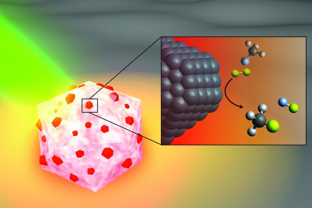
Rice University engineers have created a light-powered catalyst that can break the strong chemical bonds in fluorocarbons, a group of synthetic materials that includes persistent environmental pollutants.

A June 22, 2020 Rice University news release (also on EurekAlert), which originated the news item, describes the work in greater detail,

In a study published this month in Nature Catalysis, Rice nanophotonics pioneer Naomi Halas and collaborators at the University of California, Santa Barbara (UCSB) and Princeton University showed that tiny spheres of aluminum dotted with specks of palladium could break carbon-fluorine (C-F) bonds via a catalytic process known as hydrodefluorination in which a fluorine atom is replaced by an atom of hydrogen.

The strength and stability of C-F bonds are behind some of the 20th century’s most recognizable chemical brands, including Teflon, Freon and Scotchgard. But the strength of those bonds can be problematic when fluorocarbons get into the air, soil and water. Chlorofluorocarbons, or CFCs, for example, were banned by international treaty in the 1980s after they were found to be destroying Earth’s protective ozone layer, and other fluorocarbons were on the list of “forever chemicals” targeted by a 2001 treaty.

“The hardest part about remediating any of the fluorine-containing compounds is breaking the C-F bond; it requires a lot of energy,” said Halas, an engineer and chemist whose Laboratory for Nanophotonics (LANP) specializes in creating and studying nanoparticles that interact with light.

Over the past five years, Halas and colleagues have pioneered methods for making “antenna-reactor” catalysts that spur or speed up chemical reactions. While catalysts are widely used in industry, they are typically used in energy-intensive processes that require high temperature, high pressure or both. For example, a mesh of catalytic material is inserted into a high-pressure vessel at a chemical plant, and natural gas or another fossil fuel is burned to heat the gas or liquid that’s flowed through the mesh. LANP’s antenna-reactors dramatically improve energy efficiency by capturing light energy and inserting it directly at the point of the catalytic reaction.

In the Nature Catalysis study, the energy-capturing antenna is an aluminum particle smaller than a living cell, and the reactors are islands of palladium scattered across the aluminum surface. The energy-saving feature of antenna-reactor catalysts is perhaps best illustrated by another of Halas’ previous successes: solar steam. In 2012, her team showed its energy-harvesting particles could instantly vaporize water molecules near their surface, meaning Halas and colleagues could make steam without boiling water. To drive home the point, they showed they could make steam from ice-cold water.

The antenna-reactor catalyst design allows Halas’ team to mix and match metals that are best suited for capturing light and catalyzing reactions in a particular context. The work is part of the green chemistry movement toward cleaner, more efficient chemical processes, and LANP has previously demonstrated catalysts for producing ethylene and syngas and for splitting ammonia to produce hydrogen fuel.

Study lead author Hossein Robatjazi, a Beckman Postdoctoral Fellow at UCSB who earned his Ph.D. from Rice in 2019, conducted the bulk of the research during his graduate studies in Halas’ lab. He said the project also shows the importance of interdisciplinary collaboration.

“I finished the experiments last year, but our experimental results had some interesting features, changes to the reaction kinetics under illumination, that raised an important but interesting question: What role does light play to promote the C-F breaking chemistry?” he said.

The answers came after Robatjazi arrived for his postdoctoral experience at UCSB. He was tasked with developing a microkinetics model, and a combination of insights from the model and from theoretical calculations performed by collaborators at Princeton helped explain the puzzling results.

“With this model, we used the perspective from surface science in traditional catalysis to uniquely link the experimental results to changes to the reaction pathway and reactivity under the light,” he said.

The demonstration experiments on fluoromethane could be just the beginning for the C-F breaking catalyst.

“This general reaction may be useful for remediating many other types of fluorinated molecules,” Halas said.

Caption: An artist’s illustration of the light-activated antenna-reactor catalyst Rice University engineers designed to break carbon-fluorine bonds in fluorocarbons. The aluminum portion of the particle (white and pink) captures energy from light (green), activating islands of palladium catalysts (red). In the inset, fluoromethane molecules (top) comprised of one carbon atom (black), three hydrogen atoms (grey) and one fluorine atom (light blue) react with deuterium (yellow) molecules near the palladium surface (black), cleaving the carbon-fluorine bond to produce deuterium fluoride (right) and monodeuterated methane (bottom). Credit: H. Robatjazi/Rice University

Here’s a link to and a citation for the paper,

Plasmon-driven carbon–fluorine (C(sp3)–F) bond activation with mechanistic insights into hot-carrier-mediated pathways by Hossein Robatjazi, Junwei Lucas Bao, Ming Zhang, Linan Zhou, Phillip Christopher, Emily A. Carter, Peter Nordlander & Naomi J. Halas. Nature Catalysis (2020) DOI: https://doi.org/10.1038/s41929-020-0466-5 Published: 08 June 2020

This paper is behind a paywall.

Northwestern University (Illinois, US) brings soft robots to ‘life’

This June 22, 2020 news item on ScienceDaily reveals how scientists are getting soft robots to mimic living creatures,

Northwestern University researchers have developed a family of soft materials that imitates living creatures.

When hit with light, the film-thin materials come alive — bending, rotating and even crawling on surfaces.

A June 22, 2020 Northwestern University news release (also on EurekAlert) by Amanda Morris, which originated the news item, delves further into the details,

Called “robotic soft matter by the Northwestern team,” the materials move without complex hardware, hydraulics or electricity. The researchers believe the lifelike materials could carry out many tasks, with potential applications in energy, environmental remediation and advanced medicine.

“We live in an era in which increasingly smarter devices are constantly being developed to help us manage our everyday lives,” said Northwestern’s Samuel I. Stupp, who led the experimental studies. “The next frontier is in the development of new science that will bring inert materials to life for our benefit — by designing them to acquire capabilities of living creatures.”

The research will be published on June 22 [2020] in the journal Nature Materials.

Stupp is the Board of Trustees Professor of Materials Science and Engineering, Chemistry, Medicine and Biomedical Engineering at Northwestern and director of the Simpson Querrey Institute He has appointments in the McCormick School of Engineering, Weinberg College of Arts and Sciences and Feinberg School of Medicine. George Schatz, the Charles E. and Emma H. Morrison Professor of Chemistry in Weinberg, led computer simulations of the materials’ lifelike behaviors. Postdoctoral fellow Chuang Li and graduate student Aysenur Iscen, from the Stupp and Schatz laboratories, respectively, are co-first authors of the paper.

Although the moving material seems miraculous, sophisticated science is at play. Its structure comprises nanoscale peptide assemblies that drain water molecules out of the material. An expert in materials chemistry, Stupp linked the peptide arrays to polymer networks designed to be chemically responsive to blue light.

When light hits the material, the network chemically shifts from hydrophilic (attracts water) to hydrophobic (resists water). As the material expels the water through its peptide “pipes,” it contracts — and comes to life. When the light is turned off, water re-enters the material, which expands as it reverts to a hydrophilic structure.

This is reminiscent of the reversible contraction of muscles, which inspired Stupp and his team to design the new materials.

“From biological systems, we learned that the magic of muscles is based on the connection between assemblies of small proteins and giant protein polymers that expand and contract,” Stupp said. “Muscles do this using a chemical fuel rather than light to generate mechanical energy.”

For Northwestern’s bio-inspired material, localized light can trigger directional motion. In other words, bending can occur in different directions, depending on where the light is located. And changing the direction of the light also can force the object to turn as it crawls on a surface.

Stupp and his team believe there are endless possible applications for this new family of materials. With the ability to be designed in different shapes, the materials could play a role in a variety of tasks, ranging from environmental clean-up to brain surgery.

“These materials could augment the function of soft robots needed to pick up fragile objects and then release them in a precise location,” he said. “In medicine, for example, soft materials with ‘living’ characteristics could bend or change shape to retrieve blood clots in the brain after a stroke. They also could swim to clean water supplies and sea water or even undertake healing tasks to repair defects in batteries, membranes and chemical reactors.”

Fascinating, eh? No batteries, no power source, just light to power movement. For the curious, here’s a link to and a citation for the paper,

Supramolecular–covalent hybrid polymers for light-activated mechanical actuation by Chuang Li, Aysenur Iscen, Hiroaki Sai, Kohei Sato, Nicholas A. Sather, Stacey M. Chin, Zaida Álvarez, Liam C. Palmer, George C. Schatz & Samuel I. Stupp. Nature Materials (2020) DOI: https://doi.org/10.1038/s41563-020-0707-7 Published: 22 June 2020

This paper is behind a paywall.

Sugar in your bones might be better for you than you think

These days sugar is often  viewed as leading to health problems but there is an instance where it may be useful—bone regeneration. From a June 19, 2017 news item on Nanowerk (Note: A link has been removed),

There hasn’t been a gold standard for how orthopaedic spine surgeons promote new bone growth in patients, but now Northwestern University scientists have designed a bioactive nanomaterial that is so good at stimulating bone regeneration it could become the method surgeons prefer.

While studied in an animal model of spinal fusion, the method for promoting new bone growth could translate readily to humans, the researchers say, where an aging but active population in the U.S. is increasingly receiving this surgery to treat pain due to disc degeneration, trauma and other back problems. Many other procedures could benefit from the nanomaterial, ranging from repair of bone trauma to treatment of bone cancer to bone growth for dental implants.

“Regenerative medicine can improve quality of life by offering less invasive and more successful approaches to promoting bone growth,” said Samuel I. Stupp, who developed the new nanomaterial. “Our method is very flexible and could be adapted for the regeneration of other tissues, including muscle, tendons and cartilage.”

Stupp is director of Northwestern’s Simpson Querrey Institute for BioNanotechnology and the Board of Trustees Professor of Materials Science and Engineering, Chemistry, Medicine and Biomedical Engineering.

For the interdisciplinary study, Stupp collaborated with Dr. Wellington K. Hsu, associate professor of orthopaedic surgery, and Erin L. K. Hsu, research assistant professor of orthopaedic surgery, both at Northwestern University Feinberg School of Medicine. The husband-and-wife team is working to improve clinically employed methods of bone regeneration.

Sugar molecules on the surface of the nanomaterial provide its regenerative power. The researchers studied in vivo the effect of the “sugar-coated” nanomaterial on the activity of a clinically used growth factor, called bone morphogenetic protein 2 (BMP-2). They found the amount of protein needed for a successful spinal fusion was reduced to an unprecedented level: 100 times less of BMP-2 was needed. This is very good news, because the growth factor is known to cause dangerous side effects when used in the amounts required to regenerate high-quality bone, and it is expensive as well.

A June 19, 2017 Northwestern University news release by Megan Fellman, which originated the news item, tells the rest of the story,

Stupp’s biodegradable nanomaterial functions as an artificial extracellular matrix, which mimics what cells in the body usually interact with in their surroundings. BMP-2 activates certain types of stem cells and signals them to become bone cells. The Northwestern matrix, which consists of tiny nanoscale filaments, binds the protein by molecular design in the way that natural sugars bind it in our bodies and then slowly releases it when needed, instead of in one early burst, which can contribute to side effects.

To create the nanostructures, the research team led by Stupp synthesized a specific type of sugar that closely resembles those used by nature to activate BMP-2 when cell signaling is necessary for bone growth. Rapidly moving flexible sugar molecules displayed on the surface of the nanostructures “grab” the protein in a specific spot that is precisely the same one used in biological systems when it is time to deploy the signal. This potentiates the bone-growing signals to a surprising level that surpasses even the naturally occurring sugar polymers in our bodies.

In nature, the sugar polymers are known as sulfated polysaccharides, which have super-complex structures impossible to synthesize at the present time with chemical techniques. Hundreds of proteins in biological systems are known to have specific domains to bind these sugar polymers in order to activate signals. Such proteins include those involved in the growth of blood vessels, cell recruitment and cell proliferation, all very important biologically in tissue regeneration. Therefore, the approach of the Stupp team could be extended to other regenerative targets.

Spinal fusion is a common surgical procedure that joins adjacent vertebra together using a bone graft and growth factors to promote new bone growth, which stabilizes the spine. The bone used in the graft can come from the patient’s pelvis — an invasive procedure — or from a bone bank.

“There is a real need for a clinically efficacious, safe and cost-effective way to form bone,” said Wellington Hsu, a spine surgeon. “The success of this nanomaterial makes me excited that every spine surgeon may one day subscribe to this method for bone graft. Right now, if you poll an audience of spine surgeons, you will get 15 to 20 different answers on what they use for bone graft. We need to standardize choice and improve patient outcomes.”

In the in vivo portion of the study, the nanomaterial was delivered to the spine using a collagen sponge. This is the way surgeons currently deliver BMP-2 clinically to promote bone growth.

The Northwestern research team plans to seek approval from the Food and Drug Administration to launch a clinical trial studying the nanomaterial for bone regeneration in humans.

“We surgeons are looking for optimal carriers for growth factors and cells,” Wellington Hsu said. “With its numerous binding sites, the long filaments of this new nanomaterial is more successful than existing carriers in releasing the growth factor when the body is ready. Timing is critical for success in bone regeneration.”

In the new nanomaterial, the sugars are displayed in a scaffold built from self-assembling molecules known as peptide amphiphiles, first developed by Stupp 15 years ago. These synthetic molecules have been essential in his work on regenerative medicine.

“We focused on bone regeneration to demonstrate the power of the sugar nanostructure to provide a big signaling boost,” Stupp said. “With small design changes, the method could be used with other growth factors for the regeneration of all kinds of tissues. One day we may be able to fully do away with the use of growth factors made by recombinant biotechnology and instead empower the natural ones in our bodies.”

Here’s a link to and a citation for the paper,

Sulfated glycopeptide nanostructures for multipotent protein activation by Sungsoo S. Lee, Timmy Fyrner, Feng Chen, Zaida Álvarez, Eduard Sleep, Danielle S. Chun, Joseph A. Weiner, Ralph W. Cook, Ryan D. Freshman, Michael S. Schallmo, Karina M. Katchko, Andrew D. Schneider, Justin T. Smith, Chawon Yun, Gurmit Singh, Sohaib Z. Hashmi, Mark T. McClendon, Zhilin Yu, Stuart R. Stock, Wellington K. Hsu, Erin L. Hsu, & Samuel I. Stupp. Nature Nanotechnology 12, 821–829 (2017) doi:10.1038/nnano.2017.109 Published online 19 June 2017

This paper is behind a paywall.