Tag Archives: University of California at Berkeley (UC Berkeley)

A graphene ‘camera’ and your beating heart: say cheese

Comparing it to a ‘camera’, even with the quotes, is a bit of a stretch for my taste but I can’t come up with a better comparison. Here’s a video so you can judge for yourself,

Caption: This video repeats three times the graphene camera images of a single beat of an embryonic chicken heart. The images, separated by 5 milliseconds, were measured by a laser bouncing off a graphene sheet lying beneath the heart. The images are about 2 millimeters on a side. Credit: UC Berkeley images by Halleh Balch, Alister McGuire and Jason Horng

A June 16, 2021 news item on ScienceDaily announces the research,

Bay Area [San Francisco, California] scientists have captured the real-time electrical activity of a beating heart, using a sheet of graphene to record an optical image — almost like a video camera — of the faint electric fields generated by the rhythmic firing of the heart’s muscle cells.

A University of California at Berkeley (UC Berkeley) June 16, 2021 news release (also on EurekAlert) by Robert Sanders, which originated the news item, provides more detail,

The graphene camera represents a new type of sensor useful for studying cells and tissues that generate electrical voltages, including groups of neurons or cardiac muscle cells. To date, electrodes or chemical dyes have been used to measure electrical firing in these cells. But electrodes and dyes measure the voltage at one point only; a graphene sheet measures the voltage continuously over all the tissue it touches.

The development, published online last week in the journal Nano Letters, comes from a collaboration between two teams of quantum physicists at the University of California, Berkeley, and physical chemists at Stanford University.

“Because we are imaging all cells simultaneously onto a camera, we don’t have to scan, and we don’t have just a point measurement. We can image the entire network of cells at the same time,” said Halleh Balch, one of three first authors of the paper and a recent Ph.D. recipient in UC Berkeley’s Department of Physics.

While the graphene sensor works without having to label cells with dyes or tracers, it can easily be combined with standard microscopy to image fluorescently labeled nerve or muscle tissue while simultaneously recording the electrical signals the cells use to communicate.

“The ease with which you can image an entire region of a sample could be especially useful in the study of neural networks that have all sorts of cell types involved,” said another first author of the study, Allister McGuire, who recently received a Ph.D. from Stanford and. “If you have a fluorescently labeled cell system, you might only be targeting a certain type of neuron. Our system would allow you to capture electrical activity in all neurons and their support cells with very high integrity, which could really impact the way that people do these network level studies.”

Graphene is a one-atom thick sheet of carbon atoms arranged in a two-dimensional hexagonal pattern reminiscent of honeycomb. The 2D structure has captured the interest of physicists for several decades because of its unique electrical properties and robustness and its interesting optical and optoelectronic properties.

“This is maybe the first example where you can use an optical readout of 2D materials to measure biological electrical fields,” said senior author Feng Wang, UC Berkeley professor of physics. “People have used 2D materials to do some sensing with pure electrical readout before, but this is unique in that it works with microscopy so that you can do parallel detection.”

The team calls the tool a critically coupled waveguide-amplified graphene electric field sensor, or CAGE sensor.

“This study is just a preliminary one; we want to showcase to biologists that there is such a tool you can use, and you can do great imaging. It has fast time resolution and great electric field sensitivity,” said the third first author, Jason Horng, a UC Berkeley Ph.D. recipient who is now a postdoctoral fellow at the National Institute of Standards and Technology. “Right now, it is just a prototype, but in the future, I think we can improve the device.”

Graphene is sensitive to electric fields

Ten years ago, Wang discovered that an electric field affects how graphene reflects or absorbs light. Balch and Horng exploited this discovery in designing the graphene camera. They obtained a sheet of graphene about 1 centimeter on a side produced by chemical vapor deposition in the lab of UC Berkeley physics professor Michael Crommie and placed on it a live heart from a chicken embryo, freshly extracted from a fertilized egg. These experiments were performed in the Stanford lab of Bianxiao Cui, who develops nanoscale tools to study electrical signaling in neurons and cardiac cells.

The team showed that when the graphene was tuned properly, the electrical signals that flowed along the surface of the heart during a beat were sufficient to change the reflectance of the graphene sheet.

“When cells contract, they fire action potentials that generate a small electric field outside of the cell,” Balch said. “The absorption of graphene right under that cell is modified, so we will see a change in the amount of light that comes back from that position on the large area of graphene.”

In initial studies, however, Horng found that the change in reflectance was too small to detect easily. An electric field reduces the reflectance of graphene by at most 2%; the effect was much less from changes in the electric field when the heart muscle cells fired an action potential.

Together, Balch, Horng and Wang found a way to amplify this signal by adding a thin waveguide below graphene, forcing the reflected laser light to bounce internally about 100 times before escaping. This made the change in reflectance detectable by a normal optical video camera.

“One way of thinking about it is that the more times that light bounces off of graphene as it propagates through this little cavity, the more effects that light feels from graphene’s response, and that allows us to obtain very, very high sensitivity to electric fields and voltages down to microvolts,” Balch said.

The increased amplification necessarily lowers the resolution of the image, but at 10 microns, it is more than enough to study cardiac cells that are several tens of microns across, she said.

Another application, McGuire said, is to test the effect of drug candidates on heart muscle before these drugs go into clinical trials to see whether, for example, they induce an unwanted arrhythmia. To demonstrate this, he and his colleagues observed the beating chicken heart with CAGE and an optical microscope while infusing it with a drug, blebbistatin, that inhibits the muscle protein myosin. They observed the heart stop beating, but CAGE showed that the electrical signals were unaffected.

Because graphene sheets are mechanically tough, they could also be placed directly on the surface of the brain to get a continuous measure of electrical activity — for example, to monitor neuron firing in the brains of those with epilepsy or to study fundamental brain activity. Today’s electrode arrays measure activity at a few hundred points, not continuously over the brain surface.

“One of the things that is amazing to me about this project is that electric fields mediate chemical interactions, mediate biophysical interactions — they mediate all sorts of processes in the natural world — but we never measure them. We measure current, and we measure voltage,” Balch said. “The ability to actually image electric fields gives you a look at a modality that you previously had little insight into.”

Here’s a link to and a citation for the paper,

Graphene Electric Field Sensor Enables Single Shot Label-Free Imaging of Bioelectric Potentials by Halleh B. Balch, Allister F. McGuire, Jason Horng, Hsin-Zon Tsai, Kevin K. Qi, Yi-Shiou Duh, Patrick R. Forrester, Michael F. Crommie, Bianxiao Cui, and Feng Wang. Nano Lett. 2021, XXXX, XXX, XXX-XXX OI: https://doi.org/10.1021/acs.nanolett.1c00543 Publication Date: June 8, 2021 © 2021 American Chemical Society

This paper is behind a paywall.

US Food and Drug Administration (FDA) gives first authorization for CRISPR (clustered regularly interspersed short palindromic repeats) use in COVID-19 crisis

Clustered regularly interspersed short palindromic repeats (CRISPR) gene editing has been largely confined to laboratory use or tested in agricultural trials. I believe that is true worldwide excepting the CRISPR twin scandal. (There are numerous postings about the CRISPR twins here including a Nov. 28, 2018 post, a May 17, 2019 post, and a June 20, 2019 post. Update: It was reported (3rd. para.) in December 2019 that He had been sentenced to three years jail time.)

Connie Lin in a May 7, 2020 article for Fast Company reports on this surprising decision by the US Food and Drug Administration (FDA), Note: A link has been removed),

The U.S. Food and Drug Administration has granted Emergency Use Authorization to a COVID-19 test that uses controversial gene-editing technology CRISPR.

This marks the first time CRISPR has been authorized by the FDA, although only for the purpose of detecting the coronavirus, and not for its far more contentious applications. The new test kit, developed by Cambridge, Massachusetts-based Sherlock Biosciences, will be deployed in laboratories certified to carry out high-complexity procedures and is “rapid,” returning results in about an hour as opposed to those that rely on the standard polymerase chain reaction method, which typically requires six hours.

The announcement was made in the FDA’s Coronavirus (COVID-19) Update: May 7, 2020 Daily Roundup (4th item in the bulleted list), Or, you can read the May 6, 2020 letter (PDF) sent to John Vozella of Sherlock Biosciences by the FDA.

As well, there’s the May 7, 2020 Sherlock BioSciences news release (the most informative of the lot),

Sherlock Biosciences, an Engineering Biology company dedicated to making diagnostic testing better, faster and more affordable, today announced the company has received Emergency Use Authorization (EUA) from the U.S. Food and Drug Administration (FDA) for its Sherlock™ CRISPR SARS-CoV-2 kit for the detection of the virus that causes COVID-19, providing results in approximately one hour.

“While it has only been a little over a year since the launch of Sherlock Biosciences, today we have made history with the very first FDA-authorized use of CRISPR technology, which will be used to rapidly identify the virus that causes COVID-19,” said Rahul Dhanda, co-founder, president and CEO of Sherlock Biosciences. “We are committed to providing this initial wave of testing kits to physicians, laboratory experts and researchers worldwide to enable them to assist frontline workers leading the charge against this pandemic.”

The Sherlock™ CRISPR SARS-CoV-2 test kit is designed for use in laboratories certified under the Clinical Laboratory Improvement Amendments of 1988 (CLIA), 42 U.S.C. §263a, to perform high complexity tests. Based on the SHERLOCK method, which stands for Specific High-sensitivity Enzymatic Reporter unLOCKing, the kit works by programming a CRISPR molecule to detect the presence of a specific genetic signature – in this case, the genetic signature for SARS-CoV-2 – in a nasal swab, nasopharyngeal swab, oropharyngeal swab or bronchoalveolar lavage (BAL) specimen. When the signature is found, the CRISPR enzyme is activated and releases a detectable signal. In addition to SHERLOCK, the company is also developing its INSPECTR™ platform to create an instrument-free, handheld test – similar to that of an at-home pregnancy test – that utilizes Sherlock Biosciences’ Synthetic Biology platform to provide rapid detection of a genetic match of the SARS-CoV-2 virus.

“When our lab collaborated with Dr. Feng Zhang’s team to develop SHERLOCK, we believed that this CRISPR-based diagnostic method would have a significant impact on global health,” said James J. Collins, co-founder and board member of Sherlock Biosciences and Termeer Professor of Medical Engineering and Science for MIT’s Institute for Medical Engineering and Science (IMES) and Department of Biological Engineering. “During what is a major healthcare crisis across the globe, we are heartened that the first FDA-authorized use of CRISPR will aid in the fight against this global COVID-19 pandemic.”

Access to rapid diagnostics is critical for combating this pandemic and is a primary focus for Sherlock Biosciences co-founder and board member, David R. Walt, Ph.D., who co-leads the Mass [Massachusetts] General Brigham Center for COVID Innovation.

“SHERLOCK enables rapid identification of a single alteration in a DNA or RNA sequence in a single molecule,” said Dr. Walt. “That precision, coupled with its capability to be deployed to multiplex over 100 targets or as a simple point-of-care system, will make it a critical addition to the arsenal of rapid diagnostics already being used to detect COVID-19.”

This development is particularly interesting since there was a major intellectual property dispute over CRISPR between the Broad Institute (a Harvard University and Massachusetts Institute of Technology [MIT] joint initiative), and the University of California at Berkeley (UC Berkeley). The Broad Institute mostly won in the first round of the patent fight, as I noted in a March 15, 2017 post but, as far as I’m aware, UC Berkeley is still disputing that decision.

In the period before receiving authorization, it appears that Sherlock Biosciences was doing a little public relations and ‘consciousness raising’ work. Here’s a sample from a May 5, 2020 article by Sharon Begley for STAT (Note: Links have been removed),

The revolutionary genetic technique better known for its potential to cure thousands of inherited diseases could also solve the challenge of Covid-19 diagnostic testing, scientists announced on Tuesday. A team headed by biologist Feng Zhang of the McGovern Institute at MIT and the Broad Institute has repurposed the genome-editing tool CRISPR into a test able to quickly detect as few as 100 coronavirus particles in a swab or saliva sample.

Crucially, the technique, dubbed a “one pot” protocol, works in a single test tube and does not require the many specialty chemicals, or reagents, whose shortage has hampered the rollout of widespread Covid-19 testing in the U.S. It takes about an hour to get results, requires minimal handling, and in preliminary studies has been highly accurate, Zhang told STAT. He and his colleagues, led by the McGovern’s Jonathan Gootenberg and Omar Abudayyeh, released the protocol on their STOPCovid.science website.

Because the test has not been approved by the Food and Drug Administration, it is only for research purposes for now. But minutes before speaking to STAT on Monday, Zhang and his colleagues were on a conference call with FDA officials about what they needed to do to receive an “emergency use authorization” that would allow clinical use of the test. The FDA has used EUAs to fast-track Covid-19 diagnostics as well as experimental therapies, including remdesivir, after less extensive testing than usually required.

For an EUA, the agency will require the scientists to validate the test, which they call STOPCovid, on dozens to hundreds of samples. Although “it is still early in the process,” Zhang said, he and his colleagues are confident enough in its accuracy that they are conferring with potential commercial partners who could turn the test into a cartridge-like device, similar to a pregnancy test, enabling Covid-19 testing at doctor offices and other point-of-care sites.

“It could potentially even be used at home or at workplaces,” Zhang said. “It’s inexpensive, does not require a lab, and can return results within an hour using a paper strip, not unlike a pregnancy test. This helps address the urgent need for widespread, accurate, inexpensive, and accessible Covid-19 testing.” Public health experts say the availability of such a test is one of the keys to safely reopening society, which will require widespread testing, and then tracing and possibly isolating the contacts of those who test positive.

If you have time, do read Begley’s in full.

Gecko-like toes needed for climbing robots

Caption: The spotted belly of a Tokay gecko used by UC Berkeley biologists to understand how the animal’s five sticky toes help it climb on many types of surface. Credit: Yi Song

Those are fabulous toes. Geckos and the fine hairs on their toes have been of great interest to researchers looking to increase qualities of adhesion for all kinds of purposes including for robots that climb. The latest foray into the research suggests that it’s not just the fine hairs found on gecko toes that are important.

A May 8, 2020 news item on ScienceDaily makes the proclamation,

Robots with toes? Experiments suggest that climbing robots could benefit from having flexible, hairy toes, like those of geckos, that can adjust quickly to accommodate shifting weight and slippery surfaces.

Biologists from the University of California, Berkeley, and Nanjing University of Aeronautics and Astronautics observed geckos running horizontally along walls to learn how they use their five toes to compensate for different types of surfaces without slowing down.

Close-up look at the toe pads of a Tokay gecko. They have about 15,000 hairs per foot, each of which has split ends that maximize contact with the surface and support the animal’s weight by interacting with surface molecules via van der Waals forces. (Photo by Yi Song)

You can find that image and more embedded in the May 8, 2020 University of California at Berkeley news release (also on EurekAlert) by Robert Sanders. The news release delves further into the work

“The research helped answer a fundamental question: Why have many toes?” said Robert Full, UC Berkeley professor of integrative biology.

As his previous research showed, geckos’ toes can stick to the smoothest surfaces through the use of intermolecular forces, and uncurl and peel in milliseconds. Their toes have up to 15,000 hairs per foot, and each hair has “an awful case of split ends, with as many as a thousand nano-sized tips that allow close surface contact,” he said.

These discoveries have spawned research on new types of adhesives that use intermolecular forces, or van der Waals forces, to stick almost anywhere, even underwater.

One puzzle, he said, is that gecko toes only stick in one direction. They grab when pulled in one direction, but release when peeled in the opposite direction. Yet, geckos move agilely in any orientation.

To determine how geckos have learned to deal with shifting forces as they move on different surfaces, Yi Song, a UC Berkeley visiting student from Nanjing, China, ran geckos sideways along a vertical wall while making high-speed video recordings to show the orientation of their toes. The sideways movement allowed him to distinguish downward gravity from forward running forces to best test the idea of toe compensation.

Using a technique called frustrated total internal reflection, Song, also measured the area of contact of each toe. The technique made the toes light up when they touched a surface.

To the researcher’s surprise, geckos ran sideways just as fast as they climbed upward, easily and quickly realigning their toes against gravity. The toes of the front and hind top feet during sideways wall-running shifted upward and acted just like toes of the front feet during climbing.

To further explore the value of adjustable toes, researchers added slippery patches and strips, as well as irregular surfaces. To deal with these hazards, geckos took advantage of having multiple, soft toes. The redundancy allowed toes that still had contact with the surface to reorient and distribute the load, while the softness let them conform to rough surfaces.

“Toes allowed agile locomotion by distributing control among multiple, compliant, redundant structures that mitigate the risks of moving on challenging terrain,” Full said. “Distributed control shows how biological adhesion can be deployed more effectively and offers design ideas for new robot feet, novel grippers and unique manipulators.”

The team, which also includes Zhendong Dai and Zhouyi Wang of the College of Mechanical and Electrical Engineering at Nanjing University of Aeronautics and Astronautics, published its findings this week in the journal Proceedings of the Royal Society B.

Here’s a link to and a citation for the paper,

Role of multiple, adjustable toes in distributed control shown by sideways wall-running in geckos by Yi Song, Zhendong Dai, Zhouyi Wang, and Robert J. Full. Proceedings of the Royal Society B; Biological Sciences 29 April 2020 Volume 287Issue 1926 DOI: https://doi.org/10.1098/rspb.2020.0123 Published [online]:06 May 2020

This paper is open access.

Of puke, CRISPR, fruit flies, and monarch butterflies

I’ve never seen an educational institution use a somewhat vulgar slang term such as ‘puke’ before. Especially not in a news release. You’ll find that elsewhere online ‘puke’ has been replaced, in the headline, with the more socially acceptable ‘vomit’.

Since I wanted to catch this historic moment amid concerns that the original version of the news release will disappear, I’m including the entire news release as i saw it on EurekAlert.com (from an October 2, 2019 University of California at Berkeley news release),

News Release 2-Oct-2019

CRISPRed fruit flies mimic monarch butterfly — and could make you puke
Scientists recreate in flies the mutations that let monarch butterfly eat toxic milkweed with impunity

University of California – Berkeley

The fruit flies in Noah Whiteman’s lab may be hazardous to your health.

Whiteman and his University of California, Berkeley, colleagues have turned perfectly palatable fruit flies — palatable, at least, to frogs and birds — into potentially poisonous prey that may cause anything that eats them to puke. In large enough quantities, the flies likely would make a human puke, too, much like the emetic effect of ipecac syrup.

That’s because the team genetically engineered the flies, using CRISPR-Cas9 gene editing, to be able to eat milkweed without dying and to sequester its toxins, just as America’s most beloved butterfly, the monarch, does to deter predators.

This is the first time anyone has recreated in a multicellular organism a set of evolutionary mutations leading to a totally new adaptation to the environment — in this case, a new diet and new way of deterring predators.

Like monarch caterpillars, the CRISPRed fruit fly maggots thrive on milkweed, which contains toxins that kill most other animals, humans included. The maggots store the toxins in their bodies and retain them through metamorphosis, after they turn into adult flies, which means the adult “monarch flies” could also make animals upchuck.

The team achieved this feat by making three CRISPR edits in a single gene: modifications identical to the genetic mutations that allow monarch butterflies to dine on milkweed and sequester its poison. These mutations in the monarch have allowed it to eat common poisonous plants other insects could not and are key to the butterfly’s thriving presence throughout North and Central America.

Flies with the triple genetic mutation proved to be 1,000 times less sensitive to milkweed toxin than the wild fruit fly, Drosophila melanogaster.

Whiteman and his colleagues will describe their experiment in the Oct. 2 [2019] issue of the journal Nature.

Monarch flies

The UC Berkeley researchers created these monarch flies to establish, beyond a shadow of a doubt, which genetic changes in the genome of monarch butterflies were necessary to allow them to eat milkweed with impunity. They found, surprisingly, that only three single-nucleotide substitutions in one gene are sufficient to give fruit flies the same toxin resistance as monarchs.

“All we did was change three sites, and we made these superflies,” said Whiteman, an associate professor of integrative biology. “But to me, the most amazing thing is that we were able to test evolutionary hypotheses in a way that has never been possible outside of cell lines. It would have been difficult to discover this without having the ability to create mutations with CRISPR.”

Whiteman’s team also showed that 20 other insect groups able to eat milkweed and related toxic plants – including moths, beetles, wasps, flies, aphids, a weevil and a true bug, most of which sport the color orange to warn away predators – independently evolved mutations in one, two or three of the same amino acid positions to overcome, to varying degrees, the toxic effects of these plant poisons.

In fact, his team reconstructed the one, two or three mutations that led to each of the four butterfly and moth lineages, each mutation conferring some resistance to the toxin. All three mutations were necessary to make the monarch butterfly the king of milkweed.
Resistance to milkweed toxin comes at a cost, however. Monarch flies are not as quick to recover from upsets, such as being shaken — a test known as “bang” sensitivity.

“This shows there is a cost to mutations, in terms of recovery of the nervous system and probably other things we don’t know about,” Whiteman said. “But the benefit of being able to escape a predator is so high … if it’s death or toxins, toxins will win, even if there is a cost.”

Plant vs. insect

Whiteman is interested in the evolutionary battle between plants and parasites and was intrigued by the evolutionary adaptations that allowed the monarch to beat the milkweed’s toxic defense. He also wanted to know whether other insects that are resistant — though all less resistant than the monarch — use similar tricks to disable the toxin.

“Since plants and animals first invaded land 400 million years ago, this coevolutionary arms race is thought to have given rise to a lot of the plant and animal diversity that we see, because most animals are insects, and most insects are herbivorous: they eat plants,” he said.

Milkweeds and a variety of other plants, including foxglove, the source of digitoxin and digoxin, contain related toxins — called cardiac glycosides — that can kill an elephant and any creature with a beating heart. Foxglove’s effect on the heart is the reason that an extract of the plant, in the genus Digitalis, has been used for centuries to treat heart conditions, and why digoxin and digitoxin are used today to treat congestive heart failure.

These plants’ bitterness alone is enough to deter most animals, but a small minority of insects, including the monarch (Danaus plexippus) and its relative, the queen butterfly (Danaus gilippus), have learned to love milkweed and use it to repel predators.

Whiteman noted that the monarch is a tropical lineage that invaded North America after the last ice age, in part enabled by the three mutations that allowed it to eat a poisonous plant other animals could not, giving it a survival edge and a natural defense against predators.

“The monarch resists the toxin the best of all the insects, and it has the biggest population size of any of them; it’s all over the world,” he said.

The new paper reveals that the mutations had to occur in the right sequence, or else the flies would never have survived the three separate mutational events.

Thwarting the sodium pump

The poisons in these plants, most of them a type of cardenolide, interfere with the sodium/potassium pump (Na+/K+-ATPase) that most of the body’s cells use to move sodium ions out and potassium ions in. The pump creates an ion imbalance that the cell uses to its favor. Nerve cells, for example, transmit signals along their elongated cell bodies, or axons, by opening sodium and potassium gates in a wave that moves down the axon, allowing ions to flow in and out to equilibrate the imbalance. After the wave passes, the sodium pump re-establishes the ionic imbalance.

Digitoxin, from foxglove, and ouabain, the main toxin in milkweed, block the pump and prevent the cell from establishing the sodium/potassium gradient. This throws the ion concentration in the cell out of whack, causing all sorts of problems. In animals with hearts, like birds and humans, heart cells begin to beat so strongly that the heart fails; the result is death by cardiac arrest.

Scientists have known for decades how these toxins interact with the sodium pump: they bind the part of the pump protein that sticks out through the cell membrane, clogging the channel. They’ve even identified two specific amino acid changes or mutations in the protein pump that monarchs and the other insects evolved to prevent the toxin from binding.

But Whiteman and his colleagues weren’t satisfied with this just so explanation: that insects coincidentally developed the same two identical mutations in the sodium pump 14 separate times, end of story. With the advent of CRISPR-Cas9 gene editing in 2012, coinvented by UC Berkeley’s Jennifer Doudna, Whiteman and colleagues Anurag Agrawal of Cornell University and Susanne Dobler of the University of Hamburg in Germany applied to the Templeton Foundation for a grant to recreate these mutations in fruit flies and to see if they could make the flies immune to the toxic effects of cardenolides.

Seven years, many failed attempts and one new grant from the National Institutes of Health later, along with the dedicated CRISPR work of GenetiVision of Houston, Texas, they finally achieved their goal. In the process, they discovered a third critical, compensatory mutation in the sodium pump that had to occur before the last and most potent resistance mutation would stick. Without this compensatory mutation, the maggots died.

Their detective work required inserting single, double and triple mutations into the fruit fly’s own sodium pump gene, in various orders, to assess which ones were necessary. Insects having only one of the two known amino acid changes in the sodium pump gene were best at resisting the plant poisons, but they also had serious side effects — nervous system problems — consistent with the fact that sodium pump mutations in humans are often associated with seizures. However, the third, compensatory mutation somehow reduces the negative effects of the other two mutations.

“One substitution that evolved confers weak resistance, but it is always present and allows for substitutions that are going to confer the most resistance,” said postdoctoral fellow Marianna Karageorgi, a geneticist and evolutionary biologist. “This substitution in the insect unlocks the resistance substitutions, reducing the neurological costs of resistance. Because this trait has evolved so many times, we have also shown that this is not random.”

The fact that one compensatory mutation is required before insects with the most resistant mutation could survive placed a constraint on how insects could evolve toxin resistance, explaining why all 21 lineages converged on the same solution, Whiteman said. In other situations, such as where the protein involved is not so critical to survival, animals might find different solutions.

“This helps answer the question, ‘Why does convergence evolve sometimes, but not other times?'” Whiteman said. “Maybe the constraints vary. That’s a simple answer, but if you think about it, these three mutations turned a Drosophila protein into a monarch one, with respect to cardenolide resistance. That’s kind of remarkable.”

###

The research was funded by the Templeton Foundation and the National Institutes of Health. Co-authors with Whiteman and Agrawal are co-first authors Marianthi Karageorgi of UC Berkeley and Simon Groen, now at New York University; Fidan Sumbul and Felix Rico of Aix-Marseille Université in France; Julianne Pelaez, Kirsten Verster, Jessica Aguilar, Susan Bernstein, Teruyuki Matsunaga and Michael Astourian of UC Berkeley; Amy Hastings of Cornell; and Susanne Dobler of Universität Hamburg in Germany.

Robert Sanders’ Oct. 2, 2019′ news release for the University of California at Berkeley (it’s also been republished as an Oct. 2, 2019 news item on ScienceDaily) has had its headline changed to ‘vomit’ but you’ll find the more vulgar word remains in two locations of the second paragraph of the revised new release.

If you have time, go to the news release on the University of California at Berkeley website just to admire the images that have been embedded in the news release. Here’s one,

Caption: A Drosophila melanogaster “monarch fly” with mutations introduced by CRISPR-Cas9 genome editing (V111, S119 and H122) to the sodium potassium pump, on a wing of a monarch butterfly (Danaus plexippus). Credit & Ccpyright: Julianne Pelaez

Here’s a link to and a citation for the paper,

Genome editing retraces the evolution of toxin resistance in the monarch butterfly by Marianthi Karageorgi, Simon C. Groen, Fidan Sumbul, Julianne N. Pelaez, Kirsten I. Verster, Jessica M. Aguilar, Amy P. Hastings, Susan L. Bernstein, Teruyuki Matsunaga, Michael Astourian, Geno Guerra, Felix Rico, Susanne Dobler, Anurag A. Agrawal & Noah K. Whiteman. Nature (2019) DOI: https://doi.org/10.1038/s41586-019-1610-8 Published 02 October 2019

This paper is behind a paywall.

Words about a word

I’m glad they changed the headline and substituted vomit for puke. I think we need vulgar and/or taboo words to release anger or disgust or other difficult emotions. Incorporating those words into standard language deprives them of that power.

The last word: Genetivision

The company mentioned in the new release, Genetivision, is the place to go for transgenic flies. Here’s a sampling from the their Testimonials webpage,

GenetiVision‘s service has been excellent in the quality and price. The timeliness of its international service has been a big plus. We are very happy with its consistent service and the flies it generates.”
Kwang-Wook Choi, Ph.D.
Department of Biological Sciences
Korea Advanced Institute of Science and Technology


“We couldn’t be happier with GenetiVision. Great prices on both standard P and PhiC31 transgenics, quick turnaround time, and we’re still batting 1000 with transformant success. We used to do our own injections but your service makes it both faster and more cost-effective. Thanks for your service!”
Thomas Neufeld, Ph.D.
Department of Genetics, Cell Biology and Development
University of Minnesota

You can find out more here at the Genetivision website.

Audio map of 24 emotions

Caption: Audio map of vocal bursts across 24 emotions. To visit the online map and hear the sounds, go to https://s3-us-west-1.amazonaws.com/vocs/map.html# and move the cursor across the map. Credit: Courtesy of Alan Cowen

The real map, not the the image of the map you see above, offers a disconcerting (for me, anyway) experience. Especially since I’ve just finished reading Lisa Feldman Barrett’s 2017 book, How Emotions are Made, where she presents her theory of ‘constructed emotion. (There’s more about ‘constructed emotion’ later in this post.)

Moving on to the story about the ‘auditory emotion map’ in the headline, a February 4, 2019 University of California at Berkeley news release by Yasmin Anwar (also on EurekAlert but published on Feb. 5, 2019) describes the work,

Ooh, surprise! Those spontaneous sounds we make to express everything from elation (woohoo) to embarrassment (oops) say a lot more about what we’re feeling than previously understood, according to new research from the University of California, Berkeley.

Proving that a sigh is not just a sigh [a reference to the song, As Time Goes By? The lyric is “a kiss is still a kiss, a sigh is just a sigh …”], UC Berkeley scientists conducted a statistical analysis of listener responses to more than 2,000 nonverbal exclamations known as “vocal bursts” and found they convey at least 24 kinds of emotion. Previous studies of vocal bursts set the number of recognizable emotions closer to 13.

The results, recently published online in the American Psychologist journal, are demonstrated in vivid sound and color on the first-ever interactive audio map of nonverbal vocal communication.

“This study is the most extensive demonstration of our rich emotional vocal repertoire, involving brief signals of upwards of two dozen emotions as intriguing as awe, adoration, interest, sympathy and embarrassment,” said study senior author Dacher Keltner, a psychology professor at UC Berkeley and faculty director of the Greater Good Science Center, which helped support the research.

For millions of years, humans have used wordless vocalizations to communicate feelings that can be decoded in a matter of seconds, as this latest study demonstrates.

“Our findings show that the voice is a much more powerful tool for expressing emotion than previously assumed,” said study lead author Alan Cowen, a Ph.D. student in psychology at UC Berkeley.

On Cowen’s audio map, one can slide one’s cursor across the emotional topography and hover over fear (scream), then surprise (gasp), then awe (woah), realization (ohhh), interest (ah?) and finally confusion (huh?).

Among other applications, the map can be used to help teach voice-controlled digital assistants and other robotic devices to better recognize human emotions based on the sounds we make, he said.

As for clinical uses, the map could theoretically guide medical professionals and researchers working with people with dementia, autism and other emotional processing disorders to zero in on specific emotion-related deficits.

“It lays out the different vocal emotions that someone with a disorder might have difficulty understanding,” Cowen said. “For example, you might want to sample the sounds to see if the patient is recognizing nuanced differences between, say, awe and confusion.”

Though limited to U.S. responses, the study suggests humans are so keenly attuned to nonverbal signals – such as the bonding “coos” between parents and infants – that we can pick up on the subtle differences between surprise and alarm, or an amused laugh versus an embarrassed laugh.

For example, by placing the cursor in the embarrassment region of the map, you might find a vocalization that is recognized as a mix of amusement, embarrassment and positive surprise.

A tour through amusement reveals the rich vocabulary of laughter and a spin through the sounds of adoration, sympathy, ecstasy and desire may tell you more about romantic life than you might expect,” said Keltner.

Researchers recorded more than 2,000 vocal bursts from 56 male and female professional actors and non-actors from the United States, India, Kenya and Singapore by asking them to respond to emotionally evocative scenarios.

Next, more than 1,000 adults recruited via Amazon’s Mechanical Turk online marketplace listened to the vocal bursts and evaluated them based on the emotions and meaning they conveyed and whether the tone was positive or negative, among several other characteristics.

A statistical analysis of their responses found that the vocal bursts fit into at least two dozen distinct categories including amusement, anger, awe, confusion, contempt, contentment, desire, disappointment, disgust, distress, ecstasy, elation, embarrassment, fear, interest, pain, realization, relief, sadness, surprise (positive) surprise (negative), sympathy and triumph.

For the second part of the study, researchers sought to present real-world contexts for the vocal bursts. They did this by sampling YouTube video clips that would evoke the 24 emotions established in the first part of the study, such as babies falling, puppies being hugged and spellbinding magic tricks.

This time, 88 adults of all ages judged the vocal bursts extracted from YouTube videos. Again, the researchers were able to categorize their responses into 24 shades of emotion. The full set of data were then organized into a semantic space onto an interactive map.

“These results show that emotional expressions color our social interactions with spirited declarations of our inner feelings that are difficult to fake, and that our friends, co-workers, and loved ones rely on to decipher our true commitments,” Cowen said.

The writer assumes that emotions are pre-existing. Somewhere, there’s happiness, sadness, anger, etc. It’s the pre-existence that Lisa Feldman Barret challenges with her theory that we construct our emotions (from her Wikipedia entry),

She highlights differences in emotions between different cultures, and says that emotions “are not triggered; you create them. They emerge as a combination of the physical properties of your body, a flexible brain that wires itself to whatever environment it develops in, and your culture and upbringing, which provide that environment.”

You can find Barrett’s December 6, 2017 TED talk here wheres she explains her theory in greater detail. One final note about Barrett, she was born and educated in Canada and now works as a Professor of Psychology at Northeastern University, with appointments at Harvard Medical School and Massachusetts General Hospital at Northeastern University in Boston, Massachusetts; US.

A February 7, 2019 by Mark Wilson for Fast Company delves further into the 24 emotion audio map mentioned at the outset of this posting (Note: Links have been removed),

Fear, surprise, awe. Desire, ecstasy, relief.

These emotions are not distinct, but interconnected, across the gradient of human experience. At least that’s what a new paper from researchers at the University of California, Berkeley, Washington University, and Stockholm University proposes. The accompanying interactive map, which charts the sounds we make and how we feel about them, will likely persuade you to agree.

At the end of his article, Wilson also mentions the Dalai Lama and his Atlas of Emotions, a data visualization project, (featured in Mark Wilson’s May 13, 2016 article for Fast Company). It seems humans of all stripes are interested in emotions.

Here’s a link to and a citation for the paper about the audio map,

Mapping 24 emotions conveyed by brief human vocalization by Cowen, Alan S;, Elfenbein, Hillary Ange;, Laukka, Petri; Keltner, Dacher. American Psychologist, Dec 20, 2018, No Pagination Specified DOI: 10.1037/amp0000399


This paper is behind a paywall.

Xenotransplantation—organs for transplantation in human patients—it’s a business and a science

The last time (June 18, 2018 post) I mentioned xenotransplantation (transplanting organs from one species into another species; see more here), it was in the context of an art/sci (or sciart) event coming to Vancouver (Canada).,

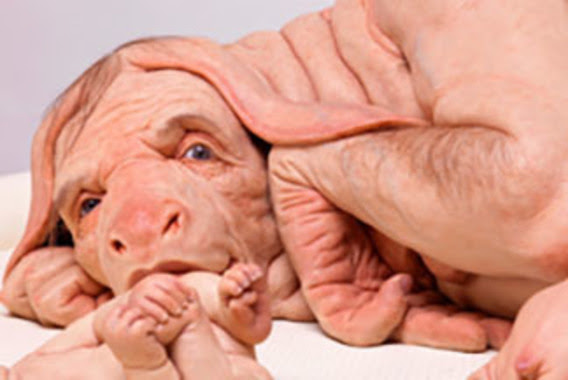
Patricia Piccinini’s Curious Imaginings Courtesy: Vancouver Biennale [downloaded from http://dailyhive.com/vancouver/vancouver-biennale-unsual-public-art-2018/]

The latest edition of the Vancouver Biennale was featured in a June 6, 2018 news item on the Daily Hive (Vancouver),

Melbourne artist Patricia Piccinini’s Curious Imaginings is expected to be one of the most talked about installations of the exhibit. Her style of “oddly captivating, somewhat grotesque, human-animal hybrid creature” is meant to be shocking and thought-provoking.

Piccinini’s interactive [emphasis mine] experience will “challenge us to explore the social impacts of emerging biotechnology and our ethical limits in an age where genetic engineering and digital technologies are already pushing the boundaries of humanity.”

Piccinini’s work will be displayed in the 105-year-old Patricia Hotel in Vancouver’s Strathcona neighbourhood. The 90-day ticketed exhibition [emphasis mine] is scheduled to open this September [2018].

(The show opens on Sept. 14, 2018.)

At the time, I had yet to stumble across Ingfei Chen’s thoughtful dive into the topic in her May 9, 2018 article for Slate.com,

In the United States, the clock is ticking for more than 114,700 adults and children waiting for a donated kidney or other lifesaving organ, and each day, nearly 20 of them die. Researchers are devising a new way to grow human organs inside other animals, but the method raises potentially thorny ethical issues. Other conceivable futuristic techniques sound like dystopian science fiction. As we envision an era of regenerative medicine decades from now, how far is society willing to go to solve the organ shortage crisis?

I found myself pondering this question after a discussion about the promises of stem cell technologies veered from the intriguing into the bizarre. I was interviewing bioengineer Zev Gartner, co-director and research coordinator of the Center for Cellular Construction at the University of California, San Francisco, about so-called organoids, tiny clumps of organlike tissue that can self-assemble from human stem cells in a Petri dish. These tissue bits are lending new insights into how our organs form and diseases take root. Some researchers even hope they can nurture organoids into full-size human kidneys, pancreases, and other organs for transplantation.

Certain organoid experiments have recently set off alarm bells, but when I asked Gartner about it, his radar for moral concerns was focused elsewhere. For him, the “really, really thought-provoking” scenarios involve other emerging stem cell–based techniques for engineering replacement organs for people, he told me. “Like blastocyst complementation,” he said.

Never heard of it? Neither had I. Turns out it’s a powerful new genetic engineering trick that researchers hope to use for growing human organs inside pigs or sheep—organs that could be genetically personalized for transplant patients, in theory avoiding immune-system rejection problems. The science still has many years to go, but if it pans out, it could be one solution to the organ shortage crisis. However, the prospect of creating hybrid animals with human parts and killing them to harvest organs has already raised a slew of ethical questions. In 2015, the National Institutes of Health placed a moratorium on federal funding of this nascent research area while it evaluated and discussed the issues.

As Gartner sees it, the debate over blastocyst complementation research—work that he finds promising—is just one of many conversations that society needs to have about the ethical and social costs and benefits of future technologies for making lifesaving transplant organs. “There’s all these weird ways that we could go about doing this,” he said, with a spectrum of imaginable approaches that includes organoids, interspecies organ farming, and building organs from scratch using 3D bioprinters. But even if it turns out we can produce human organs in these novel ways, the bigger issue, in each technological instance, may be whether we should.

Gartner crystallized things with a downright creepy example: “We know that the best bioreactor for tissues and organs for humans are human beings,” he said. Hypothetically, “the best way to get you a new heart would be to clone you, grow up a copy of yourself, and take the heart out.” [emphasis mine] Scientists could probably produce a cloned person with the technologies we already have, if money and ethics were of no concern. “But we don’t want to go there, right?” he added in the next breath. “The ethics involved in doing it are not compatible with who we want to be as a society.”

This sounds like Gartner may have been reading some science fiction, specifically, Lois McMaster Bujold and her Barrayar series where she often explored the ethics and possibilities of bioengineering. At this point, some of her work seems eerily prescient.

As for Chen’s article, I strongly encourage you to read it in its entirety if you have the time.

Medicine, healing, and big money

At about the same time, there was a May 31, 2018 news item on phys.org offering a perspective from some of the leaders in the science and the business (Note: Links have been removed),

Over the past few years, researchers led by George Church have made important strides toward engineering the genomes of pigs to make their cells compatible with the human body. So many think that it’s possible that, with the help of CRISPR technology, a healthy heart for a patient in desperate need might one day come from a pig.

“It’s relatively feasible to change one gene in a pig, but to change many dozens—which is quite clear is the minimum here—benefits from CRISPR,” an acronym for clustered regularly interspaced short palindromic repeats, said Church, the Robert Winthrop Professor of Genetics at Harvard Medical School (HMS) and a core faculty member of Harvard’s Wyss Institute for Biologically Inspired Engineering. Xenotransplantation is “one of few” big challenges (along with gene drives and de-extinction, he said) “that really requires the ‘oomph’ of CRISPR.”

To facilitate the development of safe and effective cells, tissues, and organs for future medical transplantation into human patients, Harvard’s Office of Technology Development has granted a technology license to the Cambridge biotech startup eGenesis.

Co-founded by Church and former HMS doctoral student Luhan Yang in 2015, eGenesis announced last year that it had raised $38 million to advance its research and development work. At least eight former members of the Church lab—interns, doctoral students, postdocs, and visiting researchers—have continued their scientific careers as employees there.

“The Church Lab is well known for its relentless pursuit of scientific achievements so ambitious they seem improbable—and, indeed, [for] its track record of success,” said Isaac Kohlberg, Harvard’s chief technology development officer and senior associate provost. “George deserves recognition too for his ability to inspire passion and cultivate a strong entrepreneurial drive among his talented research team.”

The license from Harvard OTD covers a powerful set of genome-engineering technologies developed at HMS and the Wyss Institute, including access to foundational intellectual property relating to the Church Lab’s 2012 breakthrough use of CRISPR, led by Yang and Prashant Mali, to edit the genome of human cells. Subsequent innovations that enabled efficient and accurate editing of numerous genes simultaneously are also included. The license is exclusive to eGenesis but limited to the field of xenotransplantation.

A May 30, 2018 Harvard University news release by Caroline Petty, which originated the news item, explores some of the issues associated with incubating humans organs in other species,

The prospect of using living, nonhuman organs, and concerns over the infectiousness of pathogens either present in the tissues or possibly formed in combination with human genetic material, have prompted the Food and Drug Administration to issue detailed guidance on xenotransplantation research and development since the mid-1990s. In pigs, a primary concern has been that porcine endogenous retroviruses (PERVs), strands of potentially pathogenic DNA in the animals’ genomes, might infect human patients and eventually cause disease. [emphases mine]

That’s where the Church lab’s CRISPR expertise has enabled significant advances. In 2015, the lab published important results in the journal Science, successfully demonstrating the use of genome engineering to eliminate all 62 PERVs in porcine cells. Science later called it “the most widespread CRISPR editing feat to date.”

In 2017, with collaborators at Harvard, other universities, and eGenesis, Church and Yang went further. Publishing again in Science, they first confirmed earlier researchers’ fears: Porcine cells can, in fact, transmit PERVs into human cells, and those human cells can pass them on to other, unexposed human cells. (It is still unknown under what circumstances those PERVs might cause disease.) In the same paper, they corrected the problem, announcing the embryogenesis and birth of 37 PERV-free pigs. [Note: My July 17, 2018 post features research which suggests CRISPR-Cas9 gene editing may cause greater genetic damage than had been thought.]

“Taken together, those innovations were stunning,” said Vivian Berlin, director of business development in OTD, who manages the commercialization strategy for much of Harvard’s intellectual property in the life sciences. “That was the foundation they needed, to convince both the scientific community and the investment community that xenotransplantation might become a reality.”

“After hundreds of tests, this was a critical milestone for eGenesis — and the entire field — and represented a key step toward safe organ transplantation from pigs,” said Julie Sunderland, interim CEO of eGenesis. “Building on this study, we hope to continue to advance the science and potential of making xenotransplantation a safe and routine medical procedure.”

Genetic engineering may undercut human diseases, but also could help restore extinct species, researcher says. [Shades of the Jurassic Park movies!]

It’s not, however, the end of the story: An immunological challenge remains, which eGenesis will need to address. The potential for a patient’s body to outright reject transplanted tissue has stymied many previous attempts at xenotransplantation. Church said numerous genetic changes must be achieved to make porcine organs fully compatible with human patients. Among these are edits to several immune functions, coagulation functions, complements, and sugars, as well as the PERVs.

“Trying the straight transplant failed almost immediately, within hours, because there’s a huge mismatch in the carbohydrates on the surface of the cells, in particular alpha-1-3-galactose, and so that was a showstopper,” Church explained. “When you delete that gene, which you can do with conventional methods, you still get pretty fast rejection, because there are a lot of other aspects that are incompatible. You have to take care of each of them, and not all of them are just about removing things — some of them you have to humanize. There’s a great deal of subtlety involved so that you get normal pig embryogenesis but not rejection.

“Putting it all together into one package is challenging,” he concluded.

In short, it’s the next big challenge for CRISPR.

Not unexpectedly, there is no mention of the CRISPR patent fight between Harvard/MIT’s (Massachusetts Institute of Technology) Broad Institute and the University of California at Berkeley (UC Berkeley). My March 15, 2017 posting featured an outcome where the Broad Institute won the first round of the fight. As I recall, it was a decision based on the principles associated with King Solomon, i.e., the US Patent Office, divided the baby and UCBerkeley got the less important part of the baby. As you might expect the decision has been appealed. In an April 30, 2018 piece, Scientific American reprinted an article about the latest round in the fight written by Sharon Begley for STAT (Note: Links have been removed),

All You Need to Know for Round 2 of the CRISPR Patent Fight

It’s baaaaack, that reputation-shredding, stock-moving fight to the death over key CRISPR patents. On Monday morning in Washington, D.C., the U.S. Court of Appeals for the Federal Circuit will hear oral arguments in University of California v. Broad Institute. Questions?

How did we get here? The patent office ruled in February 2017 that the Broad’s 2014 CRISPR patent on using CRISPR-Cas9 to edit genomes, based on discoveries by Feng Zhang, did not “interfere” with a patent application by UC based on the work of UC Berkeley’s Jennifer Doudna. In plain English, that meant the Broad’s patent, on using CRISPR-Cas9 to edit genomes in eukaryotic cells (all animals and plants, but not bacteria), was different from UC’s, which described Doudna’s experiments using CRISPR-Cas9 to edit DNA in a test tube—and it was therefore valid. The Patent Trial and Appeal Board concluded that when Zhang got CRISPR-Cas9 to work in human and mouse cells in 2012, it was not an obvious extension of Doudna’s earlier research, and that he had no “reasonable expectation of success.” UC appealed, and here we are.

For anyone who may not realize what the stakes are for these institutions, Linda Williams in a March 16, 1999 article for the LA Times had this to say about universities, patents, and money,

The University of Florida made about $2 million last year in royalties on a patent for Gatorade Thirst Quencher, a sports drink that generates some $500 million to $600 million a year in revenue for Quaker Oats Co.

The payments place the university among the top five in the nation in income from patent royalties.

Oh, but if some people on the Gainesville, Fla., campus could just turn back the clock. “If we had done Gatorade right, we would be getting $5 or $6 million (a year),” laments Donald Price, director of the university’s office of corporate programs. “It is a classic example of how not to handle a patent idea,” he added.

Gatorade was developed in 1965 when many universities were ill equipped to judge the commercial potential of ideas emerging from their research labs. Officials blew the university’s chance to control the Gatorade royalties when they declined to develop a professor’s idea.

The Gatorade story does not stop there and, even though it’s almost 20 years old, this article stands the test of time. I strongly encourage you to read it if the business end of patents and academia interest you or if you would like to develop more insight into the Broad Institute/UC Berkeley situation.

Getting back to the science, there is that pesky matter of diseases crossing over from one species to another. While, Harvard and eGenesis claim a victory in this area, it seems more work needs to be done.

Infections from pigs

An August 29, 2018 University of Alabama at Birmingham news release (also on EurekAlert) by Jeff Hansen, describes the latest chapter in the quest to provide more organs for transplantion,

A shortage of organs for transplantation — including kidneys and hearts — means that many patients die while still on waiting lists. So, research at the University of Alabama at Birmingham and other sites has turned to pig organs as an alternative. [emphasis mine]

Using gene-editing, researchers have modified such organs to prevent rejection, and research with primates shows the modified pig organs are well-tolerated.

An added step is needed to ensure the safety of these inter-species transplants — sensitive, quantitative assays for viruses and other infectious microorganisms in donor pigs that potentially could gain access to humans during transplantation.

The U.S. Food and Drug Administration requires such testing, prior to implantation, of tissues used for xenotransplantation from animals to humans. It is possible — though very unlikely — that an infectious agent in transplanted tissues could become an emerging infectious disease in humans.

In a paper published in Xenotransplantation, Mark Prichard, Ph.D., and colleagues at UAB have described the development and testing of 30 quantitative assays for pig infectious agents. These assays had sensitivities similar to clinical lab assays for viral loads in human patients. After validation, the UAB team also used the assays on nine sows and 22 piglets delivered from the sows through caesarian section.

“Going forward, ensuring the safety of these organs is of paramount importance,” Prichard said. “The use of highly sensitive techniques to detect potential pathogens will help to minimize adverse events in xenotransplantation.”

“The assays hold promise as part of the screening program to identify suitable donor animals, validate and release transplantable organs for research purposes, and monitor transplant recipients,” said Prichard, a professor in the UAB Department of Pediatrics and director of the Department of Pediatrics Molecular Diagnostics Laboratory.

The UAB researchers developed quantitative polymerase chain reaction, or qPCR, assays for 28 viruses sometimes found in pigs and two groups of mycoplasmas. They established reproducibility, sensitivity, specificity and lower limit of detection for each assay. All but three showed features of good quantitative assays, and the lower limit of detection values ranged between one and 16 copies of the viral or bacterial genetic material.

Also, the pig virus assays did not give false positives for some closely related human viruses.

As a start to understanding the infectious disease load in normal healthy animals and ensuring the safety of pig tissues used in xenotransplantation research, the researchers then screened blood, nasal swab and stool specimens from nine adult sows and 22 of their piglets delivered by caesarian section.

Mycoplasma species and two distinct herpesviruses were the most commonly detected microorganisms. Yet 14 piglets that were delivered from three sows infected with either or both herpesviruses were not infected with the herpesviruses, showing that transmission of these viruses from sow to the caesarian-delivery piglet was inefficient.

Prichard says the assays promise to enhance the safety of pig tissues for xenotransplantation, and they will also aid evaluation of human specimens after xenotransplantation.

The UAB researchers say they subsequently have evaluated more than 300 additional specimens, and that resulted in the detection of most of the targets. “The detection of these targets in pig specimens provides reassurance that the analytical methods are functioning as designed,” said Prichard, “and there is no a priori reason some targets might be more difficult to detect than others with the methods described here.”

As is my custom, here’s a link to and a citation for the paper,

Xenotransplantation panel for the detection of infectious agents in pigs by Caroll B. Hartline, Ra’Shun L. Conner, Scott H. James, Jennifer Potter, Edward Gray, Jose Estrada, Mathew Tector, A. Joseph Tector, Mark N. Prichard. Xenotransplantaion Volume 25, Issue 4 July/August 2018 e12427 DOI: https://doi.org/10.1111/xen.12427 First published: 18 August 2018

This paper is open access.

All this leads to questions about chimeras. If a pig is incubating organs with human cells it’s a chimera but then means the human receiving the organ becomes a chimera too. (For an example, see my Dec. 22, 2013 posting where there’s mention of a woman who received a trachea from a pig. Scroll down about 30% of the way.)

What is it to be human?

A question much beloved of philosophers and others, the question seems particularly timely with xenotransplantion and other developments such neuroprosthetics (cyborgs) and neuromorphic computing (brainlike computing).

As I’ve noted before, although not recently, popular culture offers a discourse on these issues. Take a look at the superhero movies and the way in which enhanced humans and aliens are presented. For example, X-Men comics and movies present mutants (humans with enhanced abilities) as despised and rejected. Video games (not really my thing but there is the Deus Ex series which has as its hero, a cyborg also offer insight into these issues.

Other than popular culture and in the ‘bleeding edge’ arts community, I can’t recall any public discussion on these matters arising from the extraordinary set of technologies which are being deployed or prepared for deployment in the foreseeable future.

(If you’re in Vancouver (Canada) from September 14 – December 15, 2018, you may want to check out Piccinini’s work. Also, there’s ” NCSU [North Carolina State University] Libraries, NC State’s Genetic Engineering and Society (GES) Center, and the Gregg Museum of Art & Design have issued a public call for art for the upcoming exhibition Art’s Work in the Age of Biotechnology: Shaping our Genetic Futures.” from my Sept. 6, 2018 posting. Deadline: Oct. 1, 2018.)

At a guess, there will be pushback from people who have no interest in debating what it is to be human as they already know, and will find these developments, when they learn about them, to be horrifying and unnatural.