Tag Archives: Harvard Medical School (HMS)

Reversing lower limb paralysis

This regenerative treatment is at a very early stage, which means the Swiss researchers have tried it on mice as you can see in the following video (runtime: 2 mins. 15 secs.). Towards the end of the video, researcher Grégoire Courtine cautions there are many hurdles before this could be used in humans, if ever,

A September 22, 2023 Ecole Polytechnique Fédérale de Lausanne (EPFL) press release (also on EurekAlert but published September 21, 2023) by Emmanuel Barraud, describes the work in more detail,

When the spinal cords of mice and humans are partially damaged, the initial paralysis is followed by the extensive, spontaneous recovery of motor function. However, after a complete spinal cord injury, this natural repair of the spinal cord doesn’t occur and there is no recovery. Meaningful recovery after severe injuries requires strategies that promote the regeneration of nerve fibers, but the requisite conditions for these strategies to successfully restore motor function have remained elusive.

“Five years ago, we demonstrated that nerve fibers can be regenerated across anatomically complete spinal cord injuries,” says Mark Anderson, a senior author of the study. “But we also realized this wasn’t enough to restore motor function, as the new fibers failed to connect to the right places on the other side of the lesion.” Anderson is the director of Central Nervous System Regeneration at .NeuroRestore and a scientist at the Wyss Center for Bio and Neuroengineering.

Working in tandem with peers at UCLA [University of California at Los Angeles] and Harvard Medical School, the scientists used state-of-the-art equipment at EPFL’s Campus Biotech facilities in Geneva to run in-depth analyses and identity which type of neuron is involved in natural spinal-cord repair after partial spinal cord injury. “Our observations using single-cell nuclear RNA sequencing not only exposed the specific axons that must regenerate, but also revealed that these axons must reconnect to their natural targets to restore motor function,” says Jordan Squair, the study’s first author. The team’s findings appear in the 22 September 2023 issue of Science.

Towards a combination of approaches

Their discovery informed the design of a multipronged gene therapy. The scientists activated growth programs in the identified neurons in mice to regenerate their nerve fibers, upregulated specific proteins to support the neurons’ growth through the lesion core, and administered guidance molecules to attract the regenerating nerve fibers to their natural targets below the injury. “We were inspired by nature when we designed a therapeutic strategy that replicates the spinal-cord repair mechanisms occurring spontaneously after partial injuries,” says Squair.

Mice with anatomically complete spinal cord injuries regained the ability to walk, exhibiting gait patterns that resembled those quantified in mice that resumed walking naturally after partial injuries. This observation revealed a previously unknown condition for regenerative therapies to be successful in restoring motor function after neurotrauma. “We expect that our gene therapy will act synergistically with our other procedures involving electrical stimulation of the spinal cord,” says Grégoire Courtine, a senior author of the study who also heads .NeuroRestore together with Jocelyne Bloch. “We believe a complete solution for treating spinal cord injury will require both approaches – gene therapy to regrow relevant nerve fibers, and spinal stimulation to maximize the ability of both these fibers and the spinal cord below the injury to produce movement.”

While many obstacles must still be overcome before this gene therapy can be applied in humans, the scientists have taken the first steps towards developing the technology necessary to achieve this feat in the years to come.

Here’s a link to and a citation for the paper,

Recovery of walking after paralysis by regenerating characterized neurons to their natural target region by Jordan W. Squair, Marco Milano, Alexandra de Coucy, Matthieu Gautier, Michael A. Skinnider, Nicholas D. James, Newton Cho, Anna Lasne, Claudia Kathe,Thomas H. Hutson, Steven Ceto, Laetitia Baud, Katia Galan, Viviana Aureli, Achilleas Laskaratos, Quentin Barraud, Timothy J. Deming, Richie E. Kohman, Bernard L. Schneider, Zhigang He, Jocelyne Bloch, Michael V. Sofroniew, Gregoire Courtine, and Mark A. Anderson. Science 21 Sep 2023 Vol 381, Issue 6664 pp. 1338-1345 DOI: 10.1126/science.adi641

This paper is behind a paywall.

This March 25, 2015 posting, “Spinal cords, brains, implants, and remote control,” features some research from EPFL researchers whose names you might recognize from this posting’s research paper.

Mentioned in the press release, the Swiss research centre website for NeuroRestore is here.

Xenotransplantation—organs for transplantation in human patients—it’s a business and a science

The last time (June 18, 2018 post) I mentioned xenotransplantation (transplanting organs from one species into another species; see more here), it was in the context of an art/sci (or sciart) event coming to Vancouver (Canada).,
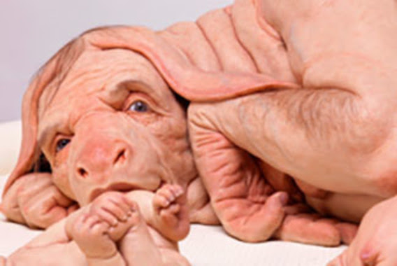
Patricia Piccinini’s Curious Imaginings Courtesy: Vancouver Biennale [downloaded from http://dailyhive.com/vancouver/vancouver-biennale-unsual-public-art-2018/]

The latest edition of the Vancouver Biennale was featured in a June 6, 2018 news item on the Daily Hive (Vancouver),

Melbourne artist Patricia Piccinini’s Curious Imaginings is expected to be one of the most talked about installations of the exhibit. Her style of “oddly captivating, somewhat grotesque, human-animal hybrid creature” is meant to be shocking and thought-provoking.

Piccinini’s interactive [emphasis mine] experience will “challenge us to explore the social impacts of emerging biotechnology and our ethical limits in an age where genetic engineering and digital technologies are already pushing the boundaries of humanity.”

Piccinini’s work will be displayed in the 105-year-old Patricia Hotel in Vancouver’s Strathcona neighbourhood. The 90-day ticketed exhibition [emphasis mine] is scheduled to open this September [2018].

(The show opens on Sept. 14, 2018.)

At the time, I had yet to stumble across Ingfei Chen’s thoughtful dive into the topic in her May 9, 2018 article for Slate.com,

In the United States, the clock is ticking for more than 114,700 adults and children waiting for a donated kidney or other lifesaving organ, and each day, nearly 20 of them die. Researchers are devising a new way to grow human organs inside other animals, but the method raises potentially thorny ethical issues. Other conceivable futuristic techniques sound like dystopian science fiction. As we envision an era of regenerative medicine decades from now, how far is society willing to go to solve the organ shortage crisis?

I found myself pondering this question after a discussion about the promises of stem cell technologies veered from the intriguing into the bizarre. I was interviewing bioengineer Zev Gartner, co-director and research coordinator of the Center for Cellular Construction at the University of California, San Francisco, about so-called organoids, tiny clumps of organlike tissue that can self-assemble from human stem cells in a Petri dish. These tissue bits are lending new insights into how our organs form and diseases take root. Some researchers even hope they can nurture organoids into full-size human kidneys, pancreases, and other organs for transplantation.

Certain organoid experiments have recently set off alarm bells, but when I asked Gartner about it, his radar for moral concerns was focused elsewhere. For him, the “really, really thought-provoking” scenarios involve other emerging stem cell–based techniques for engineering replacement organs for people, he told me. “Like blastocyst complementation,” he said.

Never heard of it? Neither had I. Turns out it’s a powerful new genetic engineering trick that researchers hope to use for growing human organs inside pigs or sheep—organs that could be genetically personalized for transplant patients, in theory avoiding immune-system rejection problems. The science still has many years to go, but if it pans out, it could be one solution to the organ shortage crisis. However, the prospect of creating hybrid animals with human parts and killing them to harvest organs has already raised a slew of ethical questions. In 2015, the National Institutes of Health placed a moratorium on federal funding of this nascent research area while it evaluated and discussed the issues.

As Gartner sees it, the debate over blastocyst complementation research—work that he finds promising—is just one of many conversations that society needs to have about the ethical and social costs and benefits of future technologies for making lifesaving transplant organs. “There’s all these weird ways that we could go about doing this,” he said, with a spectrum of imaginable approaches that includes organoids, interspecies organ farming, and building organs from scratch using 3D bioprinters. But even if it turns out we can produce human organs in these novel ways, the bigger issue, in each technological instance, may be whether we should.

Gartner crystallized things with a downright creepy example: “We know that the best bioreactor for tissues and organs for humans are human beings,” he said. Hypothetically, “the best way to get you a new heart would be to clone you, grow up a copy of yourself, and take the heart out.” [emphasis mine] Scientists could probably produce a cloned person with the technologies we already have, if money and ethics were of no concern. “But we don’t want to go there, right?” he added in the next breath. “The ethics involved in doing it are not compatible with who we want to be as a society.”

This sounds like Gartner may have been reading some science fiction, specifically, Lois McMaster Bujold and her Barrayar series where she often explored the ethics and possibilities of bioengineering. At this point, some of her work seems eerily prescient.

As for Chen’s article, I strongly encourage you to read it in its entirety if you have the time.

Medicine, healing, and big money

At about the same time, there was a May 31, 2018 news item on phys.org offering a perspective from some of the leaders in the science and the business (Note: Links have been removed),

Over the past few years, researchers led by George Church have made important strides toward engineering the genomes of pigs to make their cells compatible with the human body. So many think that it’s possible that, with the help of CRISPR technology, a healthy heart for a patient in desperate need might one day come from a pig.

“It’s relatively feasible to change one gene in a pig, but to change many dozens—which is quite clear is the minimum here—benefits from CRISPR,” an acronym for clustered regularly interspaced short palindromic repeats, said Church, the Robert Winthrop Professor of Genetics at Harvard Medical School (HMS) and a core faculty member of Harvard’s Wyss Institute for Biologically Inspired Engineering. Xenotransplantation is “one of few” big challenges (along with gene drives and de-extinction, he said) “that really requires the ‘oomph’ of CRISPR.”

To facilitate the development of safe and effective cells, tissues, and organs for future medical transplantation into human patients, Harvard’s Office of Technology Development has granted a technology license to the Cambridge biotech startup eGenesis.

Co-founded by Church and former HMS doctoral student Luhan Yang in 2015, eGenesis announced last year that it had raised $38 million to advance its research and development work. At least eight former members of the Church lab—interns, doctoral students, postdocs, and visiting researchers—have continued their scientific careers as employees there.

“The Church Lab is well known for its relentless pursuit of scientific achievements so ambitious they seem improbable—and, indeed, [for] its track record of success,” said Isaac Kohlberg, Harvard’s chief technology development officer and senior associate provost. “George deserves recognition too for his ability to inspire passion and cultivate a strong entrepreneurial drive among his talented research team.”

The license from Harvard OTD covers a powerful set of genome-engineering technologies developed at HMS and the Wyss Institute, including access to foundational intellectual property relating to the Church Lab’s 2012 breakthrough use of CRISPR, led by Yang and Prashant Mali, to edit the genome of human cells. Subsequent innovations that enabled efficient and accurate editing of numerous genes simultaneously are also included. The license is exclusive to eGenesis but limited to the field of xenotransplantation.

A May 30, 2018 Harvard University news release by Caroline Petty, which originated the news item, explores some of the issues associated with incubating humans organs in other species,

The prospect of using living, nonhuman organs, and concerns over the infectiousness of pathogens either present in the tissues or possibly formed in combination with human genetic material, have prompted the Food and Drug Administration to issue detailed guidance on xenotransplantation research and development since the mid-1990s. In pigs, a primary concern has been that porcine endogenous retroviruses (PERVs), strands of potentially pathogenic DNA in the animals’ genomes, might infect human patients and eventually cause disease. [emphases mine]

That’s where the Church lab’s CRISPR expertise has enabled significant advances. In 2015, the lab published important results in the journal Science, successfully demonstrating the use of genome engineering to eliminate all 62 PERVs in porcine cells. Science later called it “the most widespread CRISPR editing feat to date.”

In 2017, with collaborators at Harvard, other universities, and eGenesis, Church and Yang went further. Publishing again in Science, they first confirmed earlier researchers’ fears: Porcine cells can, in fact, transmit PERVs into human cells, and those human cells can pass them on to other, unexposed human cells. (It is still unknown under what circumstances those PERVs might cause disease.) In the same paper, they corrected the problem, announcing the embryogenesis and birth of 37 PERV-free pigs. [Note: My July 17, 2018 post features research which suggests CRISPR-Cas9 gene editing may cause greater genetic damage than had been thought.]

“Taken together, those innovations were stunning,” said Vivian Berlin, director of business development in OTD, who manages the commercialization strategy for much of Harvard’s intellectual property in the life sciences. “That was the foundation they needed, to convince both the scientific community and the investment community that xenotransplantation might become a reality.”

“After hundreds of tests, this was a critical milestone for eGenesis — and the entire field — and represented a key step toward safe organ transplantation from pigs,” said Julie Sunderland, interim CEO of eGenesis. “Building on this study, we hope to continue to advance the science and potential of making xenotransplantation a safe and routine medical procedure.”

Genetic engineering may undercut human diseases, but also could help restore extinct species, researcher says. [Shades of the Jurassic Park movies!]

It’s not, however, the end of the story: An immunological challenge remains, which eGenesis will need to address. The potential for a patient’s body to outright reject transplanted tissue has stymied many previous attempts at xenotransplantation. Church said numerous genetic changes must be achieved to make porcine organs fully compatible with human patients. Among these are edits to several immune functions, coagulation functions, complements, and sugars, as well as the PERVs.

“Trying the straight transplant failed almost immediately, within hours, because there’s a huge mismatch in the carbohydrates on the surface of the cells, in particular alpha-1-3-galactose, and so that was a showstopper,” Church explained. “When you delete that gene, which you can do with conventional methods, you still get pretty fast rejection, because there are a lot of other aspects that are incompatible. You have to take care of each of them, and not all of them are just about removing things — some of them you have to humanize. There’s a great deal of subtlety involved so that you get normal pig embryogenesis but not rejection.

“Putting it all together into one package is challenging,” he concluded.

In short, it’s the next big challenge for CRISPR.

Not unexpectedly, there is no mention of the CRISPR patent fight between Harvard/MIT’s (Massachusetts Institute of Technology) Broad Institute and the University of California at Berkeley (UC Berkeley). My March 15, 2017 posting featured an outcome where the Broad Institute won the first round of the fight. As I recall, it was a decision based on the principles associated with King Solomon, i.e., the US Patent Office, divided the baby and UCBerkeley got the less important part of the baby. As you might expect the decision has been appealed. In an April 30, 2018 piece, Scientific American reprinted an article about the latest round in the fight written by Sharon Begley for STAT (Note: Links have been removed),

All You Need to Know for Round 2 of the CRISPR Patent Fight

It’s baaaaack, that reputation-shredding, stock-moving fight to the death over key CRISPR patents. On Monday morning in Washington, D.C., the U.S. Court of Appeals for the Federal Circuit will hear oral arguments in University of California v. Broad Institute. Questions?

How did we get here? The patent office ruled in February 2017 that the Broad’s 2014 CRISPR patent on using CRISPR-Cas9 to edit genomes, based on discoveries by Feng Zhang, did not “interfere” with a patent application by UC based on the work of UC Berkeley’s Jennifer Doudna. In plain English, that meant the Broad’s patent, on using CRISPR-Cas9 to edit genomes in eukaryotic cells (all animals and plants, but not bacteria), was different from UC’s, which described Doudna’s experiments using CRISPR-Cas9 to edit DNA in a test tube—and it was therefore valid. The Patent Trial and Appeal Board concluded that when Zhang got CRISPR-Cas9 to work in human and mouse cells in 2012, it was not an obvious extension of Doudna’s earlier research, and that he had no “reasonable expectation of success.” UC appealed, and here we are.

For anyone who may not realize what the stakes are for these institutions, Linda Williams in a March 16, 1999 article for the LA Times had this to say about universities, patents, and money,

The University of Florida made about $2 million last year in royalties on a patent for Gatorade Thirst Quencher, a sports drink that generates some $500 million to $600 million a year in revenue for Quaker Oats Co.

The payments place the university among the top five in the nation in income from patent royalties.

Oh, but if some people on the Gainesville, Fla., campus could just turn back the clock. “If we had done Gatorade right, we would be getting $5 or $6 million (a year),” laments Donald Price, director of the university’s office of corporate programs. “It is a classic example of how not to handle a patent idea,” he added.

Gatorade was developed in 1965 when many universities were ill equipped to judge the commercial potential of ideas emerging from their research labs. Officials blew the university’s chance to control the Gatorade royalties when they declined to develop a professor’s idea.

The Gatorade story does not stop there and, even though it’s almost 20 years old, this article stands the test of time. I strongly encourage you to read it if the business end of patents and academia interest you or if you would like to develop more insight into the Broad Institute/UC Berkeley situation.

Getting back to the science, there is that pesky matter of diseases crossing over from one species to another. While, Harvard and eGenesis claim a victory in this area, it seems more work needs to be done.

Infections from pigs

An August 29, 2018 University of Alabama at Birmingham news release (also on EurekAlert) by Jeff Hansen, describes the latest chapter in the quest to provide more organs for transplantion,

A shortage of organs for transplantation — including kidneys and hearts — means that many patients die while still on waiting lists. So, research at the University of Alabama at Birmingham and other sites has turned to pig organs as an alternative. [emphasis mine]

Using gene-editing, researchers have modified such organs to prevent rejection, and research with primates shows the modified pig organs are well-tolerated.

An added step is needed to ensure the safety of these inter-species transplants — sensitive, quantitative assays for viruses and other infectious microorganisms in donor pigs that potentially could gain access to humans during transplantation.

The U.S. Food and Drug Administration requires such testing, prior to implantation, of tissues used for xenotransplantation from animals to humans. It is possible — though very unlikely — that an infectious agent in transplanted tissues could become an emerging infectious disease in humans.

In a paper published in Xenotransplantation, Mark Prichard, Ph.D., and colleagues at UAB have described the development and testing of 30 quantitative assays for pig infectious agents. These assays had sensitivities similar to clinical lab assays for viral loads in human patients. After validation, the UAB team also used the assays on nine sows and 22 piglets delivered from the sows through caesarian section.

“Going forward, ensuring the safety of these organs is of paramount importance,” Prichard said. “The use of highly sensitive techniques to detect potential pathogens will help to minimize adverse events in xenotransplantation.”

“The assays hold promise as part of the screening program to identify suitable donor animals, validate and release transplantable organs for research purposes, and monitor transplant recipients,” said Prichard, a professor in the UAB Department of Pediatrics and director of the Department of Pediatrics Molecular Diagnostics Laboratory.

The UAB researchers developed quantitative polymerase chain reaction, or qPCR, assays for 28 viruses sometimes found in pigs and two groups of mycoplasmas. They established reproducibility, sensitivity, specificity and lower limit of detection for each assay. All but three showed features of good quantitative assays, and the lower limit of detection values ranged between one and 16 copies of the viral or bacterial genetic material.

Also, the pig virus assays did not give false positives for some closely related human viruses.

As a start to understanding the infectious disease load in normal healthy animals and ensuring the safety of pig tissues used in xenotransplantation research, the researchers then screened blood, nasal swab and stool specimens from nine adult sows and 22 of their piglets delivered by caesarian section.

Mycoplasma species and two distinct herpesviruses were the most commonly detected microorganisms. Yet 14 piglets that were delivered from three sows infected with either or both herpesviruses were not infected with the herpesviruses, showing that transmission of these viruses from sow to the caesarian-delivery piglet was inefficient.

Prichard says the assays promise to enhance the safety of pig tissues for xenotransplantation, and they will also aid evaluation of human specimens after xenotransplantation.

The UAB researchers say they subsequently have evaluated more than 300 additional specimens, and that resulted in the detection of most of the targets. “The detection of these targets in pig specimens provides reassurance that the analytical methods are functioning as designed,” said Prichard, “and there is no a priori reason some targets might be more difficult to detect than others with the methods described here.”

As is my custom, here’s a link to and a citation for the paper,

Xenotransplantation panel for the detection of infectious agents in pigs by Caroll B. Hartline, Ra’Shun L. Conner, Scott H. James, Jennifer Potter, Edward Gray, Jose Estrada, Mathew Tector, A. Joseph Tector, Mark N. Prichard. Xenotransplantaion Volume 25, Issue 4 July/August 2018 e12427 DOI: https://doi.org/10.1111/xen.12427 First published: 18 August 2018

This paper is open access.

All this leads to questions about chimeras. If a pig is incubating organs with human cells it’s a chimera but then means the human receiving the organ becomes a chimera too. (For an example, see my Dec. 22, 2013 posting where there’s mention of a woman who received a trachea from a pig. Scroll down about 30% of the way.)

What is it to be human?

A question much beloved of philosophers and others, the question seems particularly timely with xenotransplantion and other developments such neuroprosthetics (cyborgs) and neuromorphic computing (brainlike computing).

As I’ve noted before, although not recently, popular culture offers a discourse on these issues. Take a look at the superhero movies and the way in which enhanced humans and aliens are presented. For example, X-Men comics and movies present mutants (humans with enhanced abilities) as despised and rejected. Video games (not really my thing but there is the Deus Ex series which has as its hero, a cyborg also offer insight into these issues.

Other than popular culture and in the ‘bleeding edge’ arts community, I can’t recall any public discussion on these matters arising from the extraordinary set of technologies which are being deployed or prepared for deployment in the foreseeable future.

(If you’re in Vancouver (Canada) from September 14 – December 15, 2018, you may want to check out Piccinini’s work. Also, there’s ” NCSU [North Carolina State University] Libraries, NC State’s Genetic Engineering and Society (GES) Center, and the Gregg Museum of Art & Design have issued a public call for art for the upcoming exhibition Art’s Work in the Age of Biotechnology: Shaping our Genetic Futures.” from my Sept. 6, 2018 posting. Deadline: Oct. 1, 2018.)

At a guess, there will be pushback from people who have no interest in debating what it is to be human as they already know, and will find these developments, when they learn about them, to be horrifying and unnatural.

Santiago Ramón y Cajal and the butterflies of the soul

The Cajal exhibit of drawings was here in Vancouver (Canada) this last fall (2017) and I still carry the memory of that glorious experience (see my Sept. 11, 2017 posting for more about the show and associated events). It seems Cajal’s drawings had a similar response in New York city, from a January 18, 2018 article by Roberta Smith for the New York Times,

It’s not often that you look at an exhibition with the help of the very apparatus that is its subject. But so it is with “The Beautiful Brain: The Drawings of Santiago Ramón y Cajal” at the Grey Art Gallery at New York University, one of the most unusual, ravishing exhibitions of the season.

The show finished its run on March 31, 2018 and is now on its way to the Massachusetts Institute of Technology (MIT) in Boston, Massachusetts for its opening on May 3, 2018. It looks like they have an exciting lineup of events to go along with the exhibit (from MIT’s The Beautiful Brain: The Drawings of Santiago Ramón y Cajal exhibit and event page),

SUMMER PROGRAMS

ONGOING

Spotlight Tours
Explorations led by local and Spanish scientists, artists, and entrepreneurs who will share their unique perspectives on particular aspects of the exhibition. (2:00 pm on select Tuesdays and Saturdays)

Tue, May 8 – Mark Harnett, Fred and Carole Middleton Career Development Professor at MIT and McGovern Institute Investigator Sat, May 26 – Marion Boulicault, MIT Graduate Student and Neuroethics Fellow in the Center for Sensorimotor Neural Engineering Tue, June 5 – Kelsey Allen, Graduate researcher, MIT Center for Brains, Minds, and Machines Sat, Jun 23 – Francisco Martin-Martinez, Research Scientist in MIT’s Laboratory for Atomistic & Molecular Mechanics and President of the Spanish Foundation for Science and Technology Jul 21 – Alex Gomez-Marin, Principal Investigator of the Behavior of Organisms Laboratory in the Instituto de Neurociencias, Spain Tue, Jul 31– Julie Pryor, Director of Communications at the McGovern Institute for Brain Research at MIT Tue, Aug 28 – Satrajit Ghosh, Principal Research Scientist at the McGovern Institute for Brain Research at MIT, Assistant Professor in the Department of Otolaryngology at Harvard Medical School, and faculty member in the Speech and Hearing Biosciences and Technology program in the Harvard Division of Medical Sciences

Idea Hub
Drop in and explore expansion microscopy in our maker-space.

Visualizing Science Workshop
Experiential learning with micro-scale biological images. (pre-registration required)

Gallery Demonstrations
Researchers share the latest on neural anatomy, signal transmission, and modern imaging techniques.

EVENTS

Teen Science Café: Mindful Matters
MIT researchers studying the brain share their mind-blowing findings.

Neuron Paint Night
Create a painting of cerebral cortex neurons and learn about the EyeWire citizen science game.

Cerebral Cinema Series
Hear from researchers and then compare real science to depictions on the big screen.

Brainy Trivia
Test your brain power in a night of science trivia and short, snappy research talks.

Come back to see our exciting lineup for the fall!

If you don’t have a chance to see the show or if you’d like a preview, I encourage you to read Smith’s article as it has embedded several Cajal drawings and rendered them exceptionally well.

For those who like a little contemporary (and related) science with their art, there’s a March 30, 2018 Harvard Medical Schoo (HMS)l news release by Kevin Jang (also on EurekAlert), Note: All links save one have been removed,

Drawing of the cells of the chick cerebellum by Santiago Ramón y Cajal, from “Estructura de los centros nerviosos de las aves,” Madrid, circa 1905


Modern neuroscience, for all its complexity, can trace its roots directly to a series of pen-and-paper sketches rendered by Nobel laureate Santiago Ramón y Cajal in the late 19th and early 20th centuries.

His observations and drawings exposed the previously hidden composition of the brain, revealing neuronal cell bodies and delicate projections that connect individual neurons together into intricate networks.

As he explored the nervous systems of various organisms under his microscope, a natural question arose: What makes a human brain different from the brain of any other species?

At least part of the answer, Ramón y Cajal hypothesized, lay in a specific class of neuron—one found in a dazzling variety of shapes and patterns of connectivity, and present in higher proportions in the human brain than in the brains of other species. He dubbed them the “butterflies of the soul.”

Known as interneurons, these cells play critical roles in transmitting information between sensory and motor neurons, and, when defective, have been linked to diseases such as schizophrenia, autism and intellectual disability.

Despite more than a century of study, however, it remains unclear why interneurons are so diverse and what specific functions the different subtypes carry out.

Now, in a study published in the March 22 [2018] issue of Nature, researchers from Harvard Medical School, New York Genome Center, New York University and the Broad Institute of MIT and Harvard have detailed for the first time how interneurons emerge and diversify in the brain.

Using single-cell analysis—a technology that allows scientists to track cellular behavior one cell at a time—the team traced the lineage of interneurons from their earliest precursor states to their mature forms in mice. The researchers identified key genetic programs that determine the fate of developing interneurons, as well as when these programs are switched on or off.

The findings serve as a guide for efforts to shed light on interneuron function and may help inform new treatment strategies for disorders involving their dysfunction, the authors said.

“We knew more than 100 years ago that this huge diversity of morphologically interesting cells existed in the brain, but their specific individual roles in brain function are still largely unclear,” said co-senior author Gordon Fishell, HMS professor of neurobiology and a faculty member at the Stanley Center for Psychiatric Research at the Broad.

“Our study provides a road map for understanding how and when distinct interneuron subtypes develop, giving us unprecedented insight into the biology of these cells,” he said. “We can now investigate interneuron properties as they emerge, unlock how these important cells function and perhaps even intervene when they fail to develop correctly in neuropsychiatric disease.”

A hippocampal interneuron. Image: Biosciences Imaging Gp, Soton, Wellcome Trust via Creative CommonsA hippocampal interneuron. Image: Biosciences Imaging Gp, Soton, Wellcome Trust via Creative Commons
Origins and Fates

In collaboration with co-senior author Rahul Satija, core faculty member of the New York Genome Center, Fishell and colleagues analyzed brain regions in developing mice known to contain precursor cells that give rise to interneurons.

Using Drop-seq, a single-cell sequencing technique created by researchers at HMS and the Broad, the team profiled gene expression in thousands of individual cells at multiple time points.

This approach overcomes a major limitation in past research, which could analyze only the average activity of mixtures of many different cells.

In the current study, the team found that the precursor state of all interneurons had similar gene expression patterns despite originating in three separate brain regions and giving rise to 14 or more interneuron subtypes alone—a number still under debate as researchers learn more about these cells.

“Mature interneuron subtypes exhibit incredible diversity. Their morphology and patterns of connectivity and activity are so different from each other, but our results show that the first steps in their maturation are remarkably similar,” said Satija, who is also an assistant professor of biology at New York University.

“They share a common developmental trajectory at the earliest stages, but the seeds of what will cause them to diverge later—a handful of genes—are present from the beginning,” Satija said.

As they profiled cells at later stages in development, the team observed the initial emergence of four interneuron “cardinal” classes, which give rise to distinct fates. Cells were committed to these fates even in the early embryo. By developing a novel computational strategy to link precursors with adult subtypes, the researchers identified individual genes that were switched on and off when cells began to diversify.

For example, they found that the gene Mef2c—mutations of which are linked to Alzheimer’s disease, schizophrenia and neurodevelopmental disorders in humans—is an early embryonic marker for a specific interneuron subtype known as Pvalb neurons. When they deleted Mef2c in animal models, Pvalb neurons failed to develop.

These early genes likely orchestrate the execution of subsequent genetic subroutines, such as ones that guide interneuron subtypes as they migrate to different locations in the brain and ones that help form unique connection patterns with other neural cell types, the authors said.

The identification of these genes and their temporal activity now provide researchers with specific targets to investigate the precise functions of interneurons, as well as how neurons diversify in general, according to the authors.

“One of the goals of this project was to address an incredibly fascinating developmental biology question, which is how individual progenitor cells decide between different neuronal fates,” Satija said. “In addition to these early markers of interneuron divergence, we found numerous additional genes that increase in expression, many dramatically, at later time points.”

The association of some of these genes with neuropsychiatric diseases promises to provide a better understanding of these disorders and the development of therapeutic strategies to treat them, a particularly important notion given the paucity of new treatments, the authors said.

Over the past 50 years, there have been no fundamentally new classes of neuropsychiatric drugs, only newer versions of old drugs, the researchers pointed out.

“Our repertoire is no better than it was in the 1970s,” Fishell said.

“Neuropsychiatric diseases likely reflect the dysfunction of very specific cell types. Our study puts forward a clear picture of what cells to look at as we work to shed light on the mechanisms that underlie these disorders,” Fishell said. “What we will find remains to be seen, but we have new, strong hypotheses that we can now test.”

As a resource for the research community, the study data and software are open-source and freely accessible online.

A gallery of the drawings of Santiago Ramón y Cajal is currently on display in New York City, and will open at the MIT Museum in Boston in May 2018.

Christian Mayer, Christoph Hafemeister and Rachel Bandler served as co-lead authors on the study.

This work was supported by the National Institutes of Health (R01 NS074972, R01 NS081297, MH071679-12, DP2-HG-009623, F30MH114462, T32GM007308, F31NS103398), the European Molecular Biology Organization, the National Science Foundation and the Simons Foundation.

Here’s link to and a citation for the paper,

Developmental diversification of cortical inhibitory interneurons by Christian Mayer, Christoph Hafemeister, Rachel C. Bandler, Robert Machold, Renata Batista Brito, Xavier Jaglin, Kathryn Allaway, Andrew Butler, Gord Fishell, & Rahul Satija. Nature volume 555, pages 457–462 (22 March 2018) doi:10.1038/nature25999 Published: 05 March 2018

This paper is behind a paywall.

‘Glow in the dark’, paint-on bandage heals

Somewhat unexpectedly (to me), this research about a ‘smart’ paint-on bandage is being published by The Optical Society of America (OSA). Here’s more about the work from an Oct. 1, 2014 news item on Nanowerk,

Inspired by a desire to help wounded soldiers, an international, multidisciplinary team of researchers led by Assistant Professor Conor L. Evans at the Wellman Center for Photomedicine of Massachusetts General Hospital (MGH) and Harvard Medical School (HMS) has created a paint-on, see-through, “smart” bandage that glows to indicate a wound’s tissue oxygenation concentration. Because oxygen plays a critical role in healing, mapping these levels in severe wounds and burns can help to significantly improve the success of surgeries to restore limbs and physical functions.

An Oct. 1, 2014 OSA news release (also on EurekAlert), which originated the news item, describes the interest in oxygenation in more detail,

“Information about tissue oxygenation is clinically relevant but is often inaccessible due to a lack of accurate or noninvasive measurements,” explained lead author Zongxi Li, an HMS research fellow on Evans’ team.

Now, the “smart” bandage developed by the team provides direct, noninvasive measurement of tissue oxygenation by combining three simple, compact and inexpensive components: a bright sensor molecule with a long phosphorescence lifetime and appropriate dynamic range; a bandage material compatible with the sensor molecule that conforms to the skin’s surface to form an airtight seal; and an imaging device capable of capturing the oxygen-dependent signals from the bandage with high signal-to-noise ratio.

This work is part of the team’s long-term program “to develop a Sensing, Monitoring And Release of Therapeutics (SMART) bandage for improved care of patients with acute or chronic wounds,” says Evans …

The news release goes on to briefly explain the technology,

For starters, the bandage’s not-so-secret key ingredient is phosphors—molecules that absorb light and then emit it via a process known as phosphorescence.

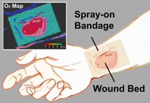
Phosphorescence is encountered by many on a daily basis—ranging from glow-in-the-dark dials on watches to t-shirt lettering. “How brightly our phosphorescent molecules emit light depends on how much oxygen is present,” said Li. “As the concentration of oxygen is reduced, the phosphors glow both longer and more brightly.” To make the bandage simple to interpret, the team also incorporated a green oxygen-insensitive reference dye, so that changes in tissue oxygenation are displayed as a green-to-red colormap.

The bandage is applied by “painting” it onto the skin’s surface as a viscous liquid, which dries to a solid thin film within a minute. Once the first layer has dried, a transparent barrier layer is then applied atop it to protect the film and slow the rate of oxygen exchange between the bandage and room air—making the bandage sensitive to the oxygen within tissue.

The final piece involves a camera-based readout device, which performs two functions: it provides a burst of excitation light that triggers the emission of the phosphors inside the bandage, and then it records the phosphors’ emission. “Depending on the camera’s configuration, we can measure either the brightness or color of the emitted light across the bandage or the change in brightness over time,” Li said. “Both of these signals can be used to create an oxygenation map.”  The emitted light from the bandage is bright enough that it can be acquired using a regular camera or smartphone—opening the possibility to a portable, field-ready device.

There are some immediate applications, as well as, plans for research that will yield applications (from the news release),

Immediate applications for the oxygen-sensing bandage include monitoring patients with a risk of developing ischemic (restricted blood supply) conditions, postoperative monitoring of skin grafts or flaps, and burn-depth determination as a guide for surgical debridement—the removal of dead or damaged tissue from the body.

“The need for a reliable, accurate and easy-to-use method of rapid assessment of blood flow to the skin for patients remains a clinical necessity,” said co-author Samuel Lin, an HMS associate professor of surgery at Beth Israel Deaconess Medical Center. “Plastic surgeons continuously monitor the state of blood flow to the skin, so the liquid-bandage oxygenation sensor is an exciting step toward improving patient care within the realm of vascular blood flow examination of the skin.”

What’s the next step for the bandage? “We’re developing brighter sensor molecules to improve the bandage’s oxygen sensing efficiency,” said Emmanuel Roussakis, another research fellow in Evans’ laboratory and co-author, who is leading the sensor development effort.  The team’s laboratory research will also focus on expanding the sensing capability of the bandage to other treatment-related parameters—such as pH, bacterial load, oxidative states and specific disease markers—and incorporating an on-demand drug release capacity.

“In the future, our goal for the bandage is to incorporate therapeutic release capabilities that allow for on-demand drug administration at a desired location,” says Evans. “It allows for the visual assessment of the wound bed, so treatment-related wound parameters are readily accessible without the need for bandage removal—preventing unnecessary wound disruption and reducing the chance for bacterial infection.”

Should you be interested, the researchers are looking for industry partners,

Beyond the lab, the team’s aim is to move this technology from the bench to the bedside, so they are actively searching for industry partners. They acknowledge research support from the Military Medical Photonics Program from the U.S. Department of Defense, and National Institutes of Health.

Here’s a link to and a citation for the paper,

Non-invasive transdermal two-dimensional mapping of cutaneous oxygenation with a rapid-drying liquid bandage by Zongxi Li, Emmanuel Roussakis, Pieter G. L. Koolen, Ahmed M. S. Ibrahim, Kuylhee Kim, Lloyd F. Rose, Jesse Wu, Alexander J. Nichols, Yunjung Baek, Reginald Birngruber, Gabriela Apiou-Sbirlea, Robina Matyal, Thomas Huang, Rodney Chan, Samuel J. Lin, and Conor L. Evans. Biomedical Optics Express, Vol. 5, Issue 11, pp. 3748-3764 (2014) http://dx.doi.org/10.1364/BOE.5.003748

This article is open access.

The researcher’s have provided an illustration of the bandage,

Caption: The transparent liquid bandage displays a quantitative, oxygenation-sensitive colormap that can be easily acquired using a simple camera or smartphone. Credit: Li/Wellman Center for Photomedicine.

Caption: The transparent liquid bandage displays a quantitative, oxygenation-sensitive colormap that can be easily acquired using a simple camera or smartphone. Credit: Li/Wellman Center for Photomedicine.