Tag Archives: cancer treatment

A new generation of xenobots made with frog cells

I meant to feature this work last year when it was first announced so I’m delighted a second chance has come around so soon after. From a March 31, 2021 news item on ScienceDaily,

Last year, a team of biologists and computer scientists from Tufts University and the University of Vermont (UVM) created novel, tiny self-healing biological machines from frog cells called “Xenobots” that could move around, push a payload, and even exhibit collective behavior in the presence of a swarm of other Xenobots.

Get ready for Xenobots 2.0.

Here’s a video of the Xenobot 2.0. It’s amazing but, for anyone who has problems with animal experimentation, this may be disturbing,


The next version of Xenobots have been created – they’re faster, live longer, and can now record information. (Source: Doug Blackiston & Emma Lederer)

A March 31, 2021 Tufts University news release by Mike Silver (also on EurekAlert and adapted and published as Scientists Create the Next Generation of Living Robots on the University of Vermont website as a UVM Today story),

The same team has now created life forms that self-assemble a body from single cells, do not require muscle cells to move, and even demonstrate the capability of recordable memory. The new generation Xenobots also move faster, navigate different environments, and have longer lifespans than the first edition, and they still have the ability to work together in groups and heal themselves if damaged. The results of the new research were published today [March 31, 2021] in Science Robotics.

Compared to Xenobots 1.0, in which the millimeter-sized automatons were constructed in a “top down” approach by manual placement of tissue and surgical shaping of frog skin and cardiac cells to produce motion, the next version of Xenobots takes a “bottom up” approach. The biologists at Tufts took stem cells from embryos of the African frog Xenopus laevis (hence the name “Xenobots”) and allowed them to self-assemble and grow into spheroids, where some of the cells after a few days differentiated to produce cilia – tiny hair-like projections that move back and forth or rotate in a specific way. Instead of using manually sculpted cardiac cells whose natural rhythmic contractions allowed the original Xenobots to scuttle around, cilia give the new spheroidal bots “legs” to move them rapidly across a surface. In a frog, or human for that matter, cilia would normally be found on mucous surfaces, like in the lungs, to help push out pathogens and other foreign material. On the Xenobots, they are repurposed to provide rapid locomotion. 

“We are witnessing the remarkable plasticity of cellular collectives, which build a rudimentary new ‘body’ that is quite distinct from their default – in this case, a frog – despite having a completely normal genome,” said Michael Levin, Distinguished Professor of Biology and director of the Allen Discovery Center at Tufts University, and corresponding author of the study. “In a frog embryo, cells cooperate to create a tadpole. Here, removed from that context, we see that cells can re-purpose their genetically encoded hardware, like cilia, for new functions such as locomotion. It is amazing that cells can spontaneously take on new roles and create new body plans and behaviors without long periods of evolutionary selection for those features.”

“In a way, the Xenobots are constructed much like a traditional robot.  Only we use cells and tissues rather than artificial components to build the shape and create predictable behavior.” said senior scientist Doug Blackiston, who co-first authored the study with research technician Emma Lederer. “On the biology end, this approach is helping us understand how cells communicate as they interact with one another during development, and how we might better control those interactions.”

While the Tufts scientists created the physical organisms, scientists at UVM were busy running computer simulations that modeled different shapes of the Xenobots to see if they might exhibit different behaviors, both individually and in groups. Using the Deep Green supercomputer cluster at UVM’s Vermont Advanced Computing Core, the team, led by computer scientists and robotics experts Josh Bongard and Sam Kriegman, simulated the Xenbots under hundreds of thousands of random environmental conditions using an evolutionary algorithm.  These simulations were used to identify Xenobots most able to work together in swarms to gather large piles of debris in a field of particles

“We know the task, but it’s not at all obvious — for people — what a successful design should look like. That’s where the supercomputer comes in and searches over the space of all possible Xenobot swarms to find the swarm that does the job best,” says Bongard. “We want Xenobots to do useful work. Right now we’re giving them simple tasks, but ultimately we’re aiming for a new kind of living tool that could, for example, clean up microplastics in the ocean or contaminants in soil.” 

It turns out, the new Xenobots are much faster and better at tasks such as garbage collection than last year’s model, working together in a swarm to sweep through a petri dish and gather larger piles of iron oxide particles. They can also cover large flat surfaces, or travel through narrow capillary tubes.

These studies also suggest that the in silico [computer] simulations could in the future optimize additional features of biological bots for more complex behaviors. One important feature added in the Xenobot upgrade is the ability to record information.

Now with memory

A central feature of robotics is the ability to record memory and use that information to modify the robot’s actions and behavior. With that in mind, the Tufts scientists engineered the Xenobots with a read/write capability to record one bit of information, using a fluorescent reporter protein called EosFP, which normally glows green. However, when exposed to light at 390nm wavelength, the protein emits red light instead. 

The cells of the frog embryos were injected with messenger RNA coding for the EosFP protein before stem cells were excised to create the Xenobots. The mature Xenobots now have a built-in fluorescent switch which can record exposure to blue light around 390nm.
The researchers tested the memory function by allowing 10 Xenobots to swim around a surface on which one spot is illuminated with a beam of 390nm light. After two hours, they found that three bots emitted red light. The rest remained their original green, effectively recording the “travel experience” of the bots.

This proof of principle of molecular memory could be extended in the future to detect and record not only light but also the presence of radioactive contamination, chemical pollutants, drugs, or a disease condition. Further engineering of the memory function could enable the recording of multiple stimuli (more bits of information) or allow the bots to release compounds or change behavior upon sensation of stimuli. 

“When we bring in more capabilities to the bots, we can use the computer simulations to design them with more complex behaviors and the ability to carry out more elaborate tasks,” said Bongard. “We could potentially design them not only to report conditions in their environment but also to modify and repair conditions in their environment.”

Xenobot, heal thyself

“The biological materials we are using have many features we would like to someday implement in the bots – cells can act like sensors, motors for movement, communication and computation networks, and recording devices to store information,” said Levin. “One thing the Xenobots and future versions of biological bots can do that their metal and plastic counterparts have difficulty doing is constructing their own body plan as the cells grow and mature, and then repairing and restoring themselves if they become damaged. Healing is a natural feature of living organisms, and it is preserved in Xenobot biology.” 

The new Xenobots were remarkably adept at healing and would close the majority of a severe full-length laceration half their thickness within 5 minutes of the injury. All injured bots were able to ultimately heal the wound, restore their shape and continue their work as before. 

Another advantage of a biological robot, Levin adds, is metabolism. Unlike metal and plastic robots, the cells in a biological robot can absorb and break down chemicals and work like tiny factories synthesizing and excreting chemicals and proteins. The whole field of synthetic biology – which has largely focused on reprogramming single celled organisms to produce useful molecules – can now be exploited in these multicellular creatures

Like the original Xenobots, the upgraded bots can survive up to ten days on their embryonic energy stores and run their tasks without additional energy sources, but they can also carry on at full speed for many months if kept in a “soup” of nutrients. 

What the scientists are really after

An engaging description of the biological bots and what we can learn from them is presented in a TED talk by Michael Levin. In his TED Talk, professor Levin describes not only the remarkable potential for tiny biological robots to carry out useful tasks in the environment or potentially in therapeutic applications, but he also points out what may be the most valuable benefit of this research – using the bots to understand how individual cells come together, communicate, and specialize to create a larger organism, as they do in nature to create a frog or human. It’s a new model system that can provide a foundation for regenerative medicine.

Xenobots and their successors may also provide insight into how multicellular organisms arose from ancient single celled organisms, and the origins of information processing, decision making and cognition in biological organisms. 

Recognizing the tremendous future for this technology, Tufts University and the University of Vermont have established the Institute for Computer Designed Organisms (ICDO), to be formally launched in the coming months, which will pull together resources from each university and outside sources to create living robots with increasingly sophisticated capabilities.

The ultimate goal for the Tufts and UVM researchers is not only to explore the full scope of biological robots they can make; it is also to understand the relationship between the ‘hardware’ of the genome and the ‘software’ of cellular communications that go into creating organized tissues, organs and limbs. Then we can gain greater control of that morphogenesis for regenerative medicine, and the treatment of cancer and diseases of aging.

Here’s a link to and a citation for the paper,

A cellular platform for the development of synthetic living machines by Douglas Blackiston, Emma Lederer, Sam Kriegman, Simon Garnier, Joshua Bongard, and Michael Levin. Science Robotics 31 Mar 2021: Vol. 6, Issue 52, eabf1571 DOI: 10.1126/scirobotics.abf1571

This paper is behind a paywall.
Probing the physical limits of plasmons in organic molecules with fewer than 50 atoms

A Sept. 5, 2018  news item on ScienceDaily introduces the work,

Rice University [Texas, US] researchers are probing the physical limits of excited electronic states called plasmons by studying them in organic molecules with fewer than 50 atoms.

A Sept. 4, 2018 Rice University news release (also on EurekAlert published on Sept. 5, 2018), which originated the news item, explains what plasmons are and why this research is being undertaken,

Plasmons are oscillations in the plasma of free electrons that constantly swirl across the surface of conductive materials like metals. In some nanomaterials, a specific color of light can resonate with the plasma and cause the electrons inside it to lose their individual identities and move as one, in rhythmic waves. Rice’s Laboratory for Nanophotonics (LANP) has pioneered a growing list of plasmonic technologies for applications as diverse as color-changing glass, molecular sensing, cancer diagnosis and treatment, optoelectronics, solar energy collection and photocatalysis.

Reporting online in the Proceedings of the National Academy of Sciences, LANP scientists detailed the results of a two-year experimental and theoretical study of plasmons in three different polycyclic aromatic hydrocarbons (PAHs). Unlike the plasmons in relatively large metal nanoparticles, which can typically be described with classical electromagnetic theory like Maxwell’s [James Clerk Maxwell] equations, the paucity of atoms in the PAHs produces plasmons that can only be understood in terms of quantum mechanics, said study co-author and co-designer Naomi Halas, the director of LANP and the lead researcher on the project.

“These PAHs are essentially scraps of graphene that contain five or six fused benzene rings surrounded by a perimeter of hydrogen atoms,” Halas said. “There are so few atoms in each that adding or removing even a single electron dramatically changes their electronic behavior.”

Halas’ team had experimentally verified the existence of molecular plasmons in several previous studies. But an investigation that combined side by side theoretical and experimental perspectives was needed, said study co-author Luca Bursi, a postdoctoral research associate and theoretical physicist in the research group of study co-designer and co-author Peter Nordlander.

“Molecular excitations are a ubiquity in nature and very well studied, especially for neutral PAHs, which have been considered as the standard of non-plasmonic excitations in the past,” Bursi said. “Given how much is already known about PAHs, they were an ideal choice for further investigation of the properties of plasmonic excitations in systems as small as actual molecules, which represent a frontier of plasmonics.”

Lead co-author Kyle Chapkin, a Ph.D. student in applied physics in the Halas research group, said, “Molecular plasmonics is a new area at the interface between plasmonics and molecular chemistry, which is rapidly evolving. When plasmonics reach the molecular scale, we lose any sharp distinction of what constitutes a plasmon and what doesn’t. We need to find a new rationale to explain this regime, which was one of the main motivations for this study.”

In their native state, the PAHs that were studied — anthanthrene, benzo[ghi]perylene and perylene — are charge-neutral and cannot be excited into a plasmonic state by the visible wavelengths of light used in Chapkin’s experiments. In their anionic form, the molecules contain an additional electron, which alters their “ground state” and makes them plasmonically active in the visible spectrum. By exciting both the native and anionic forms of the molecules and comparing precisely how they behaved as they relaxed back to their ground states, Chapkin and Bursi built a solid case that the anionic forms do support molecular plasmons in the visible spectrum.

The key, Chapkin said, was identifying a number of similarities between the behavior of known plasmonic particles and the anionic PAHs. By matching both the timescales and modes for relaxation behaviors, the LANP team built up a picture of a characteristic dynamics of low-energy plasmonic excitations in the anionic PAHs.

“In molecules, all excitations are molecular excitations, but select excited states show some characteristics that allow us to draw a parallel with the well-established plasmonic excitations in metal nanostructures,” Bursi said.

“This study offers a window on the sometimes surprising behavior of collective excitations in few-atom quantum systems,” Halas said. “What we’ve learned here will aid our lab and others in developing quantum-plasmonic approaches for ultrafast color-changing glass, molecular-scale optoelectronics and nonlinear plasmon-mediated optics.”

Here’s a link to and a citation for the paper,

Lifetime dynamics of plasmons in the few-atom limit by Kyle D. Chapkin, Luca Bursi, Grant J. Stec, Adam Lauchner, Nathaniel J. Hogan, Yao Cui, Peter Nordlander, and Naomi J. Halas. PNAS September 11, 2018 115 (37) 9134-9139; published ahead of print August 27, 2018 DOI: https://doi.org/10.1073/pnas.1805357115

This paper is behind a paywall.

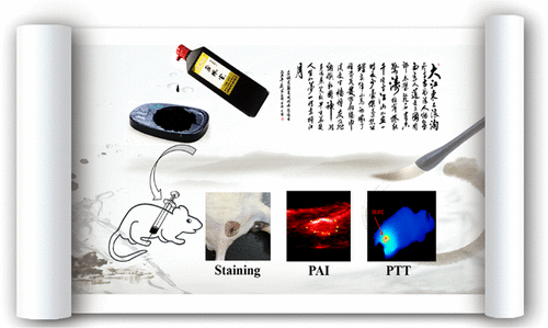
Calligraphy ink and cancer treatment

Courtesy of ACS Omega and the researchers

Nice illustration! I wish I could credit the artist. For anyone who needs a little text to make sense of it, there’s a Sept. 27, 2017 news item on Nanowerk (Note: A link has been removed),

For hundreds of years, Chinese calligraphers have used a plant-based ink to create beautiful messages and art. Now, one group reports in ACS Omega (“New Application of Old Material: Chinese Traditional Ink for Photothermal Therapy of Metastatic Lymph Nodes”) that this ink could noninvasively and effectively treat cancer cells that spread, or metastasize, to lymph nodes.

A Sept. 27, 2017 American Chemical Society (ACS) news release, which originated the news item, reveals more about the research,

As cancer cells leave a tumor, they frequently make their way to lymph nodes, which are part of the immune system. In this case, the main treatment option is surgery, but this can result in complications. Photothermal therapy (PTT) is an emerging noninvasive treatment option in which nanomaterials are injected and accumulate in cancer cells. A laser heats up the nanomaterials, and this heat kills the cells. Many of these nanomaterials are expensive, difficult-to-make and toxic. However, a traditional Chinese ink called Hu-Kaiwen ink (Hu-ink) has similar properties to the nanomaterials used in PTT. For example, they are the same color, and are both carbon-based and stable in water. So Wuli Yang and colleagues wanted to see if Hu-ink could be a good alternative material for PTT.

The researchers analyzed Hu-ink and found that it consists of nanoparticles and thin layers of carbon. When Hu-ink was heated with a laser, its temperature rose by 131 degrees Fahrenheit, much higher than current nanomaterials. Under PPT conditions, the Hu-ink killed cancer cells in a laboratory dish, but under normal conditions, the ink was non-toxic. This was also the scenario observed in mice with tumors. The researchers also noted that Hu-ink could act as a probe to locate tumors and metastases because it absorbs near-infrared light, which goes through skin.

Being a little curious about Hu-ink’s similarity to nanomaterial, I looked for more detail in the the paper (Note: Links have been removed), From the: Introduction,

Photothermal therapy (PTT) is an emerging tumor treatment strategy, which utilizes hyperthermia generated from absorbed near-infrared (NIR) light energy by photoabsorbing agents to kill tumor cells.(7-13) Different from chemotherapy, surgical treatment, and radiotherapy, PTT is noninvasive and more efficient.(7, 14, 15) In the past decade, PTT with diverse nanomaterials to eliminate cancer metastases lymph nodes has attracted extensive attention by several groups, including our group.(3, 16-20) For instance, Liu and his co-workers developed a treatment method based on PEGylated single-walled carbon nanotubes for PTT of tumor sentinel lymph nodes and achieved remarkably improved treatment effect in an animal tumor model.(21) To meet the clinical practice, the potential metastasis of deeper lymph nodes was further ablated in our previous work, using magnetic graphene oxide as a theranostic agent.(22) However, preparation of these artificial nanomaterials usually requires high cost, complicated synthetic process, and unavoidably toxic catalyst or chemicals,(23, 24) which impede their future clinical application. For the clinical application, exploring an environment-friendly material with simple preparation procedure, good biocompatibility, and excellent therapeutic efficiency is still highly desired. [emphases mine]

From the: Preparation and Characterization of Hu-Ink

To obtain an applicable sample, the condensed Hu-ink was first diluted into aqueous dispersion with a lower concentration. The obtained Hu-ink dispersion without any further treatment was black in color and stable in physiological environment, including water, phosphate-buffered saline (PBS), and Roswell Park Memorial Institute (RPMI) 1640; furthermore, no aggregation was observed even after keeping undisturbed for 3 days (Figure 2a). The nanoscaled morphology of Hu-ink was examined by transmission electron microscopy (TEM) (Figure 2b), which demonstrates that Hu-ink mainly exist in the form of small aggregates. These small aggregates consist of a few nanoparticles with diameter of about 20–50 nm. Dynamic light scattering (DLS) measurement (Figure 2c) further shows that Hu-ink aqueous dispersion possesses a hydrodynamic diameter of about 186 nm (polydispersity index: 0.18), which was a crucial prerequisite for biomedical applications.(29) In the X-ray diffraction (XRD) pattern, no other characteristic peaks are found except carbon peak (Figure S1, Supporting Information), which confirms that the main component of Hu-ink is carbon.(25) Raman spectroscopy was a common tool to characterize graphene-related materials.(30) D band (∼1300 cm–1, corresponding to the defects) and G band (∼1600 cm–1, related to the sp2 carbon sites) peaks could be observed in Figure 2d with the ratio ID/IG = 0.96, which confirms the existence of graphene sheetlike structure in Hu-ink.(31) The UV–vis–NIR spectra (Figure 2e) also revealed that Hu-ink has high absorption in the NIR region around 650–900 nm, in which hemoglobin and water, the major absorbers of biological tissue, have their lowest absorption coefficient.(32) The high NIR absorption capability of Hu-ink encouraged us to investigate its photothermal properties.(33-35) Hu-ink dispersions with different concentrations were irradiated under an 808 nm laser (the commercial and widely used wavelength in photothermal therapy).(8-13) [emphases mine]

Curiosity satisfied! For those who’d like to investigate even further, here’s a link to and a citation for the paper,

New Application of Old Material: Chinese Traditional Ink for Photothermal Therapy of Metastatic Lymph Nodes by Sheng Wang, Yongbin Cao, Qin Zhang, Haibao Peng, Lei Liang, Qingguo Li, Shun Shen, Aimaier Tuerdi, Ye Xu, Sanjun Cai, and Wuli Yang. ACS Omega, 2017, 2 (8), pp 5170–5178 DOI: 10.1021/acsomega.7b00993 Publication Date (Web): August 30, 2017

Copyright © 2017 American Chemical Society

This paper appears to be open access.

Unboiling egg technology can cut through carbon nanotubes

One of 2015’s big science stories, Flinders University’s ‘egg unboiler’ (also known as, a vortex fluidic device). has made the news again in a March 11, 2016 news item on phys.org,

Technology used by scientists to unboil an egg is being adapted to precisely cut through carbon nanotubes used in solar panel manufacturing and cancer treatment.

Scientists from Flinders University in South Australia have proven their Vortex Fluidic Device’s ability to slice through carbon nanotubes with great precision.

A March 11, 2016 story written by Caleb Radford for The Lead, which originated the news item, notes the advantages to using this technology for slicing carbon nanotubes (CNTs) and prospects for commercialization,

Device creator and Flinders University Professor Colin Raston said the carbon nanotubes could be commercialised within 12 months.

“Importantly for this technology is that we have uniformity in products,” he said.

“It opens it up for applications in drug delivery if you can get all of the carbon nanotubes to about 100 nanometres … 100 nanometres is the ideal length for getting into tumours so you can actually functionalise them to target cancer cells.

“Uniformity in products also means that you can improve the solar cell efficiency in solar cell devices.”

Flinders University scientists last year were awarded an Ig Nobel Award for creating the Vortex Fluidic Device and using it to unboil an egg.

The device can also be used to slice CNTs accurately to an average length of 170 nanometres using only water, a solvent and a laser.

It is also a simpler and cheaper process than previous methods, which resulted in random lengths that made it difficult to deliver drugs to patients and transfer electrons for solar panel manufacturing.

Flinders University PhD student Kasturi Vimalanathan, who played a key role in discovering new applications for the device, said the machines ability to cut carbon nanotubes to a similar length significantly increased the efficiency of solar cells.

“They shorten the carbon nanotubes to fit in all the chemicals so it can withstand high temperatures,” she said.

“It increases the efficiency and enhances the photoelectric conversion because they can provide a shorter transportation pathway for these electrons.

“It’s a one step method we can scale up. We can see cheaper solar panels on the back of this development.”

Here’s an image of Ralston, presumably with his Vortex Fluidic Device,

Professor Colin Raston received global attention and won an Ig Nobel prize on his way to becoming one of the biggest science stories of 2015. Courtesy Flinders University, Australia

Professor Colin Raston received global attention and won an Ig Nobel prize on his way to becoming one of the biggest science stories of 2015. Courtesy Flinders University, Australia

A Dec. 18, 2015 Flinders University blog posting announced Ralston’s Ig Nobel,

When Flinders University’s Professor Colin Raston unboiled an egg earlier this year with his ‘Vortex Fluidic Device’, in a feat previously considered impossible by science, he made TV screens and front pages all over the world, generating a veritable tsunami of ‘eggscellent’ puns.

The global impact of his achievement transformed the softly spoken South Australia Premier’s Professorial Research Fellow in Clean Technology into an internationally recognised figure overnight – and culminated in him receiving a prestigious Ig Nobel prize in September [2015].

In recognition of the massive amount of attention Professor Raston’s achievement received for Australian research, it has today been hailed as one of the top ten weird and wonderful Australian science stories of 2015 by the Australian Science and Media Centre (AusSMC).

Responding to the announcement, Professor Raston said he had been thrilled with the response to his achievement and had been ‘living the dream’ since.

“We were very interested in how the Vortex Fluidic Device might control protein folding, and the breakthrough with my collaborator at UCI, Greg Weiss, simplifies this, in a fraction of the time, minimising waste generation and energy usage. What this amounted to was unboiling an egg,” he said.

Who would have thought a device for unboiling eggs could be used to cut carbon nanotubes? Clearly, Kasturi Vimalanathan. Amazing.

For anyone interested and/or unfamiliar with the Ig Nobel prizes, there’s my Sept. 17, 2013 posting.

A city of science in Japan: Kawasaki (Kanagawa)

Happily, I’m getting more nanotechnology (for the most part) information from Japan. Given Japan’s prominence in this field of endeavour I’ve long felt FrogHeart has not adequately represented Japanese contributions. Now that I’m receiving English language translations, I hope to better address the situation.

This morning (March 26, 2015), there were two news releases from Kawasaki INnovation Gateway at SKYFRONT (KING SKYFRONT), Coastal Area International Strategy Office, Kawasaki City, Japan in my mailbox. Before getting on to the news releases, here’s a little about  the city of Kawasaki and about its innovation gateway. From the Kawasaki, Kanagawa entry in Wikipedia (Note: Links have been removed),

Kawasaki (川崎市 Kawasaki-shi?) is a city in Kanagawa Prefecture, Japan, located between Tokyo and Yokohama. It is the 9th most populated city in Japan and one of the main cities forming the Greater Tokyo Area and Keihin Industrial Area.

Kawasaki occupies a belt of land stretching about 30 kilometres (19 mi) along the south bank of the Tama River, which divides it from Tokyo. The eastern end of the belt, centered on JR Kawasaki Station, is flat and largely consists of industrial zones and densely built working-class housing, the Western end mountainous and more suburban. The coastline of Tokyo Bay is occupied by vast heavy industrial complexes built on reclaimed land.

There is a 2014 video about Kawasaki’s innovation gateway, which despite its 14 mins. 39 secs. running time I am embedding here. (Caution: They highlight their animal testing facility at some length.)

Now on to the two news releases. The first concerns research on gold nanoparticles that was published in 2014. From a March 26, 2015 Kawasaki INnovation Gateway news release,

Gold nanoparticles size up to cancer treatment

Incorporating gold nanoparticles helps optimise treatment carrier size and stability to improve delivery of cancer treatment to cells.

Treatments that attack cancer cells through the targeted silencing of cancer genes could be developed using small interfering RNA molecules (siRNA). However delivering the siRNA into the cells intact is a challenge as it is readily degraded by enzymes in the blood and small enough to be eliminated from the blood stream by kidney filtration.  Now Kazunori Kataoka at the University of Tokyo and colleagues at Tokyo Institute of Technology have designed a protective treatment delivery vehicle with optimum stability and size for delivering siRNA to cells.

The researchers formed a polymer complex with a single siRNA molecule. The siRNA-loaded complex was then bonded to a 20 nm gold nanoparticle, which thanks to advances in synthesis techniques can be produced with a reliably low size distribution. The resulting nanoarchitecture had the optimum overall size – small enough to infiltrate cells while large enough to accumulate.

In an assay containing heparin – a biological anti-coagulant with a high negative charge density – the complex was found to release the siRNA due to electrostatic interactions. However when the gold nanoparticle was incorporated the complex remained stable. Instead, release of the siRNA from the complex with the gold nanoparticle could be triggered once inside the cell by the presence of glutathione, which is present in high concentrations in intracellular fluid. The glutathione bonded with the gold nanoparticles and the complex, detaching them from each other and leaving the siRNA prone to release.

The researchers further tested their carrier in a subcutaneous tumour model. The authors concluded that the complex bonded to the gold nanoparticle “enabled the efficient tumor accumulation of siRNA and significant in vivo gene silencing effect in the tumor, demonstrating the potential for siRNA-based cancer therapies.”

The news release provides links to the March 2015 newsletter which highlights this research and to the specific article and video,

March 2015 Issue of Kawasaki SkyFront iNewsletter: http://inewsletter-king-skyfront.jp/en/

Contents

Feature video on Professor Kataoka’s research : http://inewsletter-king-skyfront.jp/en/video_feature/vol_3/feature01/

Research highlights: http://inewsletter-king-skyfront.jp/en/research_highlights/vol_3/research01/

Here’s a link to and a citation for the paper,

Precise Engineering of siRNA Delivery Vehicles to Tumors Using Polyion Complexes and Gold Nanoparticles by Hyun Jin Kim, Hiroyasu Takemoto, Yu Yi, Meng Zheng, Yoshinori Maeda, Hiroyuki Chaya, Kotaro Hayashi, Peng Mi, Frederico Pittella, R. James Christie, Kazuko Toh, Yu Matsumoto, Nobuhiro Nishiyama, Kanjiro Miyata, and Kazunori Kataoka. ACS Nano, 2014, 8 (9), pp 8979–8991 DOI: 10.1021/nn502125h Publication Date (Web): August 18, 2014
Copyright © 2014 American Chemical Society

This article is behind a paywall.

The second March 26, 2015 Kawasaki INnovation Gateway news release concerns a DNA chip and food-borne pathogens,

Rapid and efficient DNA chip technology for testing 14 major types of food borne pathogens

Conventional methods for testing food-borne pathogens is based on the cultivation of pathogens, a process that is complicated and time consuming. So there is demand for alternative methods to test for food-borne pathogens that are simpler, quick and applicable to a wide range of potential applications.

Now Toshiba Ltd and Kawasaki City Institute for Public Health have collaborated in the development of a rapid and efficient automatic abbreviated DNA detection technology that can test for 14 major types of food borne pathogens. The so called ‘DNA chip card’ employs electrochemical DNA chips and overcomes the complicated procedures associated with genetic testing of conventional methods. The ‘DNA chip card’ is expected to find applications in hygiene management in food manufacture, pharmaceuticals, and cosmetics.

Details

The so-called automatic abbreviated DNA detection technology ‘DNA chip card’ was developed by Toshiba Ltd and in a collaboration with Kawasaki City Institute for Public Health, used to simultaneously detect 14 different types of food-borne pathogens in less than 90 minutes. The detection sensitivity depends on the target pathogen and has a range of 1E+01~05 cfu/mL.

Notably, such tests would usually take 4-5 days using conventional methods based on pathogen cultivation. Furthermore, in contrast to conventional DNA protocols that require high levels of skill and expertise, the ‘DNA chip card’ only requires the operator to inject nucleic acid, thereby making the procedure easier to use and without specialized operating skills.

Examples of pathogens associated with food poisoning that were tested with the “DNA chip card”

Enterohemorrhagic Escherichia coli

Salmonella

Campylobacter

Vibrio parahaemolyticus

Shigella

Staphylococcus aureus

Enterotoxigenic Escherichia coli

Enteroaggregative Escherichia coli

Enteropathogenic Escherichia coli

Clostridium perfringens

Bacillus cereus

Yersinia

Listeria

Vibrio cholerae

I think 14 is the highest number of tests I’ve seen for one of these chips. This chip is quite an achievement.

One final bit from the news release about the DNA chip provides a brief description of the gateway and something they call King SkyFront,

About KING SKYFRONT

The Kawasaki INnovation Gateway (KING) SKYFRONT is the flagship science and technology innovation hub of Kawasaki City. KING SKYFRONT is a 40 hectare area located in the Tonomachi area of the Keihin Industrial Region that spans Tokyo and Kanagawa Prefecture and Tokyo International Airport (also often referred to as Haneda Airport).

KING SKYFRONT was launched in 2013 as a base for scholars, industrialists and government administrators to work together to devise real life solutions to global issues in the life sciences and environment.

I find this emphasis on the city interesting. It seems that cities are becoming increasingly important and active where science research and development are concerned. Europe seems to have adopted a biannual event wherein a city is declared a European City of Science in conjunction with the EuroScience Open Forum (ESOF) conferences. The first such city was Dublin in 2012 (I believe the Irish came up with the concept themselves) and was later adopted by Copenhagen for 2014. The latest city to embrace the banner will be Manchester in 2016.

Bees and smart bombs

I wasn’t expecting to think about bees and bombs again (after my July 28, 2010 posting on science knitting and yarn bombing respectively) but then this news item, Novel bee venom derivative forms a nanoparticle ‘smart bomb’ to target cancer cells, popped up on Nanowerk last week. From the news item,

The next time you are stung by a bee, here’s some consolation: a toxic protein in bee venom, when altered, significantly improves the effectiveness liposome-encapsulated drugs or dyes, such as those already used to treat or diagnose cancer. This research [Samuel Wickline], described in the August 2010 print issue of the FASEB [Federation of American Societies for Experimental Biology] Journal, shows how modified melittin may revolutionize treatments for cancer and perhaps other conditions, such as arthritis, cardiovascular disease, and serious infections.

I gather the joournal’s editor is also experiencing these coincidences,

“Our journal is abuzz in a hive of bee-related discoveries. Just last month, we published research showing for the first time how honey kills bacteria. This month, the Wickline study shows how bee venom peptides can form “smart bombs” that deliver liposomal nanoparticles directly to their target, without collateral damage,” said Gerald Weissmann, M.D., Editor-in-Chief of the FASEB Journal.

That’s a lot of military jargon.