Tag Archives: Raman spectroscopy

Canadian research into nanomaterial workplace exposure in the air and on surfaces

An August 30, 2018 news item on Nanowerk announces the report,

The monitoring of air contamination by engineered nanomaterials (ENM) is a complex process with many uncertainties and limitations owing to the presence of particles of nanometric size that are not ENMs, the lack of validated instruments for breathing zone measurements and the many indicators to be considered.

In addition, some organizations, France’s Institut national de recherche et de sécurité (INRS) and Québec’s Institut de recherche Robert-Sauvé en santé et en sécurité du travail (IRSST) among them, stress the need to also sample surfaces for ENM deposits.

In other words, to get a better picture of the risks of worker exposure, we need to fine-tune the existing methods of sampling and characterizing ENMs and develop new one. Accordingly, the main goal of this project was to develop innovative methodological approaches for detailed qualitative as well as quantitative characterization of workplace exposure to ENMs.

A PDF of the 88-page report is available in English or in French.

An August 30, 2018 (?) abstract of the IRSST report titled An Assessment of Methods of Sampling and Characterizing Engineered Nanomaterials in the Air and on Surfaces in the Workplace (2nd edition) by Maximilien Debia, Gilles L’Espérance, Cyril Catto, Philippe Plamondon, André Dufresne, Claude Ostiguy, which originated the news item, outlines what you can expect from the report,

This research project has two complementary parts: a laboratory investigation and a fieldwork component. The laboratory investigation involved generating titanium dioxide (TiO2) nanoparticles under controlled laboratory conditions and studying different sampling and analysis devices. The fieldwork comprised a series of nine interventions adapted to different workplaces and designed to test a variety of sampling devices and analytical procedures and to measure ENM exposure levels among Québec workers.

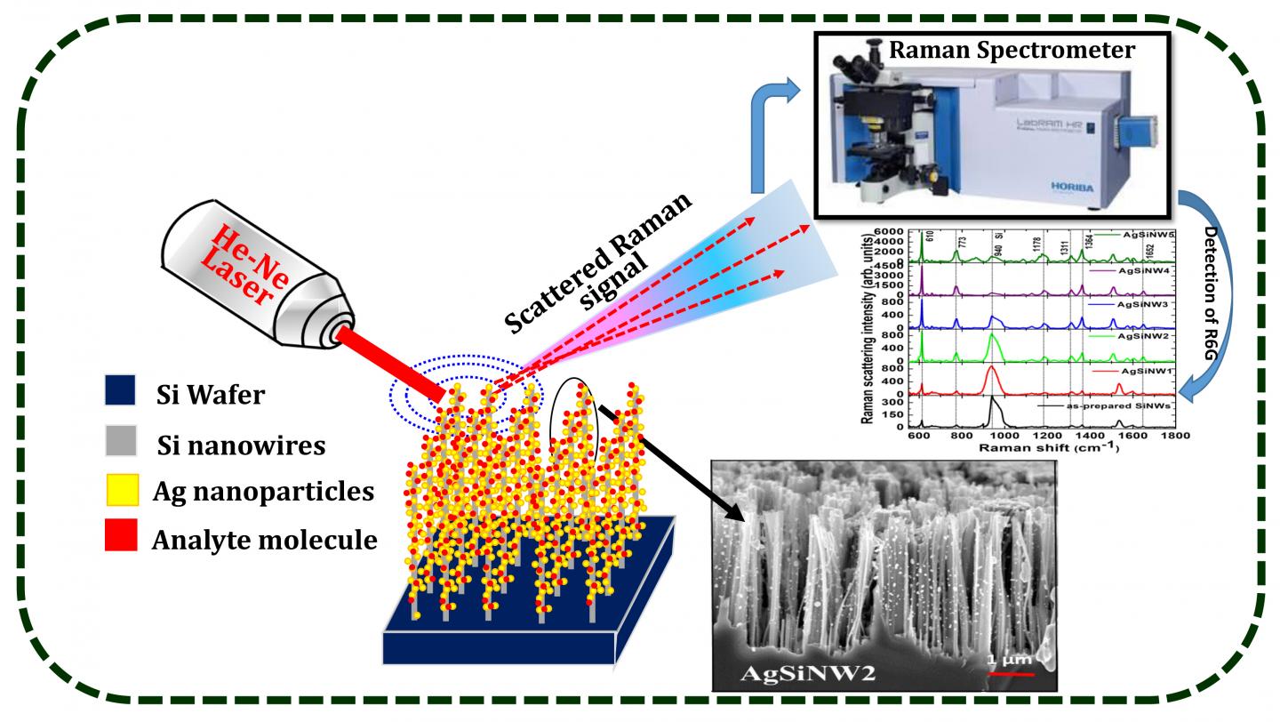
The methods for characterizing aerosols and surface deposits that were investigated include: i) measurement by direct-reading instruments (DRI), such as condensation particle counters (CPC), optical particle counters (OPC), laser photometers, aerodynamic diameter spectrometers and electric mobility spectrometer; ii) transmission electron microscopy (TEM) or scanning transmission electron microscopy (STEM) with a variety of sampling devices, including the Mini Particle Sampler® (MPS); iii) measurement of elemental carbon (EC); iv) inductively coupled plasma mass spectrometry (ICP-MS) and (v) Raman spectroscopy.

The workplace investigations covered a variety of industries (e.g., electronics, manufacturing, printing, construction, energy, research and development) and included producers as well as users or integrators of ENMs. In the workplaces investigated, we found nanometals or metal oxides (TiO2, SiO2, zinc oxides, lithium iron phosphate, titanate, copper oxides), nanoclays, nanocellulose and carbonaceous materials, including carbon nanofibers (CNF) and carbon nanotubes (CNT)—single-walled (SWCNT) as well as multiwalled (MWCNT).

The project helped to advance our knowledge of workplace assessments of ENMs by documenting specific tasks and industrial processes (e.g., printing and varnishing) as well as certain as yet little investigated ENMs (nanocellulose, for example).

Based on our investigations, we propose a strategy for more accurate assessment of ENM exposure using methods that require a minimum of preanalytical handling. The recommended strategy is a systematic two-step assessment of workplaces that produce and use ENMs. The first step involves testing with different DRIs (such as a CPC and a laser photometer) as well as sample collection and subsequent microscopic analysis (MPS + TEM/STEM) to clearly identify the work tasks that generate ENMs. The second step, once work exposure is confirmed, is specific quantification of the ENMs detected. The following findings are particularly helpful for detailed characterization of ENM exposure:

  1. The first conclusive tests of a technique using ICP-MS to quantify the metal oxide content of samples collected in the workplace
  2. The possibility of combining different sampling methods recommended by the National Institute for Occupational Safety and Health (NIOSH) to measure elemental carbon as an indicator of NTC/NFC, as well as demonstration of the limitation of this method stemming from observed interference with the black carbon particles required to synthesis carbon materials (for example, Raman spectroscopy showed that less than 6% of the particles deposited on the electron microscopy grid at one site were SWCNTs)
  3. The clear advantages of using an MPS (instead of the standard 37-mm cassettes used as sampling media for electron microscopy), which allows quantification of materials
  4. The major impact of sampling time: a long sampling time overloads electron microscopy grids and can lead to overestimation of average particle agglomerate size and underestimation of particle concentrations
  5. The feasibility and utility of surface sampling, either with sampling pumps or passively by diffusion onto the electron microscopy grids, to assess ENM dispersion in the workplace

These original findings suggest promising avenues for assessing ENM exposure, while also showing their limitations. Improvements to our sampling and analysis methods give us a better understanding of ENM exposure and help in adapting and implementing control measures that can minimize occupational exposure.

You can download the full report in either or both English and French from the ‘Nanomaterials – A Guide to Good Practices Facilitating Risk Management in the Workplace, 2nd Edition‘ webpage.

Taking spectroscopy to a new dimension with silver nanoparticles

This latest move towards better detection at the nanoscale comes from India (from a January 2, 2018 news item on ScienceDaily),

As medicine and pharmacology investigate nanoscale processes, it has become increasingly important to identify and characterize different molecules. Raman spectroscopy, a technique that leverages the scattering of laser light to identify molecules, has a limited capacity to detect molecules in diluted samples because of low signal yield.

A team of researchers from the University of Hyderabad in India has improved molecular detection at low concentration levels by arranging nanoparticles on nanowires to enhance Raman spectroscopy. Surface-enhanced Raman spectroscopy (SERS) uses electromagnetic fields to improve Raman scattering and boost sensitivity in standard dyes such as R6G by more than one billionfold.

Here’s an image illustrating the work,

Caption: Detection of a low concentration analyte molecule using silicon nanowires decorated with silver nanoparticles and surface enhanced Raman scattering measurements. Credit: V.S. Vendamani

A January 2, 2017 American Institute of Physics press release on EurekAlert, which originated the news item, explains further,

The team decorated vertically aligned silicon nanowires with varying densities of silver nanoparticles, utilizing and enhancing the structure’s 3-D shape. Their results, published in the Journal of Applied Physics, from AIP [American Institute of Physics] Publishing, show that their device was able to enhance the Raman signals for cytosine protein and ammonium perchlorate by a factor of 100,000.

“The beauty is that we can improve the density of these nanowires using simple chemistry,” said Soma Venugopal Rao, one of the paper’s authors. “If you have a large density of nanowires, you can put more silver nanoparticles into the substrate and can increase the sensitivity of the substrate.”

Applying the necessary nanostructures to SERS devices remains a challenge for the field. Building these structures in three dimensions with silicon nanowires has garnered attention for their higher surface area and superior performance, but silicon nanowires are still expensive to produce.

Instead, the team was able to find a cheaper way to make silicon nanowires and used a technique called electroless etching to make a wide range of nanowires. They “decorated” these wires with silver nanoparticles with variable and controlled densities, which increased the nanowires’ surface area.

“Optimizing these vertically aligned structures took a lot of time in the beginning,” said Nageswara Rao, another of the paper’s authors. “We increased the surface area and to do this we needed to change the aspect ratio.”

After optimizing their system to detect Rhodamine dye on a nanomolar level, these new substrates the team built enhanced Raman sensitivity by a factor of 10,000 to 100,000. The substrates detected concentrations of cytosine, a nucleotide found in DNA, and ammonium perchlorate, a molecule with potential for detecting explosives, in as dilute concentrations as 50 and 10 micromolar, respectively.

The results have given the team reason to believe that it might soon be possible to detect compounds in concentrations on the scale of nanomolar or even picomolar, Nageswara Rao said. The team’s work has opened several avenues for future research, from experimenting with different nanoparticles such as gold, increasing the sharpness of the nanowires or testing these devices across several types of molecules.

Here’s a link to and a citation for the paper,

Three-dimensional hybrid silicon nanostructures for surface enhanced Raman spectroscopy based molecular detection featured by V. S. Vendamani, S. V. S. Nageswara Rao, S. Venugopal Rao, D. Kanjilal, and A. P. Pathak. Journal of Applied Physics 123, 014301 (2018); Published Online: January 2018 https://doi.org/10.1063/1.5000994

This paper is open access.

Calligraphy ink and cancer treatment

Courtesy of ACS Omega and the researchers

Nice illustration! I wish I could credit the artist. For anyone who needs a little text to make sense of it, there’s a Sept. 27, 2017 news item on Nanowerk (Note: A link has been removed),

For hundreds of years, Chinese calligraphers have used a plant-based ink to create beautiful messages and art. Now, one group reports in ACS Omega (“New Application of Old Material: Chinese Traditional Ink for Photothermal Therapy of Metastatic Lymph Nodes”) that this ink could noninvasively and effectively treat cancer cells that spread, or metastasize, to lymph nodes.

A Sept. 27, 2017 American Chemical Society (ACS) news release, which originated the news item, reveals more about the research,

As cancer cells leave a tumor, they frequently make their way to lymph nodes, which are part of the immune system. In this case, the main treatment option is surgery, but this can result in complications. Photothermal therapy (PTT) is an emerging noninvasive treatment option in which nanomaterials are injected and accumulate in cancer cells. A laser heats up the nanomaterials, and this heat kills the cells. Many of these nanomaterials are expensive, difficult-to-make and toxic. However, a traditional Chinese ink called Hu-Kaiwen ink (Hu-ink) has similar properties to the nanomaterials used in PTT. For example, they are the same color, and are both carbon-based and stable in water. So Wuli Yang and colleagues wanted to see if Hu-ink could be a good alternative material for PTT.

The researchers analyzed Hu-ink and found that it consists of nanoparticles and thin layers of carbon. When Hu-ink was heated with a laser, its temperature rose by 131 degrees Fahrenheit, much higher than current nanomaterials. Under PPT conditions, the Hu-ink killed cancer cells in a laboratory dish, but under normal conditions, the ink was non-toxic. This was also the scenario observed in mice with tumors. The researchers also noted that Hu-ink could act as a probe to locate tumors and metastases because it absorbs near-infrared light, which goes through skin.

Being a little curious about Hu-ink’s similarity to nanomaterial, I looked for more detail in the the paper (Note: Links have been removed), From the: Introduction,

Photothermal therapy (PTT) is an emerging tumor treatment strategy, which utilizes hyperthermia generated from absorbed near-infrared (NIR) light energy by photoabsorbing agents to kill tumor cells.(7-13) Different from chemotherapy, surgical treatment, and radiotherapy, PTT is noninvasive and more efficient.(7, 14, 15) In the past decade, PTT with diverse nanomaterials to eliminate cancer metastases lymph nodes has attracted extensive attention by several groups, including our group.(3, 16-20) For instance, Liu and his co-workers developed a treatment method based on PEGylated single-walled carbon nanotubes for PTT of tumor sentinel lymph nodes and achieved remarkably improved treatment effect in an animal tumor model.(21) To meet the clinical practice, the potential metastasis of deeper lymph nodes was further ablated in our previous work, using magnetic graphene oxide as a theranostic agent.(22) However, preparation of these artificial nanomaterials usually requires high cost, complicated synthetic process, and unavoidably toxic catalyst or chemicals,(23, 24) which impede their future clinical application. For the clinical application, exploring an environment-friendly material with simple preparation procedure, good biocompatibility, and excellent therapeutic efficiency is still highly desired. [emphases mine]

From the: Preparation and Characterization of Hu-Ink

To obtain an applicable sample, the condensed Hu-ink was first diluted into aqueous dispersion with a lower concentration. The obtained Hu-ink dispersion without any further treatment was black in color and stable in physiological environment, including water, phosphate-buffered saline (PBS), and Roswell Park Memorial Institute (RPMI) 1640; furthermore, no aggregation was observed even after keeping undisturbed for 3 days (Figure 2a). The nanoscaled morphology of Hu-ink was examined by transmission electron microscopy (TEM) (Figure 2b), which demonstrates that Hu-ink mainly exist in the form of small aggregates. These small aggregates consist of a few nanoparticles with diameter of about 20–50 nm. Dynamic light scattering (DLS) measurement (Figure 2c) further shows that Hu-ink aqueous dispersion possesses a hydrodynamic diameter of about 186 nm (polydispersity index: 0.18), which was a crucial prerequisite for biomedical applications.(29) In the X-ray diffraction (XRD) pattern, no other characteristic peaks are found except carbon peak (Figure S1, Supporting Information), which confirms that the main component of Hu-ink is carbon.(25) Raman spectroscopy was a common tool to characterize graphene-related materials.(30) D band (∼1300 cm–1, corresponding to the defects) and G band (∼1600 cm–1, related to the sp2 carbon sites) peaks could be observed in Figure 2d with the ratio ID/IG = 0.96, which confirms the existence of graphene sheetlike structure in Hu-ink.(31) The UV–vis–NIR spectra (Figure 2e) also revealed that Hu-ink has high absorption in the NIR region around 650–900 nm, in which hemoglobin and water, the major absorbers of biological tissue, have their lowest absorption coefficient.(32) The high NIR absorption capability of Hu-ink encouraged us to investigate its photothermal properties.(33-35) Hu-ink dispersions with different concentrations were irradiated under an 808 nm laser (the commercial and widely used wavelength in photothermal therapy).(8-13) [emphases mine]

Curiosity satisfied! For those who’d like to investigate even further, here’s a link to and a citation for the paper,

New Application of Old Material: Chinese Traditional Ink for Photothermal Therapy of Metastatic Lymph Nodes by Sheng Wang, Yongbin Cao, Qin Zhang, Haibao Peng, Lei Liang, Qingguo Li, Shun Shen, Aimaier Tuerdi, Ye Xu, Sanjun Cai, and Wuli Yang. ACS Omega, 2017, 2 (8), pp 5170–5178 DOI: 10.1021/acsomega.7b00993 Publication Date (Web): August 30, 2017

Copyright © 2017 American Chemical Society

This paper appears to be open access.

Functional hybrid system that can connect human tissue with electronic devices

I’ve tagged this particular field of interest ‘machine/flesh’ because I find it more descriptive than ‘bio-hybrid system’ which was the term used in a Nov. 15, 2016 news item on phys.org,

One of the biggest challenges in cognitive or rehabilitation neurosciences is the ability to design a functional hybrid system that can connect and exchange information between biological systems, like neurons in the brain, and human-made electronic devices. A large multidisciplinary effort of researchers in Italy brought together physicists, chemists, biochemists, engineers, molecular biologists and physiologists to analyze the biocompatibility of the substrate used to connect these biological and human-made components, and investigate the functionality of the adhering cells, creating a living biohybrid system.

A Nov.15, 2016 American Institute of Physics news release on EurekAlert, which originated the news item, details the investigation,

In an article appearing this week in AIP Advances, from AIP Publishing, the research team used the interaction between light and matter to investigate the material properties at the molecular level using Raman spectroscopy, a technique that, until now, has been principally applied to material science. Thanks to the coupling of the Raman spectrometer with a microscope, spectroscopy becomes a useful tool for investigating micro-objects such as cells and tissues. Raman spectroscopy presents clear advantages for this type of investigation: The molecular composition and the modi?cation of subcellular compartments can be obtained in label-free conditions with non-invasive methods and under physiological conditions, allowing the investigation of a large variety of biological processes both in vitro and in vivo.

Once the biocompatibility of the substrate was analyzed and the functionality of the adhering cells investigated, the next part of this puzzle is connecting with the electronic component. In this case a memristor was used.

“Its name reveals its peculiarity (MEMory ResISTOR), it has a sort of “memory”: depending on the amount of voltage that has been applied to it in the past, it is able to vary its resistance, because of a change of its microscopic physical properties,” said Silvia Caponi, a physicist at the Italian National Research Council in Rome. By combining memristors, it is possible to create pathways within the electrical circuits that work similar to the natural synapses, which develop variable weight in their connections to reproduce the adaptive/learning mechanism. Layers of organic polymers, like polyaniline (PANI) a semiconductor polymer, also have memristive properties, allowing them to work directly with biological materials into a hybrid bio-electronic system.

“We applied the analysis on a hybrid bio-inspired device but in a prospective view, this work provides the proof of concept of an integrated study able to analyse the status of living cells in a large variety of applications that merges nanosciences, neurosciences and bioelectronics,” said Caponi. A natural long-term objective of this work would be interfacing machines and nervous systems as seamlessly as possible.

The multidisciplinary team is ready to build on this proof of principle to realize the potential of memristor networks.

“Once assured the biocompatibility of the materials on which neurons grow,” said Caponi, “we want to define the materials and their functionalization procedures to find the best configuration for the neuron-memristor interface to deliver a full working hybrid bio-memristive system.”

Caption: These are immunofluorescence analysis of SH-SY5Y cells treated for 5 days with 10uM Retinoic Acid and 50ng/ml BDNF for the next 3 days. The DAPI fluorescence stain is blue and Beta-tubulin is green. Credit: Caponi, et al.

Caption: These are immunofluorescence analysis of SH-SY5Y cells treated for 5 days with 10uM Retinoic Acid and 50ng/ml BDNF for the next 3 days. The DAPI fluorescence stain is blue and Beta-tubulin is green. Credit: Caponi, et al.

Here’s a link to and a citation for the paper,

A multidisciplinary approach to study the functional properties of neuron-like cell models constituting a living bio-hybrid system: SH-SY5Y cells adhering to PANI substrate by S. Caponi, S. Mattana, M. Ricci, K. Sagini, L. J. Juarez-Hernandez, A. M. Jimenez-Garduño, N. Cornella, L. Pasquardini, L. Urbanelli, P. Sassi, A. Morresi, C. Emiliani, D. Fioretto, M. Dalla Serra, C. Pederzolli, S. Iannotta, P. Macchi, and C. Musio. AIP Advances 6, 111303 (2016); http://dx.doi.org/10.1063/1.4966587

This paper appears to be open access.

I know something about your mummy or an ion beam microscope analyses a sarcophagus scrap

“Bearded Man, 170-180 A.D.” from the Walters Art Museum collection, object #32.6

“Bearded Man, 170-180 A.D.” from the Walters Art Museum collection, object #32.6

An Aug. 14, 2014 news item on Azonano describing this image sparks the imagination,

He looks almost Byzantine or Greek, gazing doe-eyed over the viewer’s left shoulder, his mouth forming a slight pout, like a star-struck lover or perhaps a fan of the races witnessing his favorite charioteer losing control of his horses.

In reality, he’s the “Bearded Man, 170-180 A.D.,” a Roman-Egyptian whose portrait adorned the sarcophagus sheltering his mummified remains. But the details of who he was and what he was thinking have been lost to time.

But perhaps not for much longer. A microscopic sliver of painted wood could hold the keys to unraveling the first part of this centuries-old mystery. Figuring out what kind of pigment was used (whether it was a natural matter or a synthetic pigment mixed to custom specifications), and the exact materials used to create it, could help scientists unlock his identity.

Kathleen Tuck’s Aug. 13 (?), 2014 Boise State University (Idaho, US) news release, which originated the news item, describes the nature of the research and the difficulties associated with it,

“Understanding the pigment means better understanding of the provenance of the individual” said Darryl Butt, a Boise State distinguished professor in the Department of Materials Science and Engineering and associate director of the Center for Advanced Energy Studies (CAES). “Where the pigment came from may connect it to a specific area and maybe even a family.”

For years, researchers were limited by the lack of samples large enough to be properly analyzed. But advances in the field of nanotechnology mean scientists now can work with fragments tinier than the eye can even register. Using a $1.5 million ion beam microscope at CAES, Butt — along with CAES colleagues Yaqaio Wu and Jatu Burns, and Boise State student researchers Gordon Alanko and Jennifer Watkins — is working with a sliver of the wood portrait smaller than a human hair.

The team transferred the fragment to a sample holder using a tiny deer hair called an “eyelash.” Their biggest challenge was to move it to the equipment without losing it.

So far they have extracted five needle-tip sized fragments 20 nanometers wide (a nanometer is a billionth of a meter), as well as two thin foils. From that, they have been able to analyze and map out the chemistry of the material in three dimensions.

Butt and his team are analyzing a speck of purple paint, which is significant because the blue used to blend the purple hue was a precious pigment back in the day, signaling a prominent individual.

This research is part of a larger project (from the news release),

Their data is being analyzed by researchers from the Detroit Museum of Art, where a companion to the “Bearded Man” mummy resides. It’s part of a project, titled APPEAR (Ancient Panel Paintings-Examination, Analysis, Research), a collaboration between 12 museums, including the British Museum in London and the Walters Art Museum in Baltimore, Maryland.

According to the news release there’s a personal aspect to Butt’s interest in this research, which may eventually have implications for Boise State University’s programmes,

“So far we’ve learned that the paint is a synthetic pigment,” said Butt, who as an artist in his own right often mixes his own pigments for his paintings. “These are very vibrant pigments, possibly heated in a lead crucible. People thought that process had been developed in the 1800s or so. This could prove it happened a lot earlier.

Butt got into solving art mysteries when he met Glenn Gates, a conservation scientist at the Walters Art Museum [Baltimore, Maryland] at a conference at Stanford University [California]. Both are officers of a new section of the American Ceramic Society — the Art, Archaeology and Conservation Science division.

“This research was a gamble that we [materials scientists] could do some really cool stuff,” Butt said, noting that he would love to branch out into analyzing pottery and other ancient artifacts.

While studying the provenance of Roman-Egyptian mummies is something new at Boise State, many researchers in the art, geology, history, anthropology and even English departments are involved in what Butt likes to call ‘reverse engineering’ of objects of cultural heritage.

“This particular problem, that is of understanding a particle of pigment from a 2,000-year-old sarcophagus, is a bit unique in that it highlights some of the amazing tools that we have at Boise State and at CAES that could shed new light on problems associated with understanding human history,” he said.

Butt hopes that these and similar transdisciplinary projects will open up external research opportunities for students, including creation of a “pipeline” of students who travel to various user facilities or museums to carry out interdisciplinary research.

“Envision, for example, art students studying works of art using synchrotron radiation and bright x-rays at a national laboratory, while science and engineering students use their technical skills to unravel mysteries of materials used by ancient societies in the field or held by museums,” he said.

The idea can sound far-fetched even for those who are participating in the research, although there is a certain, sound logic to transdisciplinary work between the arts and the sciences.

I was not able to find any reference to Butt’s art work online, find a published research paper or more information (website) featuring APPEAR ((Ancient Panel Paintings-Examination, Analysis, Research); admittedly, it was a brief search.

There are many techniques used to examine works of art and/or heritage. For a description of another technique, Raman spectroscopy, and its use in examining art pigments there’s my June 27, 2014 posting titled: Art (Lawren Harris and the Group of Seven), science (Raman spectroscopic examinations), and other collisions at the 2014 Canadian Chemistry Conference (part 2 of 4). Should you be interested in the entire series, additional links can be found in that posting.

Nanophotonics transforms Raman spectroscopy at Rice University (US)

This new technique for sensing molecules is intriguing. From a July 15, 2014 news item on Azonano,

Nanophotonics experts at Rice University [Texas, US] have created a unique sensor that amplifies the optical signature of molecules by about 100 billion times. Newly published tests found the device could accurately identify the composition and structure of individual molecules containing fewer than 20 atoms.

The new imaging method, which is described this week in the journal Nature Communications, uses a form of Raman spectroscopy in combination with an intricate but mass reproducible optical amplifier. Researchers at Rice’s Laboratory for Nanophotonics (LANP) said the single-molecule sensor is about 10 times more powerful that previously reported devices.

A July 15, 2014 Rice University news release (also on EurekAlert), which originated the news item, provides more detail about the research,

“Ours and other research groups have been designing single-molecule sensors for several years, but this new approach offers advantages over any previously reported method,” said LANP Director Naomi Halas, the lead scientist on the study. “The ideal single-molecule sensor would be able to identify an unknown molecule — even a very small one — without any prior information about that molecule’s structure or composition. That’s not possible with current technology, but this new technique has that potential.”

The optical sensor uses Raman spectroscopy, a technique pioneered in the 1930s that blossomed after the advent of lasers in the 1960s. When light strikes a molecule, most of its photons bounce off or pass directly through, but a tiny fraction — fewer than one in a trillion — are absorbed and re-emitted into another energy level that differs from their initial level. By measuring and analyzing these re-emitted photons through Raman spectroscopy, scientists can decipher the types of atoms in a molecule as well as their structural arrangement.

Scientists have created a number of techniques to boost Raman signals. In the new study, LANP graduate student Yu Zhang used one of these, a two-coherent-laser technique called “coherent anti-Stokes Raman spectroscopy,” or CARS. By using CARS in conjunction with a light amplifier made of four tiny gold nanodiscs, Halas and Zhang were able to measure single molecules in a powerful new way. LANP has dubbed the new technique “surface-enhanced CARS,” or SECARS.

“The two-coherent-laser setup in SECARS is important because the second laser provides further amplification,” Zhang said. “In a conventional single-laser setup, photons go through two steps of absorption and re-emission, and the optical signatures are usually amplified around 100 million to 10 billion times. By adding a second laser that is coherent with the first one, the SECARS technique employs a more complex multiphoton process.”

Zhang said the additional amplification gives SECARS the potential to address most unknown samples. That’s an added advantage over current techniques for single-molecule sensing, which generally require a prior knowledge about a molecule’s resonant frequency before it can be accurately measured.

Another key component of the SECARS process is the device’s optical amplifier, which contains four tiny gold discs in a precise diamond-shaped arrangement. The gap in the center of the four discs is about 15 nanometers wide. Owing to an optical effect called a “Fano resonance,” the optical signatures of molecules caught in that gap are dramatically amplified because of the efficient light harvesting and signal scattering properties of the four-disc structure.

Fano resonance requires a special geometric arrangement of the discs, and one of LANP’s specialties is the design, production and analysis of Fano-resonant plasmonic structures like the four-disc “quadrumer.” In previous LANP research, other geometric disc structures were used to create powerful optical processors.

Zhang said the quadrumer amplifiers are a key to SECARS, in part because they are created with standard e-beam lithographic techniques, which means they can be easily mass-produced.

“A 15-nanometer gap may sound small, but the gap in most competing devices is on the order of 1 nanometer,” Zhang said. “Our design is much more robust because even the smallest defect in a one-nanometer device can have significant effects. Moreover, the larger gap also results in a larger target area, the area where measurements take place. The target area in our device is hundreds of times larger than the target area in a one-nanometer device, and we can measure molecules anywhere in that target area, not just in the exact center.”

Halas, the Stanley C. Moore Professor in Electrical and Computer Engineering and a professor of biomedical engineering, chemistry, physics and astronomy at Rice, said the potential applications for SECARS include chemical and biological sensing as well as metamaterials research. She said scientific labs are likely be the first beneficiaries of the technology.

“Amplification is important for sensing small molecules because the smaller the molecule, the weaker the optical signature,” Halas said. “This amplification method is the most powerful yet demonstrated, and it could prove useful in experiments where existing techniques can’t provide reliable data.”

Here’s a link to and a citation for the paper,

Coherent anti-Stokes Raman scattering with single-molecule sensitivity using a plasmonic Fano resonance by Yu Zhang, Yu-Rong Zhen, Oara Neumann, Jared K. Day, Peter Nordlander & Naomi J. Halas. Nature Communications 5, Article number: 4424 doi:10.1038/ncomms5424 Published 14 July 2014

This paper is behind a paywall.

Lawren Harris (Group of Seven), art authentication, and the Canadian Conservation Institute* (addendum to four-part series)

I recently wrote an exhaustive four-part series (links at the end of this posting) featuring Raman spectroscopy testing of an authenticated (Hurdy Gurdy) and a purported (Autumn Harbour) Lawren Harris paintings. During the course of my research, I sent a query to the Canadian Conservation Institute to disprove or confirm my statements regarding Canada and its database of art pigments,

.. According to some informal sources, Canada has a very small (almost nonexistent) data bank of information about pigments used in its important paintings. For example, the federal government’s Canadian Conservation Institute has a very small database of pigments and nothing from Lawren Harris paintings [unconfirmed at time of publication; June 18, 2014 query outstanding] …

Marie-Claude Corbeil, Ph.D. Gestionnaire de la Division de la science de la conservation | Manager of Conservation Science Division, very kindly replied to my query with this on July 10, 2014 (I believe she was on holidays [en vacances] when my query was received in June),

The Canadian Conservation Institute (CCI) has been conducting research into the materials and techniques of Canadian artists (mainly 20th-century artists) since the early 1990s. Databases were created for each artists. At the moment CCI has no such database on Harris.

The CCI is the only institution in Canada carrying out this kind of research. I would add that European conservation institutes or laboratories have a long tradition of conducting this type of research focusing mainly on European art, basically because many were created long before North-American conservation institutes or laboratories were established.

… An important point to make is that scientific investigation is only one part of an authentication study. Authentication should start with stylistic study and research into the provenance of the artwork which are carried out by curators and art historians.

Regarding your question about Raman spectroscopy, I would say that Raman spectroscopy is only one of many techniques that can be used to analyse paint or any other material. At CCI we often use up to six techniques to analyse paint to obtain the full makeup of the sample including pigments, fillers and binding media. I should also add that analysis of material is carried out at CCI to answer questions related to a number of issues, including but not limited to authentication. Analysis is often carried out to understand the degradation of museum objects and works of art, or to provide information required during the course of a conservation treatment.

Thank you for this excellent explanation and for your time.

Art (Lawren Harris and the Group of Seven), science (Raman spectroscopic examinations), and other collisions at the 2014 Canadian Chemistry Conference

Part 1

Part 2

Part 3

Part 4

ETA July 14, 2014 at 1305 PDT: For those who want more information, Ms. Corbeil has provided some articles about the CCI and its Canadian Artists Painting Materials Research Project:

The Canadian Artists’ Painting Materials Project, 1992, J. M. Taylor. (PDF)

CCI 1992 Taylor

Detecting Art Fraud: Sometimes Scientific Examination Can Help, 1993. J. M. Taylor (PDF)

CCI 1993 Taylor
The Canadian Artists Painting Materials Research Project, 1995, Marie-Claude Corbeil (PDF)

CCI 1995 Corbeil

*’Istitute’ changed to ‘Institute’ on Jan. 14, 2016.

The evolution of molecules as observed with femtosecond stimulated Raman spectroscopy

A July 3, 2014 news item on Azonano features some recent research from the Université de Montréal (amongst other institutions),

Scientists don’t fully understand how ‘plastic’ solar panels work, which complicates the improvement of their cost efficiency, thereby blocking the wider use of the technology. However, researchers at the University of Montreal, the Science and Technology Facilities Council, Imperial College London and the University of Cyprus have determined how light beams excite the chemicals in solar panels, enabling them to produce charge.

A July 2, 2014 University of Montreal news release, which originated the news item, provides a fascinating description of the ultrafast laser process used to make the observations,

 “We used femtosecond stimulated Raman spectroscopy,” explained Tony Parker of the Science and Technology Facilities Council’s Central Laser Facility. “Femtosecond stimulated Raman spectroscopy is an advanced ultrafast laser technique that provides details on how chemical bonds change during extremely fast chemical reactions. The laser provides information on the vibration of the molecules as they interact with the pulses of laser light.” Extremely complicated calculations on these vibrations enabled the scientists to ascertain how the molecules were evolving. Firstly, they found that after the electron moves away from the positive centre, the rapid molecular rearrangement must be prompt and resemble the final products within around 300 femtoseconds (0.0000000000003 s). A femtosecond is a quadrillionth of a second – a femtosecond is to a second as a second is to 3.7 million years. This promptness and speed enhances and helps maintain charge separation.  Secondly, the researchers noted that any ongoing relaxation and molecular reorganisation processes following this initial charge separation, as visualised using the FSRS method, should be extremely small.

As for why the researchers’ curiosity was stimulated (from the news release),

The researchers have been investigating the fundamental beginnings of the reactions that take place that underpin solar energy conversion devices, studying the new brand of photovoltaic diodes that are based on blends of polymeric semiconductors and fullerene derivatives. Polymers are large molecules made up of many smaller molecules of the same kind – consisting of so-called ‘organic’ building blocks because they are composed of atoms that also compose molecules for life (carbon, nitrogen, sulphur). A fullerene is a molecule in the shape of a football, made of carbon. “In these and other devices, the absorption of light fuels the formation of an electron and a positive charged species. To ultimately provide electricity, these two attractive species must separate and the electron must move away. If the electron is not able to move away fast enough then the positive and negative charges simple recombine and effectively nothing changes. The overall efficiency of solar devices compares how much recombines and how much separates,” explained Sophia Hayes of the University of Cyprus, last author of the study.

… “Our findings open avenues for future research into understanding the differences between material systems that actually produce efficient solar cells and systems that should as efficient but in fact do not perform as well. A greater understanding of what works and what doesn’t will obviously enable better solar panels to be designed in the future,” said the University of Montreal’s Carlos Silva, who was senior author of the study.

Here’s a link to and a citation for the paper,

Direct observation of ultrafast long-range charge separation at polymer–fullerene heterojunctions by Françoise Provencher, Nicolas Bérubé, Anthony W. Parker, Gregory M. Greetham, Michael Towrie, Christoph Hellmann, Michel Côté, Natalie Stingelin, Carlos Silva & Sophia C. Hayes. Nature Communications 5, Article number: 4288 doi:10.1038/ncomms5288 Published 01 July 2014

This article is behind a paywall but there is a free preview available vie ReadCube Access.

Art (Lawren Harris and the Group of Seven), science (Raman spectroscopic examinations), and other collisions at the 2014 Canadian Chemistry Conference (part 4 of 4)

Cultural heritage and the importance of pigments and databases

Unlike Thom (Ian Thom, curator at the Vancouver Art Gallery), I believe that the testing was important. Knowing the spectra emitted by the pigments in Hurdy Gurdy and Autumn Harbour could help to set benchmarks for establishing the authenticity of the pigments used by artists (Harris and others) in the early part of Canada’s 20th century.

Europeans and Americans are more advanced in their use of technology as a tool in the process of authenticating, restoring, or conserving a piece of art. At the Chicago Institute of Art they identified the red pigment used in a Renoir painting as per my March 24, 2014 posting,

… The first item concerns research by Richard Van Duyne into the nature of the red paint used in one of Renoir’s paintings. A February 14, 2014 news item on Azonano describes some of the art conservation work that Van Duyne’s (nanoish) technology has made possible along with details about this most recent work,

Scientists are using powerful analytical and imaging tools to study artworks from all ages, delving deep below the surface to reveal the process and materials used by some of the world’s greatest artists.

Northwestern University chemist Richard P. Van Duyne, in collaboration with conservation scientists at the Art Institute of Chicago, has been using a scientific method he discovered nearly four decades ago to investigate masterpieces by Pierre-Auguste Renoir, Winslow Homer and Mary Cassatt.

Van Duyne recently identified the chemical components of paint, now partially faded, used by Renoir in his oil painting “Madame Léon Clapisson.” Van Duyne discovered the artist used carmine lake, a brilliant but light-sensitive red pigment, on this colorful canvas. The scientific investigation is the cornerstone of a new exhibition at the Art Institute of Chicago.

There are some similarities between the worlds of science (in this case, chemistry) and art (collectors,  institutions, curators, etc.). They are worlds where one must be very careful.

The scientists/chemists choose their words with precision while offering no certainties. Even the announcement for the discovery (by physicists) of the Higgs Boson is not described in absolute terms as I noted in my July 4, 2012 posting titled: Tears of joy as physicists announce they’re pretty sure they found the Higgs Boson. As the folks from ProsPect Scientific noted,

This is why the science must be tightly coupled with art expertise for an effective analysis.  We cannot do all of that for David [Robertson]. [He] wished to show a match between several pigments to support an interpretation that the ‘same’ paints were used. The availability of Hurdy Gurdy made this plausible because it offered a known benchmark that lessened our dependency on the databases and art-expertise. This is why Raman spectroscopy more often disproves authenticity (through pigment anachronisms). Even if all of the pigments analysed showed the same spectra we don’t know that many different painters didn’t buy the same brand of paint or that some other person didn’t take those same paints and use them for a different painting. Even if all pigments were different, that doesn’t mean Lawren Harris didn’t paint it, it just means different paints were used.

In short they proved that one of the pigments used in Autumn Harbour was also used in the authenticated Harris, Hurdy Gurdy, and the other pigment was in use at that time (early 20th century) in Canada. It doesn’t prove it’s a Harris painting but, unlike the Pollock painting where they found an anachronistic pigment, it doesn’t disprove Robertson’s contention.

To contrast the two worlds, the art world seems to revel in secrecy for its own sake while the world of science (chemistry) will suggest, hint, or hedge but never state certainties. The ProSpect* Scientific representative commented on authentication, art institutions, and databases,

We know that some art institutions are extremely cautious about any claims towards authentication, and they decline to be cited in anything other than the work they directly undertake. (One director of a well known US art institution said to me that they pointedly do not authenticate works, she offered advice on how to conduct the analysis but declined any reference to her institution.) We cannot comment on any of the business plans of any of our customers but the customers we have that use Raman spectroscopy on paintings generally build databases from their collected studies as a vital tool to their own ongoing work collecting and preserving works of art.

We don’t know of anyone with a database particular to pigments used by Canadian artists and neither did David R. We don’t know that any organization is developing such a database.The database we used is a mineral database (as pigments in the early 20th century were pre-synthetic this database contains some of the things commonly used in pigments at that time) There are databases available for many things:  many are for sale, some are protected intellectual property. We don’t have immediate access to a pigments database. Some of our art institution/museum customers are developing their own but often these are not publicly available. Raman spectroscopy is new on the scene relative to other techniques like IR and X-Ray analysis and the databases of Raman spectra are less mature.

ProSpect Scientific provided two papers which illustrate either the chemists’ approach to testing and art (RAMAN VIBRATIONAL STUDY OF PIGMENTS WITH PATRIMONIAL INTEREST FOR THE CHILEAN CULTURAL HERITAGE) and/or the art world’s approach (GENUINE OR FAKE: A MICRO-RAMAN SPECTROSCOPY STUDY OF AN ABSTRACT PAINTING ATTRIBUTED TO VASILY KANDINSKY [PDF]).

Canadian cultural heritage

Whether or not Autumn Harbour is a Lawren Harris painting may turn out to be less important than establishing a means for better authenticating, restoring, and conserving Canadian cultural heritage. (In a June 13, 2014 telephone conversation, David Robertson claims he will forward the summary version of the data from the tests to the Canadian Conservation Institute once it is received.)

If you think about it, Canadians are defined by the arts and by research. While our neighbours to the south went through a revolutionary war to declare independence, Canadians have declared independence through the visual and literary arts and the scientific research and implementation of technology (transportation and communication in the 19th and 20th centuries).

Thank you to both Tony Ma and David Robertson.

Finally, Happy Canada Day on July 1, 2014!

Part 1

Part 2

Part 3

* ‘ProsPect’ changed to ‘ProSpect’ on June 30, 2014.

ETA July 14, 2014 at 1300 hours PDT: There is now an addendum to this series, which features a reply from the Canadian Conservation Institute to a query about art pigments used by Canadian artists and access to a database of information about them.

Lawren Harris (Group of Seven), art authentication, and the Canadian Conservation Insitute (addendum to four-part series)

Art (Lawren Harris and the Group of Seven), science (Raman spectroscopic examinations), and other collisions at the 2014 Canadian Chemistry Conference (part 3 of 4)

Dramatic headlines (again)

Ignoring the results entirely, Metro News Vancouver, which favours the use of the word ‘fraud’, featured it in the headline of a second article about the testing, “Alleged Group of Seven work a fraud: VAG curator” by Thandi Fletcher (June 5, 2014 print issue); happily the online version of Fletcher’s story has had its headline changed to the more accurate: “Alleged Group of Seven painting not an authentic Lawren Harris, says Vancouver Art Gallery curator.” Fletcher’s article was updated after its initial publication with some additional text (it is worth checking out the online version even if you’re already seen the print version). There had been a second Vancouver Metro article on the testing of the authenticated painting by Nick Wells but that in common, with his June 4, 2014 article about the first test, “A fraud or a find?” is no longer available online. Note: Standard mainstream media practice is that the writer with the byline for the article is not usually the author of the article’s headline.

There are two points to be made here. First, Robertson has not attempted to represent ‘Autumn Harbour’ as an authentic Lawren Harris painting other than in a misguided headline for his 2011 news release.  From Robertson’s July 26, 2011 news release (published by Reuters and published by Market Wired) where he crossed a line by stating that Autumn Harbour is a Harris in his headline (to my knowledge the only time he’s done so),

Lost Lawren Harris Found in Bala, Ontario

Unknown 24×36 in. Canvas Piques a Storm of Controversy

VANCOUVER, BRITISH COLUMBIA–(Marketwire – July 26, 2011) –
Was Autumn Harbour painted by Lawren Harris in the fall of 1912? That summer Lawren Harris was 26 years old and had proven himself as an accomplished and professional painter. He had met J.E.H. MacDonald in November of 1911. They became fast friends and would go on to form the Group of Seven in 1920 but now in the summer of 1912 they were off on a sketching expedition to Mattawa and Temiscaming along the Quebec-Ontario border. Harris had seen the wilderness of the northern United States and Europe but this was potentially his first trip outside the confines of an urban Toronto environment into the Canadian wilderness.

By all accounts he was overwhelmed by what he saw and struggled to find new meaning in his talents that would capture these scenes in oil and canvas. There are only two small works credited to this period, archived in the McMichael gallery in Kleinburg, Ontario. Dennis Reid, Assistant Curator of the National Gallery of Canada stated in 1970 about this period: “Both Harris and (J.E.H.) MacDonald explored new approaches to handling of colour and overall design in these canvases. Harris in particular was experimenting with new methods of paint handling, and Jackson pointed out the interest of the other painters in these efforts, referring to the technique affectionately as ‘Tomato Soup’.” For most authorities the summer and fall of 1912 are simply called his ‘lost period’ because it was common for Harris to destroy, abandon or give away works that did not meet his standards. The other trait common to Harris works, is the lack of a signature and some that are signed were signed on his behalf. The most common proxy signatory was Betsy Harris, his second wife who signed canvases on his behalf when he could no longer do so.

So the question remains. Can an unsigned 24×36 in. canvas dated to 1900-1920 that was found in a curio shop in Bala, Ontario be a long lost Lawren Harris? When pictures were shown to Charles C. Hill, Curator of Canadian Art, National Gallery of Canada, he replied: “The canvas looks like no Harris I have ever seen…” A similar reply also came from Ian Thom, Head Curator for the Vancouver Art Gallery: “I do not believe that your work can be connected with Harris in any way.” [emphases mine] Yet the evidence still persists. The best example resides within the National Art Gallery. A 1919, 50.5 X 42.5 in. oil on rough canvas shows Harris’s style of under painting, broad brush strokes and stilled composition. Shacks, painted in 1919 and acquired the Gallery in 1920 is an exact technique clone of Autumn Harbour. For a list of comparisons styles with known Harris works and a full list of the collected evidence please consult www.1912lawrenharris.ca/ and see for yourself.

If Robertson was intent on perpetrating a fraud, why would he include the negative opinions from the curators or attempt to authenticate his purported Harris? The 2011 website is no longer available but Robertson has established another website, http://autumnharbour.ca/.

It’s not a crime (fraud) to have strong or fervent beliefs. After all, Robertson was the person who contacted ProSpect* Scientific to arrange for a test.

Second, Ian Thom, the VAG curator did not call ‘Autumn Harbour’ or David Robertson, a fraud. From the updated  June 5, 2014 article sporting a new headline by Thandi Fletcher,

“I do not believe that the painting … is in fact a Lawren Harris,” said Ian Thom, senior curator at the Vancouver Art Gallery, “It’s that simple.”

It seems Thom feels as strongly as Robertson does; it’s just that Thom holds an opposing opinion.

Monetary value was mentioned earlier as an incentive for Robertson’s drive to prove the authenticity of his painting, from the updated June 5, 2014 article with the new headline by Thandi Fletcher,

Still, Robertson, who has carried out his own research on the painting, said he is convinced the piece is an authentic Harris. If it were, he said it would be worth at least $3 million. [emphasis mine]

“You don’t have to have a signature on the canvas to recognize brushstroke style,” he said.

Note: In a June 13, 2014 telephone conversation, Robertson used the figure of $1M to denote his valuation of Autumn Harbour and claimed a degree in Geography with a minor in Fine Arts from the University of Waterloo. He also expressed the hope that Autumn Harbour would prove to be a* Rosetta Stone of sorts for art pigments used in the early part of the 20th century.

As for the owner of Hurdy Gurdy and the drama that preceded its test on June 4, 2014, Fletcher had this in her updated and newly titled article,

Robertson said the painting’s owner, local Vancouver businessman Tony Ma, had promised to bring the Harris original to the chemistry conference but pulled out after art curator Thom told him not to participate.

While Thom acknowledged that Ma did ask for his advice, he said he didn’t tell him to pull out of the conference.

“It was more along the lines of, ‘If I were you, I wouldn’t do it, because I don’t think it’s going to accomplish anything,’” said Thom, adding that the final decision is up to Ma. [emphasis mine]

A request for comment from Ma was not returned Wednesday [June 5, 2014].

Thom, who already examined Robertson’s painting a year ago [in 2013? then, how is he quoted in a 2011 news release?], said he has no doubt Harris did not paint it.

“The subject matter is wrong, the handling of the paint is wrong, and the type of canvas is wrong,” he said, adding that many other art experts agree with him.

Part 1

Part 2

Part 4

* ‘ProsPect’ changed to ‘ProSpect’ on June 30, 2014. Minor grammatical change made to sentence: ‘He also expressed the hope that Autumn Harbour would prove to a be of Rosetta Stone of sorts for art pigments used in the early part of the 20th century.’ to ‘He also expressed the hope that Autumn Harbour would prove to be a* Rosetta Stone of sorts for art pigments used in the early part of the 20th century.’ on July 2, 2014.

ETA July 14, 2014 at 1300 hours PDT: There is now an addendum to this series, which features a reply from the Canadian Conservation Institute to a query about art pigments used by Canadian artists and access to a database of information about them.

Lawren Harris (Group of Seven), art authentication, and the Canadian Conservation Insitute (addendum to four-part series)