Tag Archives: Yihui Zhang

Cortical spheroids (like mini-brains) could unlock (larger) brain’s mysteries

A March 19, 2021 Northwestern University news release on EurekAlert announces the creation of a device designed to monitor brain organoids (for anyone unfamiliar with brain organoids there’s more information after the news),

A team of scientists, led by researchers at Northwestern University, Shirley Ryan AbilityLab and the University of Illinois at Chicago (UIC), has developed novel technology promising to increase understanding of how brains develop, and offer answers on repairing brains in the wake of neurotrauma and neurodegenerative diseases.

Their research is the first to combine the most sophisticated 3-D bioelectronic systems with highly advanced 3-D human neural cultures. The goal is to enable precise studies of how human brain circuits develop and repair themselves in vitro. The study is the cover story for the March 19 [March 17, 2021 according to the citation] issue of Science Advances.

The cortical spheroids used in the study, akin to “mini-brains,” were derived from human-induced pluripotent stem cells. Leveraging a 3-D neural interface system that the team developed, scientists were able to create a “mini laboratory in a dish” specifically tailored to study the mini-brains and collect different types of data simultaneously. Scientists incorporated electrodes to record electrical activity. They added tiny heating elements to either keep the brain cultures warm or, in some cases, intentionally overheated the cultures to stress them. They also incorporated tiny probes — such as oxygen sensors and small LED lights — to perform optogenetic experiments. For instance, they introduced genes into the cells that allowed them to control the neural activity using different-colored light pulses.

This platform then enabled scientists to perform complex studies of human tissue without directly involving humans or performing invasive testing. In theory, any person could donate a limited number of their cells (e.g., blood sample, skin biopsy). Scientists can then reprogram these cells to produce a tiny brain spheroid that shares the person’s genetic identity. The authors believe that, by combining this technology with a personalized medicine approach using human stem cell-derived brain cultures, they will be able to glean insights faster and generate better, novel interventions.

“The advances spurred by this research will offer a new frontier in the way we study and understand the brain,” said Shirley Ryan AbilityLab’s Dr. Colin Franz, co-lead author on the paper who led the testing of the cortical spheroids. “Now that the 3-D platform has been developed and validated, we will be able to perform more targeted studies on our patients recovering from neurological injury or battling a neurodegenerative disease.”

Yoonseok Park, postdoctoral fellow at Northwestern University and co-lead author, added, “This is just the beginning of an entirely new class of miniaturized, 3-D bioelectronic systems that we can construct to expand the capacity of the regenerative medicine field. For example, our next generation of device will support the formation of even more complex neural circuits from brain to muscle, and increasingly dynamic tissues like a beating heart.”

Current electrode arrays for tissue cultures are 2-D, flat and unable to match the complex structural designs found throughout nature, such as those found in the human brain. Moreover, even when a system is 3-D, it is extremely challenging to incorporate more than one type of material into a small 3-D structure. With this advance, however, an entire class of 3-D bioelectronics devices has been tailored for the field of regenerative medicine.

“Now, with our small, soft 3-D electronics, the capacity to build devices that mimic the complex biological shapes found in the human body is finally possible, providing a much more holistic understanding of a culture,” said Northwestern’s John Rogers, who led the technology development using technology similar to that found in phones and computers. “We no longer have to compromise function to achieve the optimal form for interfacing with our biology.”

As a next step, scientists will use the devices to better understand neurological disease, test drugs and therapies that have clinical potential, and compare different patient-derived cell models. This understanding will then enable a better grasp of individual differences that may account for the wide variation of outcomes seen in neurological rehabilitation.

“As scientists, our goal is to make laboratory research as clinically relevant as possible,” said Kristen Cotton, research assistant in Dr. Franz’s lab. “This 3-D platform opens the door to new experiments, discovery and scientific advances in regenerative neurorehabilitation medicine that have never been possible.”
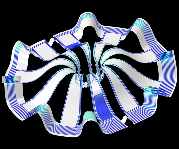
Caption: Three dimensional multifunctional neural interfaces for cortical spheroids and engineered assembloids Credit: Northwestern University

As for what brain ogranoids might be, Carl Zimmer in an Aug. 29, 2019 article for the New York Times provides an explanation,

Organoids Are Not Brains. How Are They Making Brain Waves?

Two hundred and fifty miles over Alysson Muotri’s head, a thousand tiny spheres of brain cells were sailing through space.

The clusters, called brain organoids, had been grown a few weeks earlier in the biologist’s lab here at the University of California, San Diego. He and his colleagues altered human skin cells into stem cells, then coaxed them to develop as brain cells do in an embryo.

The organoids grew into balls about the size of a pinhead, each containing hundreds of thousands of cells in a variety of types, each type producing the same chemicals and electrical signals as those cells do in our own brains.

In July, NASA packed the organoids aboard a rocket and sent them to the International Space Station to see how they develop in zero gravity.

Now the organoids were stowed inside a metal box, fed by bags of nutritious broth. “I think they are replicating like crazy at this stage, and so we’re going to have bigger organoids,” Dr. Muotri said in a recent interview in his office overlooking the Pacific.

What, exactly, are they growing into? That’s a question that has scientists and philosophers alike scratching their heads.

On Thursday, Dr. Muotri and his colleagues reported that they  have recorded simple brain waves in these organoids. In mature human brains, such waves are produced by widespread networks of neurons firing in synchrony. Particular wave patterns are linked to particular forms of brain activity, like retrieving memories and dreaming.

As the organoids mature, the researchers also found, the waves change in ways that resemble the changes in the developing brains of premature babies.

“It’s pretty amazing,” said Giorgia Quadrato, a neurobiologist at the University of Southern California who was not involved in the new study. “No one really knew if that was possible.”

But Dr. Quadrato stressed it was important not to read too much into the parallels. What she, Dr. Muotri and other brain organoid experts build are clusters of replicating brain cells, not actual brains.

If you have the time, I recommend reading Zimmer’s article in its entirety. Perhaps not coincidentally, Zimmer has an excerpt titled “Lab-Grown Brain Organoids Aren’t Alive. But They’re Not Not Alive, Either.” published in Slate.com,

From Life’s Edge: The Search For What It Means To Be Alive by Carl Zimmer, published by Dutton, an imprint of Penguin Publishing Group, a division of Penguin Random House, LLC. Copyright © 2021 by Carl Zimmer.

Cleber Trujillo led me to a windowless room banked with refrigerators, incubators, and microscopes. He extended his blue-gloved hands to either side and nearly touched the walls. “This is where we spend half our day,” he said.

In that room Trujillo and a team of graduate students raised a special kind of life. He opened an incubator and picked out a clear plastic box. Raising it above his head, he had me look up at it through its base. Inside the box were six circular wells, each the width of a cookie and filled with what looked like watered-down grape juice. In each well 100 pale globes floated, each the size of a housefly head.

Getting back to the research about monitoring brain organoids, here’s a link to and a citation for the paper about cortical spheroids,

Three-dimensional, multifunctional neural interfaces for cortical spheroids and engineered assembloids by Yoonseok Park, Colin K. Franz, Hanjun Ryu, Haiwen Luan, Kristen Y. Cotton, Jong Uk Kim, Ted S. Chung, Shiwei Zhao, Abraham Vazquez-Guardado, Da Som Yang, Kan Li, Raudel Avila, Jack K. Phillips, Maria J. Quezada, Hokyung Jang, Sung Soo Kwak, Sang Min Won, Kyeongha Kwon, Hyoyoung Jeong, Amay J. Bandodkar, Mengdi Han, Hangbo Zhao, Gabrielle R. Osher, Heling Wang, KunHyuck Lee, Yihui Zhang, Yonggang Huang, John D. Finan and John A. Rogers. Science Advances 17 Mar 2021: Vol. 7, no. 12, eabf9153 DOI: 10.1126/sciadv.abf9153

This paper appears to be open access.

According to a March 22, 2021 posting on the Shirley Riley AbilityLab website, the paper is featured on the front cover of Science Advances (vol. 7 no. 12).
On the verge of controlling neurons by wireless?

Scientists have controlled a mouse’s neurons with a wireless device (and unleashed some paranoid fantasies? well, mine if no one else’s) according to a July 16, 2015 news item on Nanowerk (Note: A link has been removed),

A study showed that scientists can wirelessly determine the path a mouse walks with a press of a button. Researchers at the Washington University School of Medicine, St. Louis, and University of Illinois, Urbana-Champaign, created a remote controlled, next-generation tissue implant that allows neuroscientists to inject drugs and shine lights on neurons deep inside the brains of mice. The revolutionary device is described online in the journal Cell (“Wireless Optofluidic Systems for Programmable In Vivo Pharmacology and Optogenetics”). Its development was partially funded by the [US] National Institutes of Health [NIH].

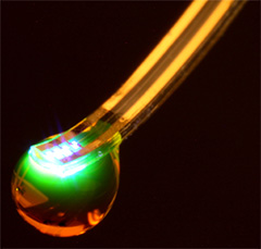
The researchers have made an image/illustration of the probe available,

Mind Bending Probe Scientists used soft materials to create a brain implant a tenth the width of a human hair that can wirelessly control neurons with lights and drugs. Courtesy of Jeong lab, University of Colorado Boulder.

A July 16, 2015 US NIH National Institute of Neurological Disorders and Stroke news release, which originated the news item, describes the study and notes that instructions for building the implant are included in the published study,

“It unplugs a world of possibilities for scientists to learn how brain circuits work in a more natural setting.” said Michael R. Bruchas, Ph.D., associate professor of anesthesiology and neurobiology at Washington University School of Medicine and a senior author of the study.

The Bruchas lab studies circuits that control a variety of disorders including stress, depression, addiction, and pain. Typically, scientists who study these circuits have to choose between injecting drugs through bulky metal tubes and delivering lights through fiber optic cables. Both options require surgery that can damage parts of the brain and introduce experimental conditions that hinder animals’ natural movements.

To address these issues, Jae-Woong Jeong, Ph.D., a bioengineer formerly at the University of Illinois at Urbana-Champaign, worked with Jordan G. McCall, Ph.D., a graduate student in the Bruchas lab, to construct a remote controlled, optofluidic implant. The device is made out of soft materials that are a tenth the diameter of a human hair and can simultaneously deliver drugs and lights.

“We used powerful nano-manufacturing strategies to fabricate an implant that lets us penetrate deep inside the brain with minimal damage,” said John A. Rogers, Ph.D., professor of materials science and engineering, University of Illinois at Urbana-Champaign and a senior author. “Ultra-miniaturized devices like this have tremendous potential for science and medicine.”

With a thickness of 80 micrometers and a width of 500 micrometers, the optofluidic implant is thinner than the metal tubes, or cannulas, scientists typically use to inject drugs. When the scientists compared the implant with a typical cannula they found that the implant damaged and displaced much less brain tissue.

The scientists tested the device’s drug delivery potential by surgically placing it into the brains of mice. In some experiments, they showed that they could precisely map circuits by using the implant to inject viruses that label cells with genetic dyes. In other experiments, they made mice walk in circles by injecting a drug that mimics morphine into the ventral tegmental area (VTA), a region that controls motivation and addiction.

The researchers also tested the device’s combined light and drug delivery potential when they made mice that have light-sensitive VTA neurons stay on one side of a cage by commanding the implant to shine laser pulses on the cells. The mice lost the preference when the scientists directed the device to simultaneously inject a drug that blocks neuronal communication. In all of the experiments, the mice were about three feet away from the command antenna.

“This is the kind of revolutionary tool development that neuroscientists need to map out brain circuit activity,” said James Gnadt, Ph.D., program director at the NIH’s National Institute of Neurological Disorders and Stroke (NINDS).  “It’s in line with the goals of the NIH’s BRAIN Initiative.”

The researchers fabricated the implant using semi-conductor computer chip manufacturing techniques. It has room for up to four drugs and has four microscale inorganic light-emitting diodes. They installed an expandable material at the bottom of the drug reservoirs to control delivery. When the temperature on an electric heater beneath the reservoir rose then the bottom rapidly expanded and pushed the drug out into the brain.

“We tried at least 30 different prototypes before one finally worked,” said Dr. McCall.

“This was truly an interdisciplinary effort,” said Dr. Jeong, who is now an assistant professor of electrical, computer, and energy engineering at University of Colorado Boulder. “We tried to engineer the implant to meet some of neurosciences greatest unmet needs.”

In the study, the scientists provide detailed instructions for manufacturing the implant.

“A tool is only good if it’s used,” said Dr. Bruchas. “We believe an open, crowdsourcing approach to neuroscience is a great way to understand normal and healthy brain circuitry.”

Here’s a link to and a citation for the paper,

Wireless Optofluidic Systems for Programmable In Vivo Pharmacology and Optogenetics by Jae-Woong Jeong, Jordan G. McCall, Gunchul Shin, Yihui Zhang, Ream Al-Hasani, Minku Kim, Shuo Li, Joo Yong Sim, Kyung-In Jang, Yan Shi, Daniel Y. Hong, Yuhao Liu, Gavin P. Schmitz, Li Xia, Zhubin He, Paul Gamble, Wilson Z. Ray, Yonggang Huang, Michael R. Bruchas, and John A. Rogers.  Cell, July 16, 2015. DOI: 10.1016/j.cell.2015.06.058

This paper is behind a paywall.

I last wrote about wireless activation of neurons in a May 28, 2014 posting which featured research at the University of Massachusetts Medical School.

E-tattoo without the nanotech

John Rogers and his team at the University of Illinois and a colleague’s (Yonggang Huang) team at Northwestern University have devised an ‘electronic tattoo’ (a soft, stick-on patch) made up from materials that anyone can purchase off-the-shelf. Rogers is known for his work with nanomaterials (my Aug. 10, 2012 posting titled ‘Surgery with fingertip control‘ mentioned a silicon nanomembrane that can be fitted onto the fingertips for possible use in surgical procedures) and with electronics (my Aug. 12, 2011 posting titled: ‘Electronic tattoos‘ mentioned his earlier attempts at developing e-tattoos).

This latest effort from Rogers and his multi-university team is mentioned in an April 4, 2014 article by Mark Wilson for Fast Company,

About a year ago, University of Illinois researcher John Rogers revealed a pretty amazing creation: a circuit that, rather than living on an inflexible board, could stick to and move with someone’s skin just like an ink stamp. But like any early research, it was mostly a proof-of-concept, and it would require relatively expensive, custom-printed electronics to work.

Today, Rogers, in conjunction with Northwestern University’s Yonggang Huang, has published details on version 2.0 in Science, revealing that this once-esoteric project has more immediate, mass market appeal.

… It means that you could create a wearable electronic that’s one-part special sticky circuit board, every other part whatever-the-hell-you-manufactured-in-China. This flexible circuit could accommodate a stock battery, an accelerometer, a Wi-Fi chip, and a Bluetooth circuitry, for instance, all living on your skin rather than inside your iPhone. And as an added bonus, it would be relatively cheap.

A University of Illinois April ?, 2014 news release describes Rogers, his multi-university team, and their current (pun intended) e-tattoo,

Engineers at the University of Illinois at Urbana-Champaign and Northwestern University have demonstrated thin, soft stick-on patches that stretch and move with the skin and incorporate commercial, off-the-shelf chip-based electronics for sophisticated wireless health monitoring.

The patches stick to the skin like a temporary tattoo and incorporate a unique microfluidic construction with wires folded like origami to allow the patch to bend and flex without being constrained by the rigid electronics components. The patches could be used for everyday health tracking – wirelessly sending updates to your cellphone or computer – and could revolutionize clinical monitoring such as EKG and EEG testing – no bulky wires, pads or tape needed.

“We designed this device to monitor human health 24/7, but without interfering with a person’s daily activity,” said Yonggang Huang, the Northwestern University professor who co-led the work with Illinois professor John A. Rogers. “It is as soft as human skin and can move with your body, but at the same time it has many different monitoring functions. What is very important about this device is it is wirelessly powered and can send high-quality data about the human body to a computer, in real time.”

The researchers did a side-by-side comparison with traditional EKG and EEG monitors and found the wireless patch performed equally to conventional sensors, while being significantly more comfortable for patients. Such a distinction is crucial for long-term monitoring, situations such as stress tests or sleep studies when the outcome depends on the patient’s ability to move and behave naturally, or for patients with fragile skin such as premature newborns.

Rogers’ group at Illinois previously demonstrated skin electronics made of very tiny, ultrathin, specially designed and printed components. While those also offer high-performance monitoring, the ability to incorporate readily available chip-based components provides many important, complementary capabilities in engineering design, at very low cost.

“Our original epidermal devices exploited specialized device geometries – super thin, structured in certain ways,” Rogers said. “But chip-scale devices, batteries, capacitors and other components must be re-formulated for these platforms. There’s a lot of value in complementing this specialized strategy with our new concepts in microfluidics and origami interconnects to enable compatibility with commercial off-the-shelf parts for accelerated development, reduced costs and expanded options in device types.”

The multi-university team turned to soft microfluidic designs to address the challenge of integrating relatively big, bulky chips with the soft, elastic base of the patch. The patch is constructed of a thin elastic envelope filled with fluid. The chip components are suspended on tiny raised support points, bonding them to the underlying patch but allowing the patch to stretch and move.

One of the biggest engineering feats of the patch is the design of the tiny, squiggly wires connecting the electronics components – radios, power inductors, sensors and more. The serpentine-shaped wires are folded like origami, so that no matter which way the patch bends, twists or stretches, the wires can unfold in any direction to accommodate the motion. Since the wires stretch, the chips don’t have to.

Skin-mounted devices could give those interested in fitness tracking a more complete and accurate picture of their activity level.

“When you measure motion on a wristwatch type device, your body is not very accurately or reliably coupled to the device,” said Rogers, a Swanlund Professor of Materials Science and Engineering at the U. of I. “Relative motion causes a lot of background noise. If you have these skin-mounted devices and an ability to locate them on multiple parts of the body, you can get a much deeper and richer set of information than would be possible with devices that are not well coupled with the skin. And that’s just the beginning of the rich range of accurate measurements relevant to physiological health that are possible when you are softly and intimately integrated onto the skin.”

The researchers hope that their sophisticated, integrated sensing systems could not only monitor health but also could help identify problems before the patient may be aware. For example, according to Rogers, data analysis could detect motions associated with Parkinson’s disease at its onset.

“The application of stretchable electronics to medicine has a lot of potential,” Huang said. “If we can continuously monitor our health with a comfortable, small device that attaches to our skin, it could be possible to catch health conditions before experiencing pain, discomfort and illness.”

Here’s a link to and a citation for the paper,

Soft Microfluidic Assemblies of Sensors, Circuits, and Radios for the Skin by Sheng Xu, Yihui Zhang, Lin Jia, Kyle E. Mathewson, Kyung-In Jang, Jeonghyun Kim, Haoran Fu, Xian Huang, Pranav Chava, Renhan Wang, Sanat Bhole, Lizhe Wang, Yoon Joo Na, Yue Guan, Matthew Flavin, Zheshen Han, Yonggang Huang, & John A. Rogers. Science 4 April 2014: Vol. 344 no. 6179 pp. 70-74 DOI: 10.1126/science.1250169

This paper is behind a paywall.

Bend it, twist it, any way you want to—a foldable lithium-ion battery

Feb. 26, 2013 news item on ScienceDaily features an extraordinary lithium-ion battery,

Northwestern University’s Yonggang Huang and the University of Illinois’ John A. Rogers are the first to demonstrate a stretchable lithium-ion battery — a flexible device capable of powering their innovative stretchable electronics.

No longer needing to be connected by a cord to an electrical outlet, the stretchable electronic devices now could be used anywhere, including inside the human body. The implantable electronics could monitor anything from brain waves to heart activity, succeeding where flat, rigid batteries would fail.

Huang and Rogers have demonstrated a battery that continues to work — powering a commercial light-emitting diode (LED) — even when stretched, folded, twisted and mounted on a human elbow. The battery can work for eight to nine hours before it needs recharging, which can be done wirelessly.

The researchers at Northwestern have produced a video where they demonstrate the battery’s ‘stretchability’,

The Northwestern University Feb. 26, 2013 news release by Megan Fellman, which originated the news item, offers this detail,

“We start with a lot of battery components side by side in a very small space, and we connect them with tightly packed, long wavy lines,” said Huang, a corresponding author of the paper. “These wires provide the flexibility. When we stretch the battery, the wavy interconnecting lines unfurl, much like yarn unspooling. And we can stretch the device a great deal and still have a working battery.”

The power and voltage of the stretchable battery are similar to a conventional lithium-ion battery of the same size, but the flexible battery can stretch up to 300 percent of its original size and still function.

Huang and Rogers have been working together for the last six years on stretchable electronics, and designing a cordless power supply has been a major challenge. Now they have solved the problem with their clever “space filling technique,” which delivers a small, high-powered battery.

For their stretchable electronic circuits, the two developed “pop-up” technology that allows circuits to bend, stretch and twist. They created an array of tiny circuit elements connected by metal wire “pop-up bridges.” When the array is stretched, the wires — not the rigid circuits — pop up.

This approach works for circuits but not for a stretchable battery. A lot of space is needed in between components for the “pop-up” interconnect to work. Circuits can be spaced out enough in an array, but battery components must be packed tightly to produce a powerful but small battery. There is not enough space between battery components for the “pop-up” technology to work.

Huang’s design solution is to use metal wire interconnects that are long, wavy lines, filling the small space between battery components. (The power travels through the interconnects.)

The unique mechanism is a “spring within a spring”: The line connecting the components is a large “S” shape and within that “S” are many smaller “S’s.” When the battery is stretched, the large “S” first stretches out and disappears, leaving a line of small squiggles. The stretching continues, with the small squiggles disappearing as the interconnect between electrodes becomes taut.

“We call this ordered unraveling,” Huang said. “And this is how we can produce a battery that stretches up to 300 percent of its original size.”

The stretching process is reversible, and the battery can be recharged wirelessly. The battery’s design allows for the integration of stretchable, inductive coils to enable charging through an external source but without the need for a physical connection.

Huang, Rogers and their teams found the battery capable of 20 cycles of recharging with little loss in capacity. The system they report in the paper consists of a square array of 100 electrode disks, electrically connected in parallel.

I’d like to see this battery actually powering a device even though the stretching is quite alluring in its way. For those who are interested here’s a citation and a link to the research paper,

Stretchable batteries with self-similar serpentine interconnects and integrated wireless recharging systems by Sheng Xu, Yihui Zhang, Jiung Cho, Juhwan Lee, Xian Huang, Lin Jia, Jonathan A. Fan, Yewang Su, Jessica Su, Huigang Zhang, Huanyu Cheng, Bingwei Lu,           Cunjiang Yu, Chi Chuang, Tae-il Kim, Taeseup Song, Kazuyo Shigeta, Sen Kang, Canan Dagdeviren, Ivan Petrov  et al.   Nature Communications 4, Article number: 1543 doi: 10.1038/ncomms2553  Published 26 February 2013

The article is behind a paywall.