Tag Archives: spinach

Spinach could help power fuel cells.

By Source (WP:NFCC#4), Fair use, https://en.wikipedia.org/w/index.php?curid=65303730

I was surprised to see a reference to the cartoon character, Popeye, in the headline (although it’s not carried forward into the text) for this October 5, 2020 news item on ScienceDaily about research into making fuel cells more efficient,

Spinach: Good for Popeye and the planet

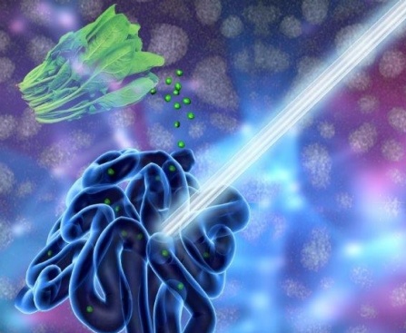
“Eat your spinach,” is a common refrain from many people’s childhoods. Spinach, the hearty, green vegetable chock full of nutrients, doesn’t just provide energy in humans. It also has potential to help power fuel cells, according to a new paper by researchers in AU’s Department of Chemistry. Spinach, when converted from its leafy, edible form into carbon nanosheets, acts as a catalyst for an oxygen reduction reaction in fuel cells and metal-air batteries.

An October 5, 2020 American University news release (also on EurekAlert) by Rebecca Basu, which originated the news item, provides more detail about the research,

An oxygen reduction reaction is one of two reactions in fuel cells and metal-air batteries and is usually the slower one that limits the energy output of these devices. Researchers have long known that certain carbon materials can catalyze the reaction. But those carbon-based catalysts don’t always perform as good or better than the traditional platinum-based catalysts. The AU researchers wanted to find an inexpensive and less toxic preparation method for an efficient catalyst by using readily available natural resources. They tackled this challenge by using spinach.

“This work suggests that sustainable catalysts can be made for an oxygen reduction reaction from natural resources,” said Prof. Shouzhong Zou, chemistry professor at AU and the paper’s lead author. “The method we tested can produce highly active, carbon-based catalysts from spinach, which is a renewable biomass. In fact, we believe it outperforms commercial platinum catalysts in both activity and stability. The catalysts are potentially applicable in hydrogen fuel cells and metal-air batteries.” Zou’s former post-doctoral students Xiaojun Liu and Wenyue Li and undergraduate student Casey Culhane are the paper’s co-authors.

Catalysts accelerate an oxygen reduction reaction to produce sufficient current and create energy. Among the practical applications for the research are fuel cells and metal-air batteries, which power electric vehicles and types of military gear. Researchers are making progress in the lab and in prototypes with catalysts derived from plants or plant products such as cattail grass or rice. Zou’s work is the first demonstration using spinach as a material for preparing oxygen reduction reaction-catalysts. Spinach is a good candidate for this work because it survives in low temperatures, is abundant and easy to grow, and is rich in iron and nitrogen that are essential for this type of catalyst.

Zou and his students created and tested the catalysts, which are spinach-derived carbon nanosheets. Carbon nanosheets are like a piece of paper with the thickness on a nanometer scale, a thousand times thinner than a piece of human hair. To create the nanosheets, the researchers put the spinach through a multi-step process that included both low- and high-tech methods, including washing, juicing and freeze-drying the spinach, manually grinding it into a fine powder with a mortar and pestle, and “doping” the resulting carbon nanosheet with extra nitrogen to improve its performance. The measurements showed that the spinach-derived catalysts performed better than platinum-based catalysts that can be expensive and lose their potency over time.

The next step for the researchers is to put the catalysts from the lab simulation into prototype devices, such as hydrogen fuel cells, to see how they perform and to develop catalysts from other plants. Zou would like to also improve sustainability by reducing the energy consumption needed for the process.

Here’s a link to and a citation for the paper,

Spinach-Derived Porous Carbon Nanosheets as High-Performance Catalysts for Oxygen Reduction Reaction by Xiaojun Liu, Casey Culhane, Wenyue Li, and Shouzhong Zou. ACS Omega 2020, 5, 38, 24367–24378 DOI: https://doi.org/10.1021/acsomega.0c02673 Publication Date:September 15, 2020 Copyright © 2020 American Chemical Society

This paper appears to be open access.

The devil’s (i.e., luciferase) in the bioluminescent plant

The American Chemical Society (ACS) and the Massachusetts Institute of Technology (MIT) have both issued news releases about the latest in bioluminescence.The researchers tested their work on watercress, a vegetable that was viewed in almost sacred terms in my family; it was not easily available in Vancouver (Canada) when I was child.

My father would hunt down fresh watercress by checking out the Chinese grocery stores. He could spot the fresh stuff from across the street while driving at 30 miles or more per hour. Spotting it entailed an immediate hunt for parking (my father hated to pay so we might have go around the block a few times or more) and a dash out of the car to ensure that he got his watercress before anyone else spotted it. These days it’s much more easily available and, thankfully, my father has passed on so he won’t have to think about glowing watercress.

Getting back to bioluninescent vegetable research, the American Chemical Society’s Dec. 13, 2017 news release on EurekAlert (and as a Dec. 13, 2017 news item on ScienceDaily) makes the announcement,

The 2009 film “Avatar” created a lush imaginary world, illuminated by magical, glowing plants. Now researchers are starting to bring this spellbinding vision to life to help reduce our dependence on artificial lighting. They report in ACS’ journal Nano Letters a way to infuse plants with the luminescence of fireflies.

Nature has produced many bioluminescent organisms, however, plants are not among them. Most attempts so far to create glowing greenery — decorative tobacco plants in particular — have relied on introducing the genes of luminescent bacteria or fireflies through genetic engineering. But getting all the right components to the right locations within the plants has been a challenge. To gain better control over where light-generating ingredients end up, Michael S. Strano and colleagues recently created nanoparticles that travel to specific destinations within plants. Building on this work, the researchers wanted to take the next step and develop a “nanobionic,” glowing plant.

The team infused watercress and other plants with three different nanoparticles in a pressurized bath. The nanoparticles were loaded with light-emitting luciferin; luciferase, which modifies luciferin and makes it glow; and coenzyme A, which boosts luciferase activity. Using size and surface charge to control where the sets of nanoparticles could go within the plant tissues, the researchers could optimize how much light was emitted. Their watercress was half as bright as a commercial 1 microwatt LED and 100,000 times brighter than genetically engineered tobacco plants. Also, the plant could be turned off by adding a compound that blocks luciferase from activating luciferin’s glow.

Here’s a video from MIT detailing their research,

A December 13, 2017 MIT news release (also on EurekAlert) casts more light on the topic (I couldn’t resist the word play),

Imagine that instead of switching on a lamp when it gets dark, you could read by the light of a glowing plant on your desk.

MIT engineers have taken a critical first step toward making that vision a reality. By embedding specialized nanoparticles into the leaves of a watercress plant, they induced the plants to give off dim light for nearly four hours. They believe that, with further optimization, such plants will one day be bright enough to illuminate a workspace.

“The vision is to make a plant that will function as a desk lamp — a lamp that you don’t have to plug in. The light is ultimately powered by the energy metabolism of the plant itself,” says Michael Strano, the Carbon P. Dubbs Professor of Chemical Engineering at MIT and the senior author of the study

This technology could also be used to provide low-intensity indoor lighting, or to transform trees into self-powered streetlights, the researchers say.

MIT postdoc Seon-Yeong Kwak is the lead author of the study, which appears in the journal Nano Letters.

Nanobionic plants

Plant nanobionics, a new research area pioneered by Strano’s lab, aims to give plants novel features by embedding them with different types of nanoparticles. The group’s goal is to engineer plants to take over many of the functions now performed by electrical devices. The researchers have previously designed plants that can detect explosives and communicate that information to a smartphone, as well as plants that can monitor drought conditions.

Lighting, which accounts for about 20 percent of worldwide energy consumption, seemed like a logical next target. “Plants can self-repair, they have their own energy, and they are already adapted to the outdoor environment,” Strano says. “We think this is an idea whose time has come. It’s a perfect problem for plant nanobionics.”

To create their glowing plants, the MIT team turned to luciferase, the enzyme that gives fireflies their glow. Luciferase acts on a molecule called luciferin, causing it to emit light. Another molecule called co-enzyme A helps the process along by removing a reaction byproduct that can inhibit luciferase activity.

The MIT team packaged each of these three components into a different type of nanoparticle carrier. The nanoparticles, which are all made of materials that the U.S. Food and Drug Administration classifies as “generally regarded as safe,” help each component get to the right part of the plant. They also prevent the components from reaching concentrations that could be toxic to the plants.

The researchers used silica nanoparticles about 10 nanometers in diameter to carry luciferase, and they used slightly larger particles of the polymers PLGA and chitosan to carry luciferin and coenzyme A, respectively. To get the particles into plant leaves, the researchers first suspended the particles in a solution. Plants were immersed in the solution and then exposed to high pressure, allowing the particles to enter the leaves through tiny pores called stomata.

Particles releasing luciferin and coenzyme A were designed to accumulate in the extracellular space of the mesophyll, an inner layer of the leaf, while the smaller particles carrying luciferase enter the cells that make up the mesophyll. The PLGA particles gradually release luciferin, which then enters the plant cells, where luciferase performs the chemical reaction that makes luciferin glow.

The researchers’ early efforts at the start of the project yielded plants that could glow for about 45 minutes, which they have since improved to 3.5 hours. The light generated by one 10-centimeter watercress seedling is currently about one-thousandth of the amount needed to read by, but the researchers believe they can boost the light emitted, as well as the duration of light, by further optimizing the concentration and release rates of the components.

Plant transformation

Previous efforts to create light-emitting plants have relied on genetically engineering plants to express the gene for luciferase, but this is a laborious process that yields extremely dim light. Those studies were performed on tobacco plants and Arabidopsis thaliana, which are commonly used for plant genetic studies. However, the method developed by Strano’s lab could be used on any type of plant. So far, they have demonstrated it with arugula, kale, and spinach, in addition to watercress.

For future versions of this technology, the researchers hope to develop a way to paint or spray the nanoparticles onto plant leaves, which could make it possible to transform trees and other large plants into light sources.

“Our target is to perform one treatment when the plant is a seedling or a mature plant, and have it last for the lifetime of the plant,” Strano says. “Our work very seriously opens up the doorway to streetlamps that are nothing but treated trees, and to indirect lighting around homes.”

The researchers have also demonstrated that they can turn the light off by adding nanoparticles carrying a luciferase inhibitor. This could enable them to eventually create plants that shut off their light emission in response to environmental conditions such as sunlight, the researchers say.

Here’s a link to and a citation for the paper,

A Nanobionic Light-Emitting Plant by Seon-Yeong Kwak, Juan Pablo Giraldo, Min Hao Wong, Volodymyr B. Koman, Tedrick Thomas Salim Lew, Jon Ell, Mark C. Weidman, Rosalie M. Sinclair, Markita P. Landry, William A. Tisdale, and Michael S. Strano. Nano Lett., 2017, 17 (12), pp 7951–7961 DOI: 10.1021/acs.nanolett.7b04369 Publication Date (Web): November 17, 2017

Copyright © 2017 American Chemical Society

This paper is behind a paywall.

Spinach and plant nanobionics

Who knew that spinach leaves could be turned into electronic devices? The answer is: engineers at the Massachusetts Institute of Technology, according to an Oct. 31, 2016 news item on phys.org,

Spinach is no longer just a superfood: By embedding leaves with carbon nanotubes, MIT engineers have transformed spinach plants into sensors that can detect explosives and wirelessly relay that information to a handheld device similar to a smartphone.

This is one of the first demonstrations of engineering electronic systems into plants, an approach that the researchers call “plant nanobionics.”

An Oct. 31, 2016 MIT news release (also on EurekAlert), which originated the news item, describes the research further (Note: Links have been removed),

“The goal of plant nanobionics is to introduce nanoparticles into the plant to give it non-native functions,” says Michael Strano, the Carbon P. Dubbs Professor of Chemical Engineering at MIT and the leader of the research team.

In this case, the plants were designed to detect chemical compounds known as nitroaromatics, which are often used in landmines and other explosives. When one of these chemicals is present in the groundwater sampled naturally by the plant, carbon nanotubes embedded in the plant leaves emit a fluorescent signal that can be read with an infrared camera. The camera can be attached to a small computer similar to a smartphone, which then sends an email to the user.

“This is a novel demonstration of how we have overcome the plant/human communication barrier,” says Strano, who believes plant power could also be harnessed to warn of pollutants and environmental conditions such as drought.

Strano is the senior author of a paper describing the nanobionic plants in the Oct. 31 [2016] issue of Nature Materials. The paper’s lead authors are Min Hao Wong, an MIT graduate student who has started a company called Plantea to further develop this technology, and Juan Pablo Giraldo, a former MIT postdoc who is now an assistant professor at the University of California at Riverside.

Environmental monitoring

Two years ago, in the first demonstration of plant nanobionics, Strano and former MIT postdoc Juan Pablo Giraldo used nanoparticles to enhance plants’ photosynthesis ability and to turn them into sensors for nitric oxide, a pollutant produced by combustion.

Plants are ideally suited for monitoring the environment because they already take in a lot of information from their surroundings, Strano says.

“Plants are very good analytical chemists,” he says. “They have an extensive root network in the soil, are constantly sampling groundwater, and have a way to self-power the transport of that water up into the leaves.”

Strano’s lab has previously developed carbon nanotubes that can be used as sensors to detect a wide range of molecules, including hydrogen peroxide, the explosive TNT, and the nerve gas sarin. When the target molecule binds to a polymer wrapped around the nanotube, it alters the tube’s fluorescence.

In the new study, the researchers embedded sensors for nitroaromatic compounds into the leaves of spinach plants. Using a technique called vascular infusion, which involves applying a solution of nanoparticles to the underside of the leaf, they placed the sensors into a leaf layer known as the mesophyll, which is where most photosynthesis takes place.

They also embedded carbon nanotubes that emit a constant fluorescent signal that serves as a reference. This allows the researchers to compare the two fluorescent signals, making it easier to determine if the explosive sensor has detected anything. If there are any explosive molecules in the groundwater, it takes about 10 minutes for the plant to draw them up into the leaves, where they encounter the detector.

To read the signal, the researchers shine a laser onto the leaf, prompting the nanotubes in the leaf to emit near-infrared fluorescent light. This can be detected with a small infrared camera connected to a Raspberry Pi, a $35 credit-card-sized computer similar to the computer inside a smartphone. The signal could also be detected with a smartphone by removing the infrared filter that most camera phones have, the researchers say.

“This setup could be replaced by a cell phone and the right kind of camera,” Strano says. “It’s just the infrared filter that would stop you from using your cell phone.”

Using this setup, the researchers can pick up a signal from about 1 meter away from the plant, and they are now working on increasing that distance.

Michael McAlpine, an associate professor of mechanical engineering at the University of Minnesota, says this approach holds great potential for engineering not only sensors but many other kinds of bionic plants that might receive radio signals or change color.

“When you have manmade materials infiltrated into a living organism, you can have plants do things that plants don’t ordinarily do,” says McAlpine, who was not involved in the research. “Once you start to think of living organisms like plants as biomaterials that can be combined with electronic materials, this is all possible.”

“A wealth of information”

In the 2014 plant nanobionics study, Strano’s lab worked with a common laboratory plant known as Arabidopsis thaliana. However, the researchers wanted to use common spinach plants for the latest study, to demonstrate the versatility of this technique. “You can apply these techniques with any living plant,” Strano says.

So far, the researchers have also engineered spinach plants that can detect dopamine, which influences plant root growth, and they are now working on additional sensors, including some that track the chemicals plants use to convey information within their own tissues.

“Plants are very environmentally responsive,” Strano says. “They know that there is going to be a drought long before we do. They can detect small changes in the properties of soil and water potential. If we tap into those chemical signaling pathways, there is a wealth of information to access.”

These sensors could also help botanists learn more about the inner workings of plants, monitor plant health, and maximize the yield of rare compounds synthesized by plants such as the Madagascar periwinkle, which produces drugs used to treat cancer.

“These sensors give real-time information from the plant. It is almost like having the plant talk to us about the environment they are in,” Wong says. “In the case of precision agriculture, having such information can directly affect yield and margins.”

Once getting over the excitement, questions spring to mind. How could this be implemented? Is somebody  going to plant a field of spinach and then embed the leaves so they can detect landmines? How will anyone know where to plant the spinach? And on a different track, is this spinach edible? I suspect that if spinach can be successfully used as a sensor, it might not be for explosives but for pollution as the researchers suggest.

Here’s a link to and a citation for the paper,

Nitroaromatic detection and infrared communication from wild-type plants using plant nanobionics by Min Hao Wong, Juan P. Giraldo, Seon-Yeong Kwak, Volodymyr B. Koman, Rosalie Sinclair, Tedrick Thomas Salim Lew, Gili Bisker, Pingwei Liu, & Michael S. Strano. Nature Materials (2016) doi:10.1038/nmat4771 Published online 31 October 2016

This paper is behind a paywall.

The last posting here which featured Strano’s research is in an Aug. 25, 2015 piece about carbon nanotubes and medical sensors.

Synthite and its new ‘nano’ line of intensely coloured natural extracts

Synthite Industries, an Indian firm, has just announced a new line of intensely coloured natural extracts  using a nanotechnology process. There’s a little more detail in an Aug. 25, 2016 news article by Robin Wyers for foodingredientsfirst.com,

Indian extracts company Synthite has introduced a new line of colors derived from a nanotechnology process that offers a much brighter and better hue and therefore requires far lower dosages in use. Vextrano is the result of incessant research and scientific deliberations with an aim to give key characteristics to spices and spice derived products at an elemental level. The purpose of the exercise is multi-faceted with a view to develop an array of novel products that can achieve customized applications in food, beverage, cosmetics and pharmaceutical industries.

Ashish Sharma (…) at Synthite briefly explained the concept to FoodIngredientsFirst: “This is a new product range which we commercialized in the market two months ago. We have bought a new plant for the production of these products. We are deriving this range from natural sources. For red colors we are using chili or paprika. For yellow, turmeric, and for green colors we are using black pepper [piperin]. …

“The key thing,” he notes, “is that when we are reducing the size of the particles to a very small level [to a particle level of 180-200 mesh], the dispersion of the light in any solvent is very good. That’s why you get the hue of the color much better.” In scientific terms, the process of maximizing the various active ingredients in a spice by reducing the size and inter molecular porosity to a feasible and ideal extent, without altering its molecular structure, leads to reduced energy consumption, waste generation and time required to achieve the end result in an application.

Sharma stresses that there are no regulatory issues around the use of this new line.  …

Synthite is just starting to roll the product out into market. …

So far, however, the product is only being sold in India, but it will be exported too, with the next promotion occurring at Fi South America, which is currently taking place in Sao Paolo, Brazil.

Vextrano is positioned as a vision for the future based on value addition to the bio-ingredients from spices. Synthite’s range includes: turmeric, spinach, piperine, marigold, paprika, black pepper, annatto and lutein.

Synthite Industries has a Wikipedia entry (Synthite Industrial Chemicals); Note: Links have been removed),

Synthite Industries Ltd (Synthite) is an Indian oleoresin extraction firm, supplying ingredients to the major food, fragrance and flavour houses. The company is based in Kochi. In 2008, it had 30% of the world’s market share,.[1][2]

The company was established in 1972 with 20 employees. It was founded by C.V. Jacob, who started the company after working in civil construction for two decades. Initially it produced industrial chemicals before shifting to oleoresins.[3] The oleoresin business was initially based on research by the Central Food Technological Research Institute in Mysore. However, the technology developed was not yet mature, and it took several years of additional research and development by Synthite to make the technology viable. It took another four years before they convinced food producers that they could produce quality products on time.[2]

By 2008, it has grown to 450 crore and 1200 employees, with a 2012 goal of 1,000 crore.[1] The company achieved this goal, with a total of 2,000 employees. The company only began selling directly to consumers in its native India in 2014.[4] Some of its major clients include Nestle, Bacardi and Pepsi.[4] The company is currently run by the founder’s son, Viju Jacob.[5]

The company produces oleoresin spices, essential oils, food colors, and sprayed products. It also has products that are organic and fair-trade. The company also has investments in realty and hospitality.[1]

You can find Synthite here but I haven’t found anything about Vextrano on that site. However, there is a LinkedIn account for Vextrano here.

Drink your spinach juice—illuminate your guts

Contrast agents used for magnetic resonance imaging, x-ray imaging, ultrasounds, and other imaging technologies are not always kind to the humans ingesting them. So, scientists at the University at Buffalo (also known as the State University of New York at Buffalo) have developed a veggie juice that does the job according to a July 11, 2016 news item on Nanowerk (Note: A link has been removed),

The pigment that gives spinach and other plants their verdant color may improve doctors’ ability to examine the human gastrointestinal tract.

That’s according to a study, published in the journal Advanced Materials (“Surfactant-Stripped Frozen Pheophytin Micelles for Multimodal Gut Imaging”), which describes how chlorophyll-based nanoparticles suspended in liquid are an effective imaging agent for the gut.

The University of Buffalo has provided an illustration of the work,

A new UB-led study suggests that chlorophyll-based nanoparticles are an effective imaging agent for the gut. The medical imaging drink, developed to diagnose and treat gastrointestinal illnesses, is made of concentrated chlorophyll, the pigment that makes spinach green. Photo illustration credit: University at Buffalo.

A new UB-led study suggests that chlorophyll-based nanoparticles are an effective imaging agent for the gut. The medical imaging drink, developed to diagnose and treat gastrointestinal illnesses, is made of concentrated chlorophyll, the pigment that makes spinach green. Photo illustration credit: University at Buffalo.

A July 11, 2016 University at Buffalo (UB) news release (also on EurekAlert) by Cory Nealon, which originated the news item, expands on the theme,

“Our work suggests that this spinach-like, nanoparticle juice can help doctors get a better look at what’s happening inside the stomach, intestines and other areas of the GI tract,” says Jonathan Lovell, PhD, assistant professor in the Department of Biomedical Engineering, a joint program between UB’s School of Engineering and Applied Sciences and the Jacobs School of Medicine and Biomedical Sciences at UB, and the study’s corresponding author.

To examine the gastrointestinal tract, doctors typically use X-rays, magnetic resonance imaging or ultrasounds, but these techniques are limited with respect to safety, accessibility and lack of adequate contrast, respectively.

Doctors also perform endoscopies, in which a tiny camera attached to a thin tube is inserted into the patient’s body. While effective, this procedure is challenging to perform in the small intestine, and it can cause infections, tears and pose other risks.

The new study, which builds upon Lovell’s previous medical imaging research, is a collaboration between researchers at UB and the University of Wisconsin-Madison. It focuses on Chlorophyll a, a pigment found in spinach and other green vegetables that is essential to photosynthesis.

In the laboratory, researchers removed magnesium from Chlorophyll a, a process which alters the pigment’s chemical structure to form another edible compound called pheophytin. Pheophytin plays an important role in photosynthesis, acting as a gatekeeper that allows electrons from sunlight to enter plants.

Next, they dissolved pheophytin in a solution of soapy substances known as surfactants. The researchers were then able to remove nearly all of the surfactants, leaving nearly pure pheophytin nanoparticles.

The drink, when tested in mice, provided imaging of the gut in three modes: photoacoustic imaging, fluorescence imaging and positron emission tomography (PET). (For PET, the researchers added to the drink Copper-64, an isotope of the metal that, in small amounts, is harmless to the human body.)

Additional studies are needed, but the drink has commercial potential because it:

·         Works in different imaging techniques.

·         Moves stably through the gut.

·         And is naturally consumed in the human diet already.

In lab tests, mice excreted 100 percent of the drink in photoacoustic and fluorescence imaging, and nearly 93 percent after the PET test.

“The veggie juice allows for techniques that are not commonly used today by doctors for imaging the gut like photoacoustic, PET, and fluorescence,” Lovell says. “And part of the appeal is the safety of the juice.”

Here’s a link to and a citation for the paper,

Surfactant-Stripped Frozen Pheophytin Micelles for Multimodal Gut Imaging by Yumiao Zhang, Depeng Wang, Shreya Goel, Boyang Sun, Upendra Chitgupi, Jumin Geng, Haiyan Sun, Todd E. Barnhart, Weibo Cai, Jun Xia, and Jonathan F. Lovell. Advanced Materials DOI: 10.1002/adma.201602373 Version of Record online: 11 JUL 2016

© 2016 WILEY-VCH Verlag GmbH & Co. KGaA, Weinheim

This paper is behind a paywall.

Spinach + Silicon = Green Power

I wouldn’t expect that anyone will be turning their spinach salads into hybrid solar cells anytime soon despite what scientists at Vanderbilt University (Tennessee) have achieved. From the Sept. 4, 2012 news release on EurekAlert,

An interdisciplinary team of researchers at Vanderbilt University have developed a way to combine the photosynthetic protein that converts light into electrochemical energy in spinach with silicon, the material used in solar cells, in a fashion that produces substantially more electrical current than has been reported by previous “biohybrid” solar cells.

Here’s an illustration of the concept provided by Vanderbilt University,

(Julie Turner/Vanderbilt)

According to the Sept. 4, 2012 Vanderbilt University news release , the researchers were trying exploit a feature of a protein found in spinach (and other plants),

More than 40 years ago, scientists discovered that one of the proteins involved in photosynthesis, called Photosystem 1 (PS1), continued to function when it was extracted from plants like spinach. Then they determined PS1 converts sunlight into electrical energy with nearly 100 percent efficiency, compared to conversion efficiencies of less than 40 percent achieved by manmade devices. This prompted various research groups around the world to begin trying to use PS1 to create more efficient solar cells.

When a PS1 protein exposed to light, it absorbs the energy in the photons and uses it to free electrons and transport them to one side of the protein. That creates regions of positive charge, called holes, which move to the opposite side of the protein.

In a leaf, all the PS1 proteins are aligned. But in the protein layer on the device, individual proteins are oriented randomly. Previous modeling work indicated that this was a major problem. When the proteins are deposited on a metallic substrate, those that are oriented in one direction provide electrons that the metal collects while those that are oriented in the opposite direction pull electrons out of the metal in order to fill the holes that they produce. As a result, they produce both positive and negative currents that cancel each other out to leave a very small net current flow.

The problem with using a metallic substrate was addressed by using and ‘doping’ silicon (from the Vanderbilt University news release),

The Vanderbilt researchers report that their PS1/silicon combination produces nearly a milliamp (850 microamps) of current per square centimeter at 0.3 volts. That is nearly two and a half times more current than the best level reported previously from a biohybrid cell. The reason this combo works so well is because the electrical properties of the silicon substrate have been tailored to fit those of the PS1 molecule. This is done by implanting electrically charge atoms in the silicon to alter its electrical properties: a process called “doping.” In this case, the protein worked extremely well with silicon doped with positive charges and worked poorly with negatively doped silicon.

To make the device, the researchers extracted PS1 from spinach into an aqueous solution and poured the mixture on the surface of a p-doped silicon wafer. Then they put the wafer in a vacuum chamber in order to evaporate the water away leaving a film of protein. They found that the optimum thickness was about one micron, about 100 PS1 molecules thick.

Here’s a graph illustrating the improvement (larger version available here),

Graph shows the dramatic increase in electrical current that Vanderbilt researchers have managed to produce from biohybrid solar cells. (Courtesy of Cliffel Lab/Vanderbilt University)

Encouraging news overall but the researchers still have more work to do (from the Vanderbilt University news release),

This combination produces current levels almost 1,000 times higher than we were able to achieve by depositing the protein on various types of metals. It also produces a modest increase in voltage,” said David Cliffel, associate professor of chemistry, who collaborated on the project with Kane Jennings, professor of chemical and biomolecular engineering.

“If we can continue on our current trajectory of increasing voltage and current levels, we could reach the range of mature solar conversion technologies in three years.”

The researchers’ next step is to build a functioning PS1-silicon solar cell using this new design. Jennings has an Environmental Protection Agency award that will allow a group of undergraduate engineering students to build the prototype. The students won the award at the National Sustainable Design Expo in April based on a solar panel that they had created using a two-year old design. With the new design, Jennings estimates that a two-foot panel could put out at least 100 milliamps at one volt – enough to power a number of different types of small electrical devices.

So, our solar cells are going to become more and more plantlike? I can certainly see the appeal if it means minimizing dependency on “rare and expensive materials like platinum or indium” as per the Vanderbilt University news release.