Tag Archives: hydrogen peroxide

Oxygen-deficient nanotitania (titanium dioxide nanoparticles) for whiter teeth without the damage

A September 8, 2021 news item on phys.org announces research that could make the process of whitening teeth safer,

Most people would like to flash a smile of pearly whites, but over time teeth can become stained by foods, beverages and some medications. Unfortunately, the high levels of hydrogen peroxide in dentists’ bleaching treatments can damage enamel and cause tooth sensitivity and gum irritation. Now, researchers reporting in ACS Applied Materials & Interfaces have developed a gel that, when exposed to near infrared (NIR) light, safely whitens teeth without the burn.

Caption: A new bleaching gel whitened tooth samples by six shades, using a low level of hydrogen peroxide (12%). Credit: Adapted from ACS Applied Materials & Interfaces 2021, DOI: 10.1021/acsami.1c06774

A September 8, 2021Amercian Chemical Society (ACS) news release (also on EurekAlert), which originated the news item, provides more detail,

The growing demand for selfie-ready smiles has made tooth whitening one of the most popular dental procedures. Treatments at a dentist’s office are effective, but they use high-concentration hydrogen peroxide (30–40%). Home bleaching products contain less peroxide (6–12%), but they usually require weeks of treatment and don’t work as well. When a bleaching gel is applied to teeth, hydrogen peroxide and peroxide-derived reactive oxygen species (mainly the hydroxyl radical) degrade pigments in stains. The hydroxyl radical is much better at doing this than hydrogen peroxide itself, so researchers have tried to improve the bleaching capacity of low-concentration hydrogen peroxide by boosting the generation of powerful hydroxyl radicals. Because previous approaches have had limitations, Xingyu Hu, Li Xie, Weidong Tian and colleagues wanted to develop a safe, effective whitening gel containing a catalyst that, when exposed to NIR light, would convert low levels of hydrogen peroxide into abundant hydroxyl radicals.

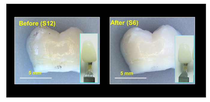
The researchers made oxygen-deficient titania nanoparticles that catalyzed hydroxyl radical production from hydrogen peroxide. Exposing the nanoparticles to NIR light increased their catalytic activity, allowing them to completely bleach tooth samples stained with orange dye, tea or red dye within 2 hours. Then, the researchers made a gel containing the nanoparticles, a carbomer gel and 12% hydrogen peroxide. They applied it to naturally stained tooth samples and treated them with NIR light for an hour. The gel bleached teeth just as well as a popular tooth whitening gel containing 40% hydrogen peroxide, with less damage to enamel. The nanoparticle system is highly promising for tooth bleaching and could also be extended to other biomedical applications, such as developing antibacterial materials, the researchers say.

The authors acknowledge funding from the National Natural Science Foundation of China, the National Key R&D Program of China and the Key Technologies R&D Program of Sichuan Province.

Here’s a link to and a citation for the paper,

Photothermal-Enhanced Fenton-like Catalytic Activity of Oxygen-Deficient Nanotitania for Efficient and Safe Tooth Whitening by Xingyu Hu, Li Xie, Zhaoyu Xu, Suru Liu, Xinzhi Tan, Ruojing Qian, Ruitao Zhang, Mingyan Jiang, Wenjia Xie, and Weidong Tian. ACS Appl. Mater. Interfaces 2021, 13, 30, 35315–35327 Publication Date: July 22, 2021 DOI: https://doi.org/10.1021/acsami.1c06774 Copyright © 2021 American Chemical Society

This paper is behind a paywall.

Human lung enzyme can degrade graphene

Caption: A human lung enzyme can biodegrade graphene. Credit: Fotolia Courtesy: Graphene Flagship

The big European Commission research programme, Grahene Flagship, has announced some new work with widespread implications if graphene is to be used in biomedical implants. From a August 23, 2018 news item on ScienceDaily,

Myeloperoxidase — an enzyme naturally found in our lungs — can biodegrade pristine graphene, according to the latest discovery of Graphene Flagship partners in CNRS, University of Strasbourg (France), Karolinska Institute (Sweden) and University of Castilla-La Mancha (Spain). Among other projects, the Graphene Flagship designs based like flexible biomedical electronic devices that will interfaced with the human body. Such applications require graphene to be biodegradable, so our body can be expelled from the body.

An August 23, 2018 Grapehene Flagship press release (mildly edited version on EurekAlert), which originated the news item, provides more detail,

To test how graphene behaves within the body, researchers analysed how it was broken down with the addition of a common human enzyme – myeloperoxidase or MPO. If a foreign body or bacteria is detected, neutrophils surround it and secrete MPO, thereby destroying the threat. Previous work by Graphene Flagship partners found that MPO could successfully biodegrade graphene oxide.

However, the structure of non-functionalized graphene was thought to be more resistant to degradation. To test this, the team looked at the effects of MPO ex vivo on two graphene forms; single- and few-layer.

Alberto Bianco, researcher at Graphene Flagship Partner CNRS, explains: “We used two forms of graphene, single- and few-layer, prepared by two different methods in water. They were then taken and put in contact with myeloperoxidase in the presence of hydrogen peroxide. This peroxidase was able to degrade and oxidise them. This was really unexpected, because we thought that non-functionalized graphene was more resistant than graphene oxide.”

Rajendra Kurapati, first author on the study and researcher at Graphene Flagship Partner CNRS, remarks how “the results emphasize that highly dispersible graphene could be degraded in the body by the action of neutrophils. This would open the new avenue for developing graphene-based materials.”

With successful ex-vivo testing, in-vivo testing is the next stage. Bengt Fadeel, professor at Graphene Flagship Partner Karolinska Institute believes that “understanding whether graphene is biodegradable or not is important for biomedical and other applications of this material. The fact that cells of the immune system are capable of handling graphene is very promising.”

Prof. Maurizio Prato, the Graphene Flagship leader for its Health and Environment Work Package said that “the enzymatic degradation of graphene is a very important topic, because in principle, graphene dispersed in the atmosphere could produce some harm. Instead, if there are microorganisms able to degrade graphene and related materials, the persistence of these materials in our environment will be strongly decreased. These types of studies are needed.” “What is also needed is to investigate the nature of degradation products,” adds Prato. “Once graphene is digested by enzymes, it could produce harmful derivatives. We need to know the structure of these derivatives and study their impact on health and environment,” he concludes.

Prof. Andrea C. Ferrari, Science and Technology Officer of the Graphene Flagship, and chair of its management panel added: “The report of a successful avenue for graphene biodegradation is a very important step forward to ensure the safe use of this material in applications. The Graphene Flagship has put the investigation of the health and environment effects of graphene at the centre of its programme since the start. These results strengthen our innovation and technology roadmap.”

Here’s a link to and a citation for the paper,

Degradation of Single‐Layer and Few‐Layer Graphene by Neutrophil Myeloperoxidase by Dr. Rajendra Kurapati, Dr. Sourav P. Mukherjee, Dr. Cristina Martín, Dr. George Bepete, Prof. Ester Vázquez, Dr. Alain Pénicaud, Prof. Dr. Bengt Fadeel, Dr. Alberto Bianco. Angewandte Chemie https://doi.org/10.1002/anie.201806906 First published: 13 July 2018

This paper is behind a paywall.

Brighten and whiten your teeth (more safely) with nanoparticles?

This is for anyone who’s ever suspected that the all the tooth brightening and whitening might not be such a good idea after all. A July 18, 2018 news item on Nanowerk announces work on what scientists hope will be a safer way to whiten teeth (Note: A link has been removed),

In the age of Instagram and Snapchat, everyone wants to have perfect pearly whites. To get a brighter smile, consumers can opt for over the counter teeth-whitening treatments or a trip to the dentist to have their teeth bleached professionally. But both types of treatments can harm teeth.

According to an article published in ACS Biomaterials Science & Engineering (“Blue-Light -Activated Nano-TiO2@PDA for Highly Effective and Nondestructive Tooth Whitening”), researchers have now developed a new, less destructive method.

A July 18, 2018 American Chemical Society (ACS) news release (also on EurekAlert), which originated the news item expands on the theme,

Teeth can become discolored on their outer surfaces when people consume colored foods and drinks, such as coffee, tea or red wine. As a result, many people turn to non-invasive whitening treatments that bleach the teeth. Currently, the most common bleaching agent is hydrogen peroxide, which steals electrons from the pigment molecules that cause teeth discoloration, and this process can be sped up by exposing teeth to blue light. But high concentrations of hydrogen peroxide can break down a tooth’s enamel, causing sensitivity or cell death. So, Xiaolei Wang, Lan Liao and colleagues wanted to see if a different blue-light-activated compound could be a safer, but still effective, alternative.

The team modified titanium dioxide nanoparticles with polydopamine (nano-TiO2@PDA) so that they could be activated with blue light. In a proof-of-concept experiment, the nano-TiO2@PDA particles were evenly coated on the surface of a tooth and irradiated with blue light. After four hours of treatment, the whitening level was similar to that obtained with hydrogen-peroxide-based agents. The group notes that no significant enamel damage was found on the surface of the tooth, and the treatment was significantly less cytotoxic than hydrogen peroxide. In addition, the nano-TiO2@PDA therapy showed antibacterial activity against certain bacteria.

Here’s a link to and a citation for the paper,

Blue-Light -Activated Nano-TiO2@PDA for Highly Effective and Nondestructive Tooth Whitening by Feng Zhang, Chongxue Wu, Ziyu Zhou, Jiaolong Wang, Weiwei Bao, Lina Dong, Zihao Zhang, Jing Ye, Lan Liao, and Xiaolei Wang. ACS Biomater. Sci. Eng., Article ASAP DOI: 10.1021/acsbiomaterials.8b00548 Publication Date (Web): June 19, 2018

Copyright © 2018 American Chemical Society

This paper is behind a paywall.

Of course, there’s always the question of what happens as we pour more and more engineered titanium dioxide nanoparticles into our bodies and ultimately into the environment.

Spinach and plant nanobionics

Who knew that spinach leaves could be turned into electronic devices? The answer is: engineers at the Massachusetts Institute of Technology, according to an Oct. 31, 2016 news item on phys.org,

Spinach is no longer just a superfood: By embedding leaves with carbon nanotubes, MIT engineers have transformed spinach plants into sensors that can detect explosives and wirelessly relay that information to a handheld device similar to a smartphone.

This is one of the first demonstrations of engineering electronic systems into plants, an approach that the researchers call “plant nanobionics.”

An Oct. 31, 2016 MIT news release (also on EurekAlert), which originated the news item, describes the research further (Note: Links have been removed),

“The goal of plant nanobionics is to introduce nanoparticles into the plant to give it non-native functions,” says Michael Strano, the Carbon P. Dubbs Professor of Chemical Engineering at MIT and the leader of the research team.

In this case, the plants were designed to detect chemical compounds known as nitroaromatics, which are often used in landmines and other explosives. When one of these chemicals is present in the groundwater sampled naturally by the plant, carbon nanotubes embedded in the plant leaves emit a fluorescent signal that can be read with an infrared camera. The camera can be attached to a small computer similar to a smartphone, which then sends an email to the user.

“This is a novel demonstration of how we have overcome the plant/human communication barrier,” says Strano, who believes plant power could also be harnessed to warn of pollutants and environmental conditions such as drought.

Strano is the senior author of a paper describing the nanobionic plants in the Oct. 31 [2016] issue of Nature Materials. The paper’s lead authors are Min Hao Wong, an MIT graduate student who has started a company called Plantea to further develop this technology, and Juan Pablo Giraldo, a former MIT postdoc who is now an assistant professor at the University of California at Riverside.

Environmental monitoring

Two years ago, in the first demonstration of plant nanobionics, Strano and former MIT postdoc Juan Pablo Giraldo used nanoparticles to enhance plants’ photosynthesis ability and to turn them into sensors for nitric oxide, a pollutant produced by combustion.

Plants are ideally suited for monitoring the environment because they already take in a lot of information from their surroundings, Strano says.

“Plants are very good analytical chemists,” he says. “They have an extensive root network in the soil, are constantly sampling groundwater, and have a way to self-power the transport of that water up into the leaves.”

Strano’s lab has previously developed carbon nanotubes that can be used as sensors to detect a wide range of molecules, including hydrogen peroxide, the explosive TNT, and the nerve gas sarin. When the target molecule binds to a polymer wrapped around the nanotube, it alters the tube’s fluorescence.

In the new study, the researchers embedded sensors for nitroaromatic compounds into the leaves of spinach plants. Using a technique called vascular infusion, which involves applying a solution of nanoparticles to the underside of the leaf, they placed the sensors into a leaf layer known as the mesophyll, which is where most photosynthesis takes place.

They also embedded carbon nanotubes that emit a constant fluorescent signal that serves as a reference. This allows the researchers to compare the two fluorescent signals, making it easier to determine if the explosive sensor has detected anything. If there are any explosive molecules in the groundwater, it takes about 10 minutes for the plant to draw them up into the leaves, where they encounter the detector.

To read the signal, the researchers shine a laser onto the leaf, prompting the nanotubes in the leaf to emit near-infrared fluorescent light. This can be detected with a small infrared camera connected to a Raspberry Pi, a $35 credit-card-sized computer similar to the computer inside a smartphone. The signal could also be detected with a smartphone by removing the infrared filter that most camera phones have, the researchers say.

“This setup could be replaced by a cell phone and the right kind of camera,” Strano says. “It’s just the infrared filter that would stop you from using your cell phone.”

Using this setup, the researchers can pick up a signal from about 1 meter away from the plant, and they are now working on increasing that distance.

Michael McAlpine, an associate professor of mechanical engineering at the University of Minnesota, says this approach holds great potential for engineering not only sensors but many other kinds of bionic plants that might receive radio signals or change color.

“When you have manmade materials infiltrated into a living organism, you can have plants do things that plants don’t ordinarily do,” says McAlpine, who was not involved in the research. “Once you start to think of living organisms like plants as biomaterials that can be combined with electronic materials, this is all possible.”

“A wealth of information”

In the 2014 plant nanobionics study, Strano’s lab worked with a common laboratory plant known as Arabidopsis thaliana. However, the researchers wanted to use common spinach plants for the latest study, to demonstrate the versatility of this technique. “You can apply these techniques with any living plant,” Strano says.

So far, the researchers have also engineered spinach plants that can detect dopamine, which influences plant root growth, and they are now working on additional sensors, including some that track the chemicals plants use to convey information within their own tissues.

“Plants are very environmentally responsive,” Strano says. “They know that there is going to be a drought long before we do. They can detect small changes in the properties of soil and water potential. If we tap into those chemical signaling pathways, there is a wealth of information to access.”

These sensors could also help botanists learn more about the inner workings of plants, monitor plant health, and maximize the yield of rare compounds synthesized by plants such as the Madagascar periwinkle, which produces drugs used to treat cancer.

“These sensors give real-time information from the plant. It is almost like having the plant talk to us about the environment they are in,” Wong says. “In the case of precision agriculture, having such information can directly affect yield and margins.”

Once getting over the excitement, questions spring to mind. How could this be implemented? Is somebody  going to plant a field of spinach and then embed the leaves so they can detect landmines? How will anyone know where to plant the spinach? And on a different track, is this spinach edible? I suspect that if spinach can be successfully used as a sensor, it might not be for explosives but for pollution as the researchers suggest.

Here’s a link to and a citation for the paper,

Nitroaromatic detection and infrared communication from wild-type plants using plant nanobionics by Min Hao Wong, Juan P. Giraldo, Seon-Yeong Kwak, Volodymyr B. Koman, Rosalie Sinclair, Tedrick Thomas Salim Lew, Gili Bisker, Pingwei Liu, & Michael S. Strano. Nature Materials (2016) doi:10.1038/nmat4771 Published online 31 October 2016

This paper is behind a paywall.

The last posting here which featured Strano’s research is in an Aug. 25, 2015 piece about carbon nanotubes and medical sensors.

Nanoparticles for breaking up plaque and preventing cavities

There may be iron in your tooth care future if a team of researchers at the University of Pennsylvania have their way. From a July 26, 2016 news item on ScienceDaily,

The bacteria that live in dental plaque and contribute to tooth decay often resist traditional antimicrobial treatment, as they can “hide” within a sticky biofilm matrix, a glue-like polymer scaffold.

A new strategy conceived by University of Pennsylvania researchers took a more sophisticated approach. Instead of simply applying an antibiotic to the teeth, they took advantage of the pH-sensitive and enzyme-like properties of iron-containing nanoparticles to catalyze the activity of hydrogen peroxide, a commonly used natural antiseptic. The activated hydrogen peroxide produced free radicals that were able to simultaneously degrade the biofilm matrix and kill the bacteria within, significantly reducing plaque and preventing the tooth decay, or cavities, in an animal model.

“Even using a very low concentration of hydrogen peroxide, the process was incredibly effective at disrupting the biofilm,” said Hyun (Michel) Koo, a professor in the Penn School of Dental Medicine’s Department of Orthodontics and divisions of Pediatric Dentistry and Community and Oral Health and the senior author of the study, which was published in the journal Biomaterials. “Adding nanoparticles increased the efficiency of bacterial killing more than 5,000-fold.”

A July 25, 2016 University of Pennsylvania news release, which originated the news item, describes the genesis of the work and provides more details about the current research (Note: A link has been removed),

The work built off a seminal finding by Gao [Lizeng Gao, a postdoctoral researcher in Koo’s lab] and colleagues, published in 2007 in Nature Nanotechnology, showing that nanoparticles, long believed to be biologically and chemically inert, could in fact possess enzyme-like properties. In that study, Gao showed that an iron oxide nanoparticle behaved similarly to a peroxidase, an enzyme found naturally that catalyzes oxidative reactions, often using hydrogen peroxide.

When Gao joined Koo’s lab in 2013, he proposed using these nanoparticles in an oral setting, as the oxidation of hydrogen peroxide produces free radicals that can kill bacteria.

“When he first presented it to me, I was very skeptical,” Koo said, “because these free radicals can also damage healthy tissue. But then he refuted that and told me this is different because the nanoparticles’ activity is dependent on pH.”

Gao had found that the nanoparticles had no catalytic activity at neutral or near-neutral pH of 6.5 or 7, physiological values typically found in blood or in a healthy mouth. But when pH was acidic, closer to 5, they become highly active and can rapidly produce free radicals.

The scenario was ideal for targeting plaque, which can produce an acidic microenvironment when exposed to sugars.

Gao and Koo reached out to Cormode [David Cormode, an assistant professor of radiology and bioengineering], who had experience working with iron oxide nanoparticles in a radiological imaging context, to help them synthesize, characterize and test the effectiveness of the nanoparticles, several forms of which are already FDA-approved for imaging in humans.

Beginning with in vitro studies, which involved growing a biofilm containing the cavity-causing bacteria Streptococcus mutans on a tooth-enamel-like surface and then exposing it to sugar, the researchers confirmed that the nanoparticles adhered to the biofilm, were retained even after treatment stopped and could effectively catalyze hydrogen peroxide in acidic conditions.

They also showed that the nanoparticles’ reaction with a 1 percent or less hydrogen peroxide solution was remarkably effective at killing bacteria, wiping out more than 99.9 percent of the S. mutans in the biofilm within five minutes, an efficacy more than 5,000 times greater than using hydrogen peroxide alone. Even more promising, they demonstrated that the treatment regimen, involving a 30-second topical treatment of the nanoparticles followed by a 30-second treatment with hydrogen peroxide, could break down the biofilm matrix components, essentially removing the protective sticky scaffold.

Moving to an animal model, they applied the nanoparticles and hydrogen peroxide topically to the teeth of rats, which can develop tooth decay when infected with S. mutans just as humans do. Twice-a-day, one-minute treatments for three weeks significantly reduced the onset and severity of carious lesions, the clinical term for tooth decay, compared to the control or treatment with hydrogen peroxide alone. The researchers observed no adverse effects on the gum or oral soft tissues from the treatment.

“It’s very promising,” said Koo. “The efficacy and toxicity need to be validated in clinical studies, but I think the potential is there.”

Among the attractive features of the platform is the fact that the components are relatively inexpensive.

“If you look at the amount you would need for a dose, you’re looking at something like 5 milligrams,” Cormode said. “It’s a tiny amount of material, and the nanoparticles are fairly easily synthesize, so we’re talking about a cost of cents per dose.”

In addition, the platform uses a concentration of hydrogen peroxide, 1 percent, which is lower than many currently available tooth-whitening systems that use 3 to 10 percent concentrations, minimizing the chance of negative side effects.

Looking ahead, Gao, Koo, Cormode and colleagues hope to continue refining and improving upon the effectiveness of the nanoparticle platform to fight biofilms.

“We’re studying the role of nanoparticle coatings, composition, size and so forth so we can engineer the particles for even better performance,” Cormode said.

The funding agencies provide a note of interest (Note: Links have been removed),

The study was funded by the International Association for Dental Research/GlaxoSmithKline Innovation in Oral Health Award, National Science Foundation and University of Pennsylvania Research Foundation.

Presumably the industry as represented by the GlaxoSmithKline Innovation in Oral Health Award is keeping a close eye on this work.

Here’s a link to and a citation for the paper,

Nanocatalysts promote Streptococcus mutans biofilm matrix degradation and enhance bacterial killing to suppress dental caries in vivo by Lizeng Gao, Yuan Liu, Dongyeop Kim, Yong Li, Geelsu Hwang, Pratap C. Naha, David P. Cormode, & Hyun Koo. Biomaterials Volume 101, September 2016, Pages 272–284 doi:10.1016/j.biomaterials.2016.05.051

This paper is behind a paywall.

Synthetic microfish (nanoengineered and 3D printed) to inspire ‘smart’ microbots

An August 26, 2015 news item on Nanowerk features some microfish (they look like sharks to me) fabricated in University of California at San Diego (UCSD) laboratories,

Nanoengineers at the University of California, San Diego used an innovative 3D printing technology they developed to manufacture multipurpose fish-shaped microrobots — called microfish — that swim around efficiently in liquids, are chemically powered by hydrogen peroxide and magnetically controlled. These proof-of-concept synthetic microfish will inspire a new generation of “smart” microrobots that have diverse capabilities such as detoxification, sensing and directed drug delivery, researchers said.

3D-printed microfish contain functional nanoparticles that enable them to be self-propelled, chemically powered and magnetically steered. The microfish are also capable of removing and sensing toxins. Image credit: J. Warner, UC San Diego Jacobs School of Engineering.

3D-printed microfish contain functional nanoparticles that enable them to be self-propelled, chemically powered and magnetically steered. The microfish are also capable of removing and sensing toxins. Image credit: J. Warner, UC San Diego Jacobs School of Engineering.

An August 25, 2015 UCSD news release (also on EurekAlert) by Liezel Labios, which originated the news item, provides more detail,

The technique used to fabricate the microfish provides numerous improvements over other methods traditionally employed to create microrobots with various locomotion mechanisms, such as microjet engines, microdrillers and microrockets. Most of these microrobots are incapable of performing more sophisticated tasks because they feature simple designs — such as spherical or cylindrical structures — and are made of homogeneous inorganic materials. In this new study, researchers demonstrated a simple way to create more complex microrobots.

By combining Chen’s 3D printing technology with Wang’s expertise in microrobots, the team was able to custom-build microfish that can do more than simply swim around when placed in a solution containing hydrogen peroxide. Nanoengineers were able to easily add functional nanoparticles into certain parts of the microfish bodies. They installed platinum nanoparticles in the tails, which react with hydrogen peroxide to propel the microfish forward, and magnetic iron oxide nanoparticles in the heads, which allowed them to be steered with magnets.

Here’s an illustration of the platinum and iron oxide microfish,

Schematic illustration of the process of functionalizing the microfish. Platinum nanoparticles are first loaded into the tail of the fish for propulsion via reaction with hydrogen peroxide. Next, iron oxide nanoparticles are loaded into the head of the fish for magnetic control. Image credit: W. Zhu and J. Li, UC San Diego Jacobs School of Engineering.

Schematic illustration of the process of functionalizing the microfish. Platinum nanoparticles are first loaded into the tail of the fish for propulsion via reaction with hydrogen peroxide. Next, iron oxide nanoparticles are loaded into the head of the fish for magnetic control. Image credit: W. Zhu and J. Li, UC San Diego Jacobs School of Engineering.

Back to the news release,

“We have developed an entirely new method to engineer nature-inspired microscopic swimmers that have complex geometric structures and are smaller than the width of a human hair. With this method, we can easily integrate different functions inside these tiny robotic swimmers for a broad spectrum of applications,” said the co-first author Wei Zhu, a nanoengineering Ph.D. student in Chen’s research group at the Jacobs School of Engineering at UC San Diego.

As a proof-of-concept demonstration, the researchers incorporated toxin-neutralizing nanoparticles throughout the bodies of the microfish. Specifically, the researchers mixed in polydiacetylene (PDA) nanoparticles, which capture harmful pore-forming toxins such as the ones found in bee venom. The researchers noted that the powerful swimming of the microfish in solution greatly enhanced their ability to clean up toxins. When the PDA nanoparticles bind with toxin molecules, they become fluorescent and emit red-colored light. The team was able to monitor the detoxification ability of the microfish by the intensity of their red glow.

“The neat thing about this experiment is that it shows how the microfish can doubly serve as detoxification systems and as toxin sensors,” said Zhu.

“Another exciting possibility we could explore is to encapsulate medicines inside the microfish and use them for directed drug delivery,” said Jinxing Li, the other co-first author of the study and a nanoengineering Ph.D. student in Wang’s research group.

For anyone curious about the new 3D printing technique, the news release provides more information about that too,

The new microfish fabrication method is based on a rapid, high-resolution 3D printing technology called microscale continuous optical printing (μCOP), which was developed in Chen’s lab. Some of the benefits of the μCOP technology are speed, scalability, precision and flexibility. Within seconds, the researchers can print an array containing hundreds of microfish, each measuring 120 microns long and 30 microns thick. This process also does not require the use of harsh chemicals. Because the μCOP technology is digitized, the researchers could easily experiment with different designs for their microfish, including shark and manta ray shapes. [emphasis mine] “With our 3D printing technology, we are not limited to just fish shapes. We can rapidly build microrobots inspired by other biological organisms such as birds,” said Zhu.

The key component of the μCOP technology is a digital micromirror array device (DMD) chip, which contains approximately two million micromirrors. Each micromirror is individually controlled to project UV light in the desired pattern (in this case, a fish shape) onto a photosensitive material, which solidifies upon exposure to UV light. The microfish are built using a photosensitive material and are constructed one layer at a time, allowing each set of functional nanoparticles to be “printed” into specific parts of the fish bodies.

“This method has made it easier for us to test different designs for these microrobots and to test different nanoparticles to insert new functional elements into these tiny structures. It’s my personal hope to further this research to eventually develop surgical microrobots that operate safer and with more precision,” said Li.

Nice to see I can recognize a shark shape when I see one. Getting back to the research, yet again, here’s a link to and a citation for the paper.

3D-Printed Artificial Microfish by Wei Zhu, Jinxing Li, Yew J. Leong, Isaac Rozen, Xin Qu, Renfeng Dong, Zhiguang Wu, Wei Gao, Peter H. Chung, Joseph Wang, and Shaochen Chen. Advanced Materials Volume 27, Issue 30, pages 4411–4417, August 12, 2015 DOI: 10.1002/adma.201501372 Article first published online: 29 JUN 2015

© 2015 WILEY-VCH Verlag GmbH & Co. KGaA, Weinheim

This paper is behind a paywall.