Tag Archives: Joseph Wang

Fridge-free COVID-19 vaccines?

COVID-19 vaccines require cold storage conditions (in some cases, extraordinarily cold storage), which pose problems with both storage and distribution.

A September 7, 2021 news item on phys.org describes research that may make vaccine distribution and storage problems a thing of the past (Note: A link has been removed),

Nanoengineers at the University of California San Diego have developed COVID-19 vaccine candidates that can take the heat. Their key ingredients? Viruses from plants or bacteria.

The new fridge-free COVID-19 vaccines are still in the early stage of development. In mice, the vaccine candidates triggered high production of neutralizing antibodies against SARS-CoV-2, the virus that causes COVID-19. If they prove to be safe and effective in people, the vaccines could be a big game changer for global distribution efforts, including those in rural areas or resource-poor communities.

A September 7, 2021 University of California at San Diego (UCSD or UC San Diego) news release (also on EurekAlert), which originated the news item, delves further into the research,

“What’s exciting about our vaccine technology is that is thermally stable, so it could easily reach places where setting up ultra-low temperature freezers, or having trucks drive around with these freezers, is not going to be possible,” said Nicole Steinmetz, a professor of nanoengineering and the director of the Center for Nano-ImmunoEngineering at the UC San Diego Jacobs School of Engineering.

The vaccines are detailed in a paper published Sept. 7 [2021] in the Journal of the American Chemical Society.

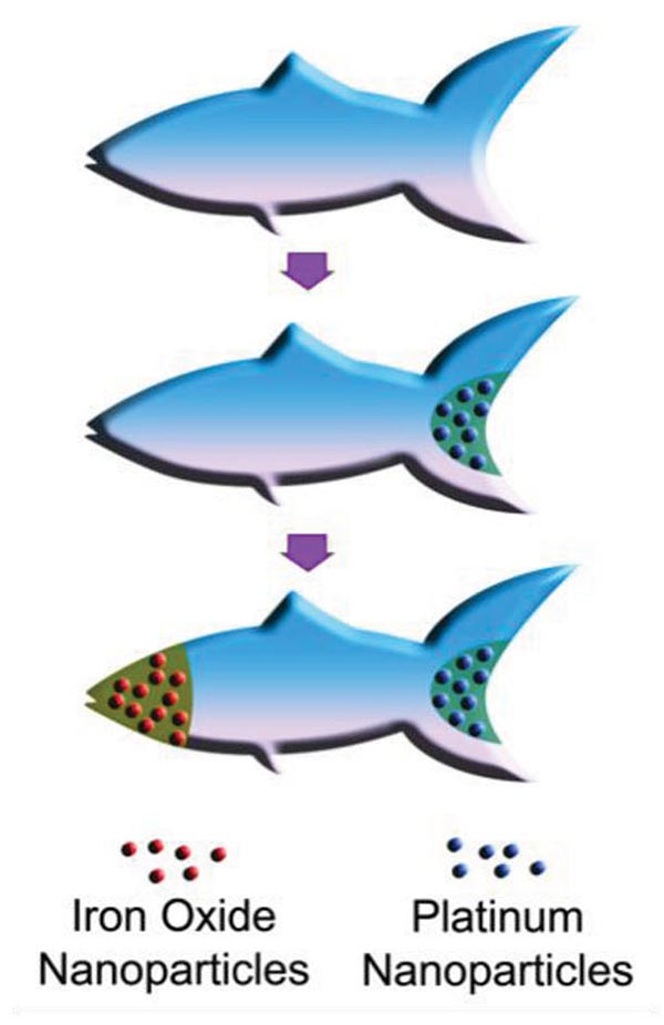
The researchers created two COVID-19 vaccine candidates. One is made from a plant virus, called cowpea mosaic virus. The other is made from a bacterial virus, or bacteriophage, called Q beta.

Both vaccines were made using similar recipes. The researchers used cowpea plants and E. coli bacteria to grow millions of copies of the plant virus and bacteriophage, respectively, in the form of ball-shaped nanoparticles. The researchers harvested these nanoparticles and then attached a small piece of the SARS-CoV-2 spike protein to the surface. The finished products look like an infectious virus so the immune system can recognize them, but they are not infectious in animals and humans. The small piece of the spike protein attached to the surface is what stimulates the body to generate an immune response against the coronavirus.

The researchers note several advantages of using plant viruses and bacteriophages to make their vaccines. For one, they can be easy and inexpensive to produce at large scales. “Growing plants is relatively easy and involves infrastructure that’s not too sophisticated,” said Steinmetz. “And fermentation using bacteria is already an established process in the biopharmaceutical industry.”

Another big advantage is that the plant virus and bacteriophage nanoparticles are extremely stable at high temperatures. As a result, the vaccines can be stored and shipped without needing to be kept cold. They also can be put through fabrication processes that use heat. The team is using such processes to package their vaccines into polymer implants and microneedle patches. These processes involve mixing the vaccine candidates with polymers and melting them together in an oven at temperatures close to 100 degrees Celsius. Being able to directly mix the plant virus and bacteriophage nanoparticles with the polymers from the start makes it easy and straightforward to create vaccine implants and patches. 

The goal is to give people more options for getting a COVID-19 vaccine and making it more accessible. The implants, which are injected underneath the skin and slowly release vaccine over the course of a month, would only need to be administered once. And the microneedle patches, which can be worn on the arm without pain or discomfort, would allow people to self-administer the vaccine.

“Imagine if vaccine patches could be sent to the mailboxes of our most vulnerable people, rather than having them leave their homes and risk exposure,” said Jon Pokorski, a professor of nanoengineering at the UC San Diego Jacobs School of Engineering, whose team developed the technology to make the implants and microneedle patches.

“If clinics could offer a one-dose implant to those who would have a really hard time making it out for their second shot, that would offer protection for more of the population and we could have a better chance at stemming transmission,” added Pokorski, who is also a founding faculty member of the university’s Institute for Materials Discovery and Design.

In tests, the team’s COVID-19 vaccine candidates were administered to mice either via implants, microneedle patches, or as a series of two shots. All three methods produced high levels of neutralizing antibodies in the blood against SARS-CoV-2.

Potential Pan-Coronavirus Vaccine

These same antibodies also neutralized against the SARS virus, the researchers found.

It all comes down to the piece of the coronavirus spike protein that is attached to the surface of the nanoparticles. One of these pieces that Steinmetz’s team chose, called an epitope, is almost identical between SARS-CoV-2 and the original SARS virus.

“The fact that neutralization is so profound with an epitope that’s so well conserved among another deadly coronavirus is remarkable,” said co-author Matthew Shin, a nanoengineering Ph.D. student in Steinmetz’s lab. “This gives us hope for a potential pan-coronavirus vaccine that could offer protection against future pandemics.”

Another advantage of this particular epitope is that it is not affected by any of the SARS-CoV-2 mutations that have so far been reported. That’s because this epitope comes from a region of the spike protein that does not directly bind to cells. This is different from the epitopes in the currently administered COVID-19 vaccines, which come from the spike protein’s binding region. This is a region where a lot of the mutations have occurred. And some of these mutations have made the virus more contagious.

Epitopes from a nonbinding region are less likely to undergo these mutations, explained Oscar Ortega-Rivera, a postdoctoral researcher in Steinmetz’s lab and the study’s first author. “Based on our sequence analyses, the epitope that we chose is highly conserved amongst the SARS-CoV-2 variants.”

This means that the new COVID-19 vaccines could potentially be effective against the variants of concern, said Ortega-Rivera, and tests are currently underway to see what effect they have against the Delta variant, for example.

Plug and Play Vaccine

Another thing that gets Steinmetz really excited about this vaccine technology is the versatility it offers to make new vaccines. “Even if this technology does not make an impact for COVID-19, it can be quickly adapted for the next threat, the next virus X,” said Steinmetz.

Making these vaccines, she says, is “plug and play:” grow plant virus or bacteriophage nanoparticles from plants or bacteria, respectively, then attach a piece of the target virus, pathogen, or biomarker to the surface.

“We use the same nanoparticles, the same polymers, the same equipment, and the same chemistry to put everything together. The only variable really is the antigen that we stick to the surface,” said Steinmetz.

The resulting vaccines do not need to be kept cold. They can be packaged into implants or microneedle patches. Or, they can be directly administered in the traditional way via shots.

Steinmetz and Pokorski’s labs have used this recipe in previous studies to make vaccine candidates for diseases like HPV and cholesterol. And now they’ve shown that it works for making COVID-19 vaccine candidates as well.

Next Steps

The vaccines still have a long way to go before they make it into clinical trials. Moving forward, the team will test if the vaccines protect against infection from COVID-19, as well as its variants and other deadly coronaviruses, in vivo.

Here’s a link to and a citation for the paper,

Trivalent Subunit Vaccine Candidates for COVID-19 and Their Delivery Devices by Oscar A. Ortega-Rivera, Matthew D. Shin, Angela Chen, Veronique Beiss, Miguel A. Moreno-Gonzalez, Miguel A. Lopez-Ramirez, Maria Reynoso, Hong Wang, Brett L. Hurst, Joseph Wang, Jonathan K. Pokorski, and Nicole F. Steinmetz. J. Am. Chem. Soc. 2021, XXXX, XXX, XXX-XXX DOI: https://doi.org/10.1021/jacs.1c06600 Publication Date:September 7, 2021 © 2021 American Chemical Society

This paper is behind a paywall.

Biohybrid cyborgs

Cyborgs are usually thought of as people who’ve been enhanced with some sort of technology, In contemporary real life that technology might be a pacemaker or hip replacement but in science fiction it’s technology such as artificial retinas (for example) that expands the range of visible light for an enhanced human.

Rarely does the topic of a microscopic life form come up in discussion about cyborgs and yet, that’s exactly what an April 3, 2019 Nanowerk spotlight article by Michael Berger describes in relationship to its use in water remediation efforts (Note: links have been removed),

Researchers often use living systems as inspiration for the design and engineering of micro- and nanoscale propulsion systems, actuators, sensors, and robots. …

“Although microrobots have recently proved successful for remediating decontaminated water at the laboratory scale, the major challenge in the field is to scale up these applications to actual environmental settings,” Professor Joseph Wang, Chair of Nanoengineering and Director, Center of Wearable Sensors at the University California San Diego, tells Nanowerk. “In order to do this, we need to overcome the toxicity of their chemical fuels, the short time span of biocompatible magnesium-based micromotors and the small domain operation of externally actuated microrobots.”

In their recent work on self-propelled biohybrid microrobots, Wang and his team were inspired by recent developments of biohybrid cyborgs that integrate self-propelling bacteria with functionalized synthetic nanostructures to transport materials.

“These tiny cyborgs are incredibly efficient for transport materials, but the limitation that we observed is that they do not provide large-scale fluid mixing,” notes Wang. ” We wanted to combine the best properties of both worlds. So, we searched for the best candidate to create a more robust biohybrid for mixing and we decided on using rotifers (Brachionus) as the engine of the cyborg.”

These marine microorganisms, which measure between 100 and 300 micrometers, are amazing creatures as they already possess sensing ability, energetic autonomy, and provide large-scale fluid mixing capability. They are also are very resilient and can survive in very harsh environments and even are one of the few organisms that have survived via asexual reproduction.

“Taking inspiration from the science fiction concept of a cybernetic organism, or cyborg – where an organism has enhanced abilities due to the integration of some artificial component – we developed a self-propelled biohybrid microrobot, that we named rotibot, employing rotifers as their engine,” says Fernando Soto, first author of a paper on this work (Advanced Functional Materials, “Rotibot: Use of Rotifers as Self-Propelling Biohybrid Microcleaners”).

This is the first demonstration of a biohybrid cyborg used for the removal and degradation of pollutants from solution. The technical breakthrough that allowed the team to achieve this task is based on a novel fabrication mechanism based on the selective accumulation of functionalized microbeads in the microorganism’s mouth: The rotifer serves not only as a transport vessel for active material or cargo but also acting as a powerful biological pump, as it creates fluid flows directed towards its mouth

Nanowerk has made this video demonstrating a rotifer available along with a description,

“The rotibot is a rotifer (a marine microorganism) that has plastic microbeads attached to the mouth, which are functionalized with pollutant-degrading enzymes. This video illustrates a free swimming rotibot mixing tracer particles in solution. “

Here’s a link to and a citation for the paper,

Rotibot: Use of Rotifers as Self‐Propelling Biohybrid Microcleaners by Fernando Soto, Miguel Angel Lopez‐Ramirez, Itthipon Jeerapan, Berta Esteban‐Fernandez de Avila, Rupesh, Kumar Mishra, Xiaolong Lu, Ingrid Chai, Chuanrui Chen, Daniel Kupor. Advanced Functional Materials DOI: https://doi.org/10.1002/adfm.201900658 First published: 28 March 2019

This paper is behind a paywall.

Berger’s April 3, 2019 Nanowerk spotlight article includes some useful images if you are interested in figuring out how these rotibots function.

Acoustic nanomotors deliver Cas9-sgRNA complex to the cell

The gene editing tool .CRISPR (clustered regularly interspaced short palindromic repeats) does feature in this story but only as a minor character; the real focus is on the delivery system. From a February 9, 2018 news item on Nanowerk ()Note: A link has been removed),

In cancer research, the “Cas-9–sgRNA” complex is an effective genomic editing tool, but its delivery across the cell membrane to the target (tumor) genome has not yet been satisfactorily solved.

American and Danish scientists have now developed an active nanomotor for the efficient transport, delivery, and release of this gene scissoring system. As detailed in their paper in the journal Angewandte Chemie (“Active Intracellular Delivery of a Cas9/sgRNA Complex Using Ultrasound-Propelled Nanomotors”), their nanovehicle is propelled towards its target by ultrasound.

The publisher (Wiley) has made this image illustrating the work available,

Courtesy: Wiley

A February 9, 2018 Wiley Publications news release (also on EurekAlert), which originated the news item, provides more information,

Genomic engineering as a promising cancer therapeutic approach has experienced a tremendous surge since the discovery of the adaptive bacterial immune defense system “CRISPR” and its potential as a gene editing tool over a decade ago. Engineered CRISPR systems for gene editing now contain two main components, a single guide RNA or sgRNA and Cas-9 nuclease. While the sgRNA guides the nuclease to the specified gene sequence, Cas-9 nuclease performs its editing with surgical efficiency. However, the delivery of the large machinery to the target genome is still problematic. The authors of the Angewandte Chemie study, Liangfang Zhang and Joseph Wang from the University of California San Diego, and their colleagues now propose ultrasound-propelled gold nanowires as an active transport/release vehicle for the Cas9-sgRNA complex over the membrane.

Gold nanowires may cross a membrane passively, but thanks to their rod- or wirelike asymmetric shape, active motion can be triggered by ultrasound. “The asymmetric shape of the gold nanowire motor, given by the fabrication process, is essential for the acoustic propulsion,” the authors remarked. They assembled the vehicle by attaching the Cas-9 protein/RNA complex to the gold nanowire through sulfide bridges. These reduceable linkages have the advantage that inside the tumor cell, the bonds would be broken by glutathione, a natural reducing compound enriched in tumor cells. The Cas9-sgRNA would be released and sent to the nucleus to do its editing work, for, example, the knockout of a gene.

As a test system, the scientists monitored the suppression of fluorescence emitted by green fluorescence protein expressing melanoma B16F10 cells. Ultrasound was applied for five minutes, which accelerated the nanomotor carrying the Cas9-sgRNA complex across the membrane, accelerating it even inside the cell, as the authors noted. Moreover, they observed their Cas9-sgRNA complex effectively suppressing fluorescence with only tiny concentrations of the complex needed.

Thus, both the effective use of an acoustic nanomotor as an active transporter and the small payload needed for efficient gene knockout are intriguing results of the study. The simplicity of the system, which uses only few and readily available components, is another remarkable achievement.

Here’s a link to and a citation for the paper,

Active Intracellular Delivery of a Cas9/sgRNA Complex Using Ultrasound-Propelled Nanomotors by Malthe Hansen-Bruhn, Dr. Berta Esteban-Fernández de Ávila, Dr. Mara Beltrán-Gastélum, Prof. Jing Zhao, Dr. Doris E. Ramírez-Herrera, Pavimol Angsantikul, Prof. Kurt Vesterager Gothelf, Prof. Liangfang Zhang, and Prof. Joseph Wang. Angewandte Chemie International Edition Vol. 57 Issue 7 DOI: 10.1002/anie.201713082 Version of Record online: 6 FEB 2018

© 2018 Wiley-VCH Verlag GmbH & Co. KGaA, Weinheim

This paper is behind a paywall.

Cleaning up carbon dioxide pollution in the oceans and elsewhere

I have a mini roundup of items (3) concerning nanotechnology and environmental applications with a special focus on carbon materials.

Carbon-capturing motors

First up, there’s a Sept. 23, 2015 news item on ScienceDaily which describes work with tiny carbon-capturing motors,

Machines that are much smaller than the width of a human hair could one day help clean up carbon dioxide pollution in the oceans. Nanoengineers at the University of California, San Diego have designed enzyme-functionalized micromotors that rapidly zoom around in water, remove carbon dioxide and convert it into a usable solid form.

The proof of concept study represents a promising route to mitigate the buildup of carbon dioxide, a major greenhouse gas in the environment, said researchers. …

A Sept 22, 2015 University of California at San Diego (UCSD) news release by Liezel Labios, which originated the news release, provides more details about the scientists’ hopes and the technology,

“We’re excited about the possibility of using these micromotors to combat ocean acidification and global warming,” said Virendra V. Singh, a postdoctoral scientist in Wang’s [nanoengineering professor and chair Joseph Wang] research group and a co-first author of this study.

In their experiments, nanoengineers demonstrated that the micromotors rapidly decarbonated water solutions that were saturated with carbon dioxide. Within five minutes, the micromotors removed 90 percent of the carbon dioxide from a solution of deionized water. The micromotors were just as effective in a sea water solution and removed 88 percent of the carbon dioxide in the same timeframe.

“In the future, we could potentially use these micromotors as part of a water treatment system, like a water decarbonation plant,” said Kevin Kaufmann, an undergraduate researcher in Wang’s lab and a co-author of the study.

The micromotors are essentially six-micrometer-long tubes that help rapidly convert carbon dioxide into calcium carbonate, a solid mineral found in eggshells, the shells of various marine organisms, calcium supplements and cement. The micromotors have an outer polymer surface that holds the enzyme carbonic anhydrase, which speeds up the reaction between carbon dioxide and water to form bicarbonate. Calcium chloride, which is added to the water solutions, helps convert bicarbonate to calcium carbonate.

The fast and continuous motion of the micromotors in solution makes the micromotors extremely efficient at removing carbon dioxide from water, said researchers. The team explained that the micromotors’ autonomous movement induces efficient solution mixing, leading to faster carbon dioxide conversion. To fuel the micromotors in water, researchers added hydrogen peroxide, which reacts with the inner platinum surface of the micromotors to generate a stream of oxygen gas bubbles that propel the micromotors around. When released in water solutions containing as little as two to four percent hydrogen peroxide, the micromotors reached speeds of more than 100 micrometers per second.

However, the use of hydrogen peroxide as the micromotor fuel is a drawback because it is an extra additive and requires the use of expensive platinum materials to build the micromotors. As a next step, researchers are planning to make carbon-capturing micromotors that can be propelled by water.

“If the micromotors can use the environment as fuel, they will be more scalable, environmentally friendly and less expensive,” said Kaufmann.

The researchers have provided an image which illustrates the carbon-capturing motors in action,

Nanoengineers have invented tiny tube-shaped micromotors that zoom around in water and efficiently remove carbon dioxide. The surfaces of the micromotors are functionalized with the enzyme carbonic anhydrase, which enables the motors to help rapidly convert carbon dioxide to calcium carbonate. Image credit: Laboratory for Nanobioelectronics, UC San Diego Jacobs School of Engineering.

Nanoengineers have invented tiny tube-shaped micromotors that zoom around in water and efficiently remove carbon dioxide. The surfaces of the micromotors are functionalized with the enzyme carbonic anhydrase, which enables the motors to help rapidly convert carbon dioxide to calcium carbonate. Image credit: Laboratory for Nanobioelectronics, UC San Diego Jacobs School of Engineering.

Here’s a link to and a citation for the paper,

Micromotor-Based Biomimetic Carbon Dioxide Sequestration: Towards Mobile Microscrubbers by Murat Uygun, Virendra V. Singh, Kevin Kaufmann, Deniz A. Uygun, Severina D. S. de Oliveira, and oseph Wang. Angewandte Chemie DOI: 10.1002/ange.201505155 Article first published online: 4 SEP 2015

© 2015 WILEY-VCH Verlag GmbH & Co. KGaA, Weinheim

This article is behind a paywall.

Carbon nanotubes for carbon dioxide capture (carbon capture)

In a Sept. 22, 2015 posting by Dexter Johnson on his Nanoclast blog (located on the IEEE [Institute for Electrical and Electronics Engineers] website) describes research where carbon nanotubes are being used for carbon capture,

Now researchers at Technische Universität Darmstadt in Germany and the Indian Institute of Technology Kanpur have found that they can tailor the gas adsorption properties of vertically aligned carbon nanotubes (VACNTs) by altering their thickness, height, and the distance between them.

“These parameters are fundamental for ‘tuning’ the hierarchical pore structure of the VACNTs,” explained Mahshid Rahimi and Deepu Babu, doctoral students at the Technische Universität Darmstadt who were the paper’s lead authors, in a press release. “This hierarchy effect is a crucial factor for getting high-adsorption capacities as well as mass transport into the nanostructure. Surprisingly, from theory and by experiment, we found that the distance between nanotubes plays a much larger role in gas adsorption than the tube diameter does.”

Dexter provides a good and brief summary of the research.

Here’s a link to and a citation for the paper,

Double-walled carbon nanotube array for CO2 and SO2 adsorption by Mahshid Rahimi, Deepu J. Babu, Jayant K. Singh, Yong-Biao Yang, Jörg J. Schneider, and Florian Müller-Plathe. J. Chem. Phys. 143, 124701 (2015); http://dx.doi.org/10.1063/1.4929609

This paper is open access.

The market for nanotechnology-enabled environmental applications

Coincident with stumbling across these two possible capture solutions, I found this Sept. 23, 2015 BCC Research news release,

A groundswell of global support for developing nanotechnology as a pollution remediation technique will continue for the foreseeable future. BCC Research reveals in its new report that this key driver, along with increasing worldwide concerns over removing pollutants and developing alternative energy sources, will drive growth in the nanotechnology environmental applications market.

The global nanotechnology market in environmental applications is expected to reach $25.7 billion by 2015 and $41.8 billion by 2020, conforming to a five-year (2015-2020) compound annual growth rate (CAGR) of 10.2%. Air remediation as a segment will reach $10.2 billion and $16.7 billion in 2015 and 2020, respectively, reflecting a five-year CAGR of 10.3%. Water remediation as a segment will grow at a five-year CAGR of 12.4% to reach $10.6 billion in 2020.

As nanoparticles push the limits and capabilities of technology, new and better techniques for pollution control are emerging. Presently, nanotechnology’s greatest potential lies in air pollution remediation.

“Nano filters could be applied to automobile tailpipes and factory smokestacks to separate out contaminants and prevent them from entering the atmosphere. In addition, nano sensors have been developed to sense toxic gas leaks at extremely low concentrations,” says BCC research analyst Aneesh Kumar. “Overall, there is a multitude of promising environmental applications for nanotechnology, with the main focus area on energy and water technologies.”

You can find links to the report, TOC (table of contents), and report overview on the BCC Research Nanotechnology in Environmental Applications: The Global Market report webpage.

Monitoring your saliva via mouth guard and smart phone

I first came across the notion that saliva instead of blood and urine could be used to assess and monitor health in a presentation abstract for the 2004 American Association for the Advancement of Science (AAAS) annual meeting held in Seattle, Washington (as per my Feb. 15, 2011 posting). There have been a few ‘saliva’ health monitoring projects mentioned here over the years but this proof-of-concept version seems like it has the potential to get to the marketplace. An August 31, 2015 news item on Nanowerk features a ‘saliva’ health monitoring project from the University of California at San Diego (UCSD),

Engineers at the University of California, San Diego, have developed a mouth guard that can monitor health markers, such as lactate, cortisol and uric acid, in saliva and transmit the information wirelessly to a smart phone, laptop or tablet.
The technology, which is at a proof-of-concept stage, could be used to monitor patients continuously without invasive procedures, as well as to monitor athletes’ performance or stress levels in soldiers and pilots. In this study, engineers focused on uric acid, which is a marker related to diabetes and to gout. Currently, the only way to monitor the levels of uric acid in a patient is to draw blood.

An August 31, 2015 UCSD news release (also on EurekAlert), which originated the news item, describes the research and the mouth guard in more detail,

In this study, researchers showed that the mouth guard sensor could offer an easy and reliable way to monitor uric acid levels. The mouth guard has been tested with human saliva but hasn’t been tested in a person’s mouth.

Researchers collected saliva samples from healthy volunteers and spread them on the sensor, which produced readings in a normal range. Next, they collected saliva from a patient who suffers from hyperuricemia, a condition characterized by an excess of uric acid in the blood. The sensor detected more than four times as much uric acid in the patient’s saliva than in the healthy volunteers.

The patient also took Allopurinol, which had been prescribed by a physician to treat their condition. Researchers were able to document a drop in the levels of uric acid over four or five days as the medication took effect. In the past, the patient would have needed blood draws to monitor levels and relied instead on symptoms to start and stop his medication.

Fabrication and design

Wang’s team created a screen-printed sensor using silver, Prussian blue ink and uricase, an enzyme that reacts with uric acid. Because saliva is extremely complex and contains many different biomarkers, researchers needed to make sure that the sensors only reacted with the uric acid. Nanoengineers set up the chemical equivalent of a two-step authentication system. The first step is a series of chemical keyholes, which ensures that only the smallest biochemicals get inside the sensor. The second step is a layer of uricase trapped in polymers, which reacts selectively with uric acid. The reaction between acid and enzyme generates hydrogen peroxide, which is detected by the Prussian blue ink.  That information is then transmitted to an electronic board as electrical signals via metallic strips that are part of the sensor.

The electronic board, developed by Mercier’s team, uses small chips that sense the output of the sensors, digitizes this output and then wirelessly transmits data to a smart phone, tablet or laptop. The entire electronic board occupies an area slightly larger than a U.S. penny.

Next steps

The next step is to embed all the electronics inside the mouth guard so that it can actually be worn. Researchers also will have to test the materials used for the sensors and electronics to make sure that they are indeed completely biocompatible. The next iteration of the mouth guard is about a year out, Mercier estimates.

“All the components are there,” he said. “It’s just a matter of refining the device and working on its stability.”

Wang and Mercier lead the Center for Wearable Sensors at UC San Diego, which has made a series of breakthroughs in the field, including temporary tattoos that monitor glucose, ultra-miniaturized energy-processing chips and pens filled with high-tech inks for Do It Yourself chemical sensors.

Here’s a link to and a citation for the paper,

Wearable salivary uric acid mouthguard biosensor with integrated wireless electronics by Jayoung Kim, Somayeh Imani, William R. de Araujo, Julian Warchall, Gabriela Valdés-Ramírez, Thiago R.L.C. Paixão, Patrick P. Mercier, & Joseph Wang. Biosensors and Bioelectronics Volume 74, 15 December 2015, Pages 1061–1068 doi:10.1016/j.bios.2015.07.039

This paper is behind a paywall.

Here’s an image of UCSD’s proposed mouth guard,

The mouth guard sensor offers an easy and reliable way to monitor uric acid levels in human saliva. Credit: Jacobs School of Engineering, UC San Diego

The mouth guard sensor offers an easy and reliable way to monitor uric acid levels in human saliva. Credit: Jacobs School of Engineering, UC San Diego

Synthetic microfish (nanoengineered and 3D printed) to inspire ‘smart’ microbots

An August 26, 2015 news item on Nanowerk features some microfish (they look like sharks to me) fabricated in University of California at San Diego (UCSD) laboratories,

Nanoengineers at the University of California, San Diego used an innovative 3D printing technology they developed to manufacture multipurpose fish-shaped microrobots — called microfish — that swim around efficiently in liquids, are chemically powered by hydrogen peroxide and magnetically controlled. These proof-of-concept synthetic microfish will inspire a new generation of “smart” microrobots that have diverse capabilities such as detoxification, sensing and directed drug delivery, researchers said.

3D-printed microfish contain functional nanoparticles that enable them to be self-propelled, chemically powered and magnetically steered. The microfish are also capable of removing and sensing toxins. Image credit: J. Warner, UC San Diego Jacobs School of Engineering.

3D-printed microfish contain functional nanoparticles that enable them to be self-propelled, chemically powered and magnetically steered. The microfish are also capable of removing and sensing toxins. Image credit: J. Warner, UC San Diego Jacobs School of Engineering.

An August 25, 2015 UCSD news release (also on EurekAlert) by Liezel Labios, which originated the news item, provides more detail,

The technique used to fabricate the microfish provides numerous improvements over other methods traditionally employed to create microrobots with various locomotion mechanisms, such as microjet engines, microdrillers and microrockets. Most of these microrobots are incapable of performing more sophisticated tasks because they feature simple designs — such as spherical or cylindrical structures — and are made of homogeneous inorganic materials. In this new study, researchers demonstrated a simple way to create more complex microrobots.

By combining Chen’s 3D printing technology with Wang’s expertise in microrobots, the team was able to custom-build microfish that can do more than simply swim around when placed in a solution containing hydrogen peroxide. Nanoengineers were able to easily add functional nanoparticles into certain parts of the microfish bodies. They installed platinum nanoparticles in the tails, which react with hydrogen peroxide to propel the microfish forward, and magnetic iron oxide nanoparticles in the heads, which allowed them to be steered with magnets.

Here’s an illustration of the platinum and iron oxide microfish,

Schematic illustration of the process of functionalizing the microfish. Platinum nanoparticles are first loaded into the tail of the fish for propulsion via reaction with hydrogen peroxide. Next, iron oxide nanoparticles are loaded into the head of the fish for magnetic control. Image credit: W. Zhu and J. Li, UC San Diego Jacobs School of Engineering.

Schematic illustration of the process of functionalizing the microfish. Platinum nanoparticles are first loaded into the tail of the fish for propulsion via reaction with hydrogen peroxide. Next, iron oxide nanoparticles are loaded into the head of the fish for magnetic control. Image credit: W. Zhu and J. Li, UC San Diego Jacobs School of Engineering.

Back to the news release,

“We have developed an entirely new method to engineer nature-inspired microscopic swimmers that have complex geometric structures and are smaller than the width of a human hair. With this method, we can easily integrate different functions inside these tiny robotic swimmers for a broad spectrum of applications,” said the co-first author Wei Zhu, a nanoengineering Ph.D. student in Chen’s research group at the Jacobs School of Engineering at UC San Diego.

As a proof-of-concept demonstration, the researchers incorporated toxin-neutralizing nanoparticles throughout the bodies of the microfish. Specifically, the researchers mixed in polydiacetylene (PDA) nanoparticles, which capture harmful pore-forming toxins such as the ones found in bee venom. The researchers noted that the powerful swimming of the microfish in solution greatly enhanced their ability to clean up toxins. When the PDA nanoparticles bind with toxin molecules, they become fluorescent and emit red-colored light. The team was able to monitor the detoxification ability of the microfish by the intensity of their red glow.

“The neat thing about this experiment is that it shows how the microfish can doubly serve as detoxification systems and as toxin sensors,” said Zhu.

“Another exciting possibility we could explore is to encapsulate medicines inside the microfish and use them for directed drug delivery,” said Jinxing Li, the other co-first author of the study and a nanoengineering Ph.D. student in Wang’s research group.

For anyone curious about the new 3D printing technique, the news release provides more information about that too,

The new microfish fabrication method is based on a rapid, high-resolution 3D printing technology called microscale continuous optical printing (μCOP), which was developed in Chen’s lab. Some of the benefits of the μCOP technology are speed, scalability, precision and flexibility. Within seconds, the researchers can print an array containing hundreds of microfish, each measuring 120 microns long and 30 microns thick. This process also does not require the use of harsh chemicals. Because the μCOP technology is digitized, the researchers could easily experiment with different designs for their microfish, including shark and manta ray shapes. [emphasis mine] “With our 3D printing technology, we are not limited to just fish shapes. We can rapidly build microrobots inspired by other biological organisms such as birds,” said Zhu.

The key component of the μCOP technology is a digital micromirror array device (DMD) chip, which contains approximately two million micromirrors. Each micromirror is individually controlled to project UV light in the desired pattern (in this case, a fish shape) onto a photosensitive material, which solidifies upon exposure to UV light. The microfish are built using a photosensitive material and are constructed one layer at a time, allowing each set of functional nanoparticles to be “printed” into specific parts of the fish bodies.

“This method has made it easier for us to test different designs for these microrobots and to test different nanoparticles to insert new functional elements into these tiny structures. It’s my personal hope to further this research to eventually develop surgical microrobots that operate safer and with more precision,” said Li.

Nice to see I can recognize a shark shape when I see one. Getting back to the research, yet again, here’s a link to and a citation for the paper.

3D-Printed Artificial Microfish by Wei Zhu, Jinxing Li, Yew J. Leong, Isaac Rozen, Xin Qu, Renfeng Dong, Zhiguang Wu, Wei Gao, Peter H. Chung, Joseph Wang, and Shaochen Chen. Advanced Materials Volume 27, Issue 30, pages 4411–4417, August 12, 2015 DOI: 10.1002/adma.201501372 Article first published online: 29 JUN 2015

© 2015 WILEY-VCH Verlag GmbH & Co. KGaA, Weinheim

This paper is behind a paywall.

ATTACH for smart clothes and personalized heating and cooling

If this research into clothing that can heat or warm you as needed sounds familiar, it is. A team out of Stanford University (US) reported on research they conducted (pun noted) using special cloth coated with metallic nanowires to achieve personalized heating and cooling (my Jan. 9, 2015 post).

Now there is a second US team, also based in southern California, working on personalized heating and cooling. Researchers at the University of California at San Diego (UCSD) have received a $2.6M grant to pursue this goal, from a June 1, 2015 news item on Nanowerk,

Imagine a fabric that will keep your body at a comfortable temperature—regardless of how hot or cold it actually is. That’s the goal of an engineering project at the University of California, San Diego, funded with a $2.6M grant from the U.S. Department of Energy’s Advanced Research Projects Agency – Energy (ARPA-E). Wearing this smart fabric could potentially reduce heating and air conditioning bills for buildings and homes.

The project, named ATTACH (Adaptive Textiles Technology with Active Cooling and Heating), is led by Joseph Wang, distinguished professor of nanoengineering at UC San Diego.

By regulating the temperature around an individual person, rather than a large room, the smart fabric could potentially cut the energy use of buildings and homes by at least 15 percent, Wang noted.

“In cases where there are only one or two people in a large room, it’s not cost-effective to heat or cool the entire room,” said Wang. “If you can do it locally, like you can in a car by heating just the car seat instead of the entire car, then you can save a lot of energy.”

A June 1, 2015 UCSD news release (also on EurekAlert), which originated the news item, describes the team’s hopes and dreams for the technology and provides some biographical information (Note: Some links have been removed),

The smart fabric will be designed to regulate the temperature of the wearer’s skin–keeping it at 93° F–by adapting to temperature changes in the room. When the room gets cooler, the fabric will become thicker. When the room gets hotter, the fabric will become thinner. To accomplish this feat, the researchers will insert polymers that expand in the cold and shrink in the heat inside the smart fabric.

“Regardless if the surrounding temperature increases or decreases, the user will still feel the same without having to adjust the thermostat,” said Wang.

“93° F is the average comfortable skin temperature for most people,” added Renkun Chen, assistant professor of mechanical and aerospace engineering at UC San Diego, and one of the collaborators on this project.

Chen’s contribution to ATTACH is to develop supplemental heating and cooling devices, called thermoelectrics, that are printable and will be incorporated into specific spots of the smart fabric. The thermoelectrics will regulate the temperature on “hot spots”–such as areas on the back and underneath the feet–that tend to get hotter than other parts of the body when a person is active.

“This is like a personalized air-conditioner and heater,” said Chen.

Saving energy

“With the smart fabric, you won’t need to heat the room as much in the winter, and you won’t need to cool the room down as much in the summer. That means less energy is consumed. Plus, you will still feel comfortable within a wider temperature range,” said Chen.

The researchers are also designing the smart fabric to power itself. The fabric will include rechargeable batteries, which will power the thermoelectrics, as well as biofuel cells that can harvest electrical power from human sweat. Plus, all of these parts–batteries, thermoelectrics and biofuel cells–will be printed using the technology developed in Wang’s lab to make printable wearable devices. These parts will also be thin, stretchable and flexible to ensure that the smart fabric is not bulky or heavy.

“We are aiming to make the smart clothing look and feel as much like the clothes that people regularly wear. It will be washable, stretchable, bendable and lightweight. We also hope to make it look attractive and fashionable to wear,” said Wang.

In terms of price, the team has not yet concluded how much the smart clothing will cost. This will depend on the scale of production, but the printing technology in Wang’s lab will offer a low-cost method to produce the parts. Keeping the costs down is a major goal, the researchers said.

The research team

Professor Joseph Wang, Department of NanoEngineering

Wang, the lead principal investigator of ATTACH, has pioneered the development of wearable printable devices, such as electrochemical sensors and temporary tattoo-based biofuel cells. He is the chair of the nanoengineering department and the director for the Center for Wearable Sensors at UC San Diego. His extensive expertise in printable, stretchable and wearable devices will be used here to make the proposed flexible biofuel cells, batteries and thermoelectrics.

Assistant Professor Renkun Chen, Department of Mechanical and Aerospace Engineering

Chen specializes in heat transfer and thermoelectrics. His research group works on physics, materials and devices related to thermal energy transport, conversion and management. His specialty in these areas will be used to develop the thermal models and the thermoelectric devices.

Associate Professor Shirley Meng, Department of NanoEngineering

Meng’s research focuses on energy storage and conversion, particularly on battery cell design and testing. At UC San Diego, she established the Laboratory for Energy Storage and Conversion and is the inaugural director for the Sustainable Power and Energy Center. Meng will develop the rechargeable batteries and will work on power integration throughout the smart fabric system.

Professor Sungho Jin, Department of Mechanical and Aerospace Engineering

Jin specializes in functional materials for applications in nanotechnology, magnetism, energy and biomedicine. He will design the self-responsive polymers that change in thickness based on changes in the surrounding temperature.

Dr. Joshua Windmiller, CEO of Electrozyme LLC

Windmiller, former Ph.D. student and postdoc in Wang’s nanoengineering lab, is an expert in printed biosensors, bioelectronics and biofuel cells. He co-founded Electrozyme LLC, a startup devoted to the development of novel biosensors for application in the personal wellness and healthcare domains. Electrozyme will serve as the industrial partner for ATTACH and will lead the efforts to test the smart fabric prototype and bring the technology into the market.

You can find out more about Electrozyme here.

Do-it-yourself sensors possible with biocatalytic pen technology

The engineers at the University of California at San Diego (UCSD) are envisioning a future where anyone can create a biosensor anywhere. From a March 3, 2015 news item on Azonano,

A new simple tool developed by nanoengineers at the University of California, San Diego, is opening the door to an era when anyone will be able to build sensors, anywhere, including physicians in the clinic, patients in their home and soldiers in the field.

The team from the University of California, San Diego, developed high-tech bio-inks that react with several chemicals, including glucose. They filled off-the-shelf ballpoint pens with the inks and were able to draw sensors to measure glucose directly on the skin and sensors to measure pollution on leaves.

A March 2, 2015 UCSD news release by Ioana Patringenaru, which originated the news item, describes the researchers’ hopes for this technology,

Skin and leaves aren’t the only media on which the pens could be used. Researchers envision sensors drawn directly on smart phones for personalized and inexpensive health monitoring or on external building walls for monitoring of toxic gas pollutants. The sensors also could be used on the battlefield to detect explosives and nerve agents.

The team, led by Joseph Wang, the chairman of the Department of NanoEngineering at the University of California, San Diego, published their findings in the Feb. 26 [2015] issue of Advanced Healthcare Materials. Wang also directs the Center for Wearable Sensors at UC San Diego.

“Our new biocatalytic pen technology, based on novel enzymatic inks, holds considerable promise for a broad range of applications on site and in the field,” Wang said.

The news release goes on to describe one of the key concerns with developing the ink,

The biggest challenge the researchers faced was making inks from chemicals and biochemicals that aren’t harmful to humans or plants; could function as the sensors’ electrodes; and retain their properties over long periods in storage and in various conditions. Researchers turned to biocompatible polyethylene glycol, which is used in several drug delivery applications, as a binder. To make the inks conductive to electric current they used graphite powder. They also added chitosan, an antibacterial agent which is used in bandages to reduce bleeding, to make sure the ink adhered to any surfaces it was used on. The inks’ recipe also includes xylitol, a sugar substitute, which helps stabilize enzymes that react with several chemicals the do-it-yourself sensors are designed to monitor.

There’s a backstory to this research,

Wang’s team has been investigating how to make glucose testing for diabetics easier for several years. The same team of engineers recently developed non-invasive glucose sensors in the form of temporary tattoos. In this study, they used pens, loaded with an ink that reacts to glucose, to draw reusable glucose-measuring sensors on a pattern printed on a transparent, flexible material which includes an electrode. Researchers then pricked a subject’s finger and put the blood sample on the sensor. The enzymatic ink reacted with glucose and the electrode recorded the measurement, which was transmitted to a glucose-measuring device. Researchers then wiped the pattern clean and drew on it again to take another measurement after the subject had eaten.

Researchers estimate that one pen contains enough ink to draw the equivalent of 500 high-fidelity glucose sensor strips. Nanoengineers also demonstrated that the sensors could be drawn directly on the skin and that they could communicate with a Bluetooth-enabled electronic device that controls electrodes called a potentiostat, to gather data.

As mentioned earlier, there are more applications being considered (from the news release),

The pens would also allow users to draw sensors that detect pollutants and potentially harmful chemicals sensors on the spot. Researchers demonstrated that this was possible by drawing a sensor on a leaf with an ink loaded with enzymes that react with phenol, an industrial chemical, which can also be found in cosmetics, including sunscreen. The leaf was then dipped in a solution of water and phenol and the sensor was connected to a pollution detector. The sensors could be modified to react with many pollutants, including heavy metals or pesticides.

Next steps include connecting the sensors wirelessly to monitoring devices and investigating how the sensors perform in difficult conditions, including extreme temperatures, varying humidity and extended exposure to sunlight.

The researchers’ have provided a picture of the pen and a leaf,

Researchers drew sensors capable of detecting pollutants on a leaf. Courtesy: University of California at San Diego

Researchers drew sensors capable of detecting pollutants on a leaf. Courtesy: University of California at San Diego

Here’s a link to and a citation for the paper,

Biocompatible Enzymatic Roller Pens for Direct Writing of Biocatalytic Materials: “Do-it-Yourself” Electrochemical Biosensors by Amay J. Bandodkar, Wenzhao Jia, Julian Ramírez, and Joseph Wang. Advanced Healthcare Materials DOI: 10.1002/adhm.201400808 Article first published online: 26 FEB 2015

© 2015 WILEY-VCH Verlag GmbH & Co. KGaA, Weinheim

This article is behind a paywall.

Tattoos that detect glucose levels

Temporary tattoos with a biomedical function are a popular topic and one of the latest detects glucose levels without subjecting a person with diabetes to pin pricks. From a Jan. 14, 2015 news item on ScienceDaily,

Scientists have developed the first ultra-thin, flexible device that sticks to skin like a rub-on tattoo and can detect a person’s glucose levels. The sensor, reported in a proof-of-concept study in the ACS [American Chemical Society] journal Analytical Chemistry, has the potential to eliminate finger-pricking for many people with diabetes.

A Jan. 14, 2015 ACS news release on EurekAlert, which originated the news item, describes the current approaches to testing glucose and the new painless technique,

Joseph Wang and colleagues in San Diego note that diabetes affects hundreds of millions of people worldwide. Many of these patients are instructed to monitor closely their blood glucose levels to manage the disease. But the standard way of checking glucose requires a prick to the finger to draw blood for testing. The pain associated with this technique can discourage people from keeping tabs on their glucose regularly. A glucose sensing wristband had been introduced to patients, but it caused skin irritation and was discontinued. Wang’s team wanted to find a better approach.

The researchers made a wearable, non-irritating platform that can detect glucose in the fluid just under the skin based on integrating glucose extraction and electrochemical biosensing. Preliminary testing on seven healthy volunteers showed it was able to accurately determine glucose levels. The researchers conclude that the device could potentially be used for diabetes management and for other conditions such as kidney disease.

There is a Jan. 14, 2015 University of California at San Diego news release (also on EurekAlert) describing the work in more detail,

Nanoengineers at the University of California, San Diego have tested a temporary tattoo that both extracts and measures the level of glucose in the fluid in between skin cells. …

The sensor was developed and tested by graduate student Amay Bandodkar and colleagues in Professor Joseph Wang’s laboratory at the NanoEngineering Department and the Center for Wearable Sensors at the Jacobs School of Engineering at UC San Diego. Bandodkar said this “proof-of-concept” tattoo could pave the way for the Center to explore other uses of the device, such as detecting other important metabolites in the body or delivering medicines through the skin.

At the moment, the tattoo doesn’t provide the kind of numerical readout that a patient would need to monitor his or her own glucose. But this type of readout is being developed by electrical and computer engineering researchers in the Center for Wearable Sensors. “The readout instrument will also eventually have Bluetooth capabilities to send this information directly to the patient’s doctor in real-time or store data in the cloud,” said Bandodkar.

The research team is also working on ways to make the tattoo last longer while keeping its overall cost down, he noted. “Presently the tattoo sensor can easily survive for a day. These are extremely inexpensive—a few cents—and hence can be replaced without much financial burden on the patient.”

The Center “envisions using these glucose tattoo sensors to continuously monitor glucose levels of large populations as a function of their dietary habits,” Bandodkar said. Data from this wider population could help researchers learn more about the causes and potential prevention of diabetes, which affects hundreds of millions of people and is one of the leading causes of death and disability worldwide.

People with diabetes often must test their glucose levels multiple times per day, using devices that use a tiny needle to extract a small blood sample from a fingertip. Patients who avoid this testing because they find it unpleasant or difficult to perform are at a higher risk for poor health, so researchers have been searching for less invasive ways to monitor glucose.

In their report in the journal Analytical Chemistry, Wang and his co-workers describe their flexible device, which consists of carefully patterned electrodes printed on temporary tattoo paper. A very mild electrical current applied to the skin for 10 minutes forces sodium ions in the fluid between skin cells to migrate toward the tattoo’s electrodes. These ions carry glucose molecules that are also found in the fluid. A sensor built into the tattoo then measures the strength of the electrical charge produced by the glucose to determine a person’s overall glucose levels.

“The concentration of glucose extracted by the non-invasive tattoo device is almost hundred times lower than the corresponding level in the human blood,” Bandodkar explained. “Thus we had to develop a highly sensitive glucose sensor that could detect such low levels of glucose with high selectivity.”

A similar device called GlucoWatch from Cygnus Inc. was marketed in 2002, but the device was discontinued because it caused skin irritation, the UC San Diego researchers note. Their proof-of-concept tattoo sensor avoids this irritation by using a lower electrical current to extract the glucose.

Wang and colleagues applied the tattoo to seven men and women between the ages of 20 and 40 with no history of diabetes. None of the volunteers reported feeling discomfort during the tattoo test, and only a few people reported feeling a mild tingling in the first 10 seconds of the test.

To test how well the tattoo picked up the spike in glucose levels after a meal, the volunteers ate a carb-rich meal of a sandwich and soda in the lab. The device performed just as well at detecting this glucose spike as a traditional finger-stick monitor.

The researchers say the device could be used to measure other important chemicals such as lactate, a metabolite analyzed in athletes to monitor their fitness. The tattoo might also someday be used to test how well a medication is working by monitoring certain protein products in the intercellular fluid, or to detect alcohol or illegal drug consumption.

This reminds me a little of the Google moonshot project concerning health diagnostics. Announced in Oct. 2014, that project involved swallowing a pill containing nanoparticles that would circulate through your body monitoring your health and recongregating at your wrist so a band worn there could display your health status (Oct. 30, 2014 article by Signe Brewster for GigaOm). Experts welcomed the funding while warning the expectations seemed unrealistic given the current state of research and technology. This temporary tattoo seems much better grounded in terms of the technology used and achievable results.

Here’s a link to and a citation for the paper,

Tattoo-Based Noninvasive Glucose Monitoring: A Proof-of-Concept Study by Amay J. Bandodkar, Wenzhao Jia, Ceren Yardımcı, Xuan Wang, Julian Ramirez, and Joseph Wang. Anal. Chem., 2015, 87 (1), pp 394–398 DOI: 10.1021/ac504300n Publication Date (Web): December 12, 2014

Copyright © 2014 American Chemical Society

This appears to be an open access paper.

My latest posting posting on medical tattoos (prior to this) is an Aug. 13, 2014 post about a wearable biobattery.

University of Toronto’s (Canada) smiley face tattoo/sensor

Researchers at the University of Toronto have created a medical sensor that can be applied to the skin like a temporary tattoo.

University of Toronto Scarborough student Vinci Hung helped create the smiley face sensor shown here in the box at upper right (photo by Ken Jones)

The Dec. 3, 2012 news item on ScienceDaily notes,

A medical sensor that attaches to the skin like a temporary tattoo could make it easier for doctors to detect metabolic problems in patients and for coaches to fine-tune athletes’ training routines. And the entire sensor comes in a thin, flexible package shaped like a smiley face.

“We wanted a design that could conceal the electrodes,” says Vinci Hung, a PhD candidate in the Department of Physical & Environmental Sciences at UTSC [University of Toronto Scarborough], who helped create the new sensor. “We also wanted to showcase the variety of designs that can be accomplished with this fabrication technique.”

The Dec. 3, 2012 University of Toronto news release by Kurt Kleiner, which originated the news item, provides details about how the sensor/tattoo is fabricated and how it functions on the skin,

The new tattoo-based solid-contact ion-selective electrode (ISE) is made using standard screen printing techniques and commercially available transfer tattoo paper, the same kind of paper that usually carries tattoos of Spiderman or Disney princesses. In the case of the smiley face sensor, the “eyes” function as the working and reference electrodes, and the “ears” are contacts to which a measurement device can connect.

The sensor Hung helped make can detect changes in the skin’s pH levels in response to metabolic stress from exertion. Similar devices, called ion-selective electrodes (ISEs), are already used by medical researchers and athletic trainers. They can give clues to underlying metabolic diseases such as Addison’s disease, or simply signal whether an athlete is fatigued or dehydrated during training. The devices are also useful in the cosmetics industry for monitoring skin secretions.

But existing devices can be bulky, or hard to keep adhered to sweating skin. The new tattoo-based sensor stayed in place during tests, and continued to work even when the people wearing them were exercising and sweating extensively. The tattoos were applied in a similar way to regular transfer tattoos, right down to using a paper towel soaked in warm water to remove the base paper.

To make the sensors, Hung and her colleagues used a standard screen printer to lay down consecutive layers of silver, carbon fibre-modified carbon and insulator inks, followed by electropolymerization of aniline to complete the sensing surface.

By using different sensing materials, the tattoos can also be modified to detect other components of sweat, such as sodium, potassium or magnesium, all of which are of potential interest to researchers in medicine and cosmetology.

You can find the reserchers’ article in the Royal Society’s Analyst journal,

Tattoo-based potentiometric ion-selective sensors for epidermal pH monitoring
Amay J. Bandodkar ,  Vinci W. S. Hung ,  Wenzhao Jia ,  Gabriela Valdés-Ramírez ,  Joshua R. Windmiller ,  Alexandra G. Martinez ,  Julian Ramírez ,  Garrett Chan ,  Kagan Kerman and Joseph Wang in Analyst, 2013,138, 123-128 DOI: 10.1039/C2AN36422K

The article is open access but you do need to register for a free account with the Royal Society’s RSC [ublishing platform.