Tag Archives: Michael Strano

Carbon nanotubes can scavenge energy from environment to generate electricity

A June 7, 2021 news item on phys.org announces research into a new method for generating electricity (Note: A link has been removed),

MIT [Massachusetts Institute of Technology] engineers have discovered a new way of generating electricity using tiny carbon particles that can create a current simply by interacting with liquid surrounding them.

The liquid, an organic solvent, draws electrons out of the particles, generating a current that could be used to drive chemical reactions or to power micro- or nanoscale robots, the researchers say.

“This mechanism is new, and this way of generating energy is completely new,” says Michael Strano, the Carbon P. Dubbs Professor of Chemical Engineering at MIT. “This technology is intriguing because all you have to do is flow a solvent through a bed of these particles. This allows you to do electrochemistry, but with no wires.”

A June 7, 2021 MIT news release (also on EurekAlert), which generated the news item, delves further into the research,

In a new study describing this phenomenon, the researchers showed that they could use this electric current to drive a reaction known as alcohol oxidation — an organic chemical reaction that is important in the chemical industry.

Strano is the senior author of the paper, which appears today [June 7, 2021] in Nature Communications. The lead authors of the study are MIT graduate student Albert Tianxiang Liu and former MIT researcher Yuichiro Kunai. Other authors include former graduate student Anton Cottrill, postdocs Amir Kaplan and Hyunah Kim, graduate student Ge Zhang, and recent MIT graduates Rafid Mollah and Yannick Eatmon.

Unique properties

The new discovery grew out of Strano’s research on carbon nanotubes — hollow tubes made of a lattice of carbon atoms, which have unique electrical properties. In 2010, Strano demonstrated, for the first time, that carbon nanotubes can generate “thermopower waves.” When a carbon nanotube is coated with layer of fuel, moving pulses of heat, or thermopower waves, travel along the tube, creating an electrical current.

That work led Strano and his students to uncover a related feature of carbon nanotubes. They found that when part of a nanotube is coated with a Teflon-like polymer, it creates an asymmetry that makes it possible for electrons to flow from the coated to the uncoated part of the tube, generating an electrical current. Those electrons can be drawn out by submerging the particles in a solvent that is hungry for electrons.

To harness this special capability, the researchers created electricity-generating particles by grinding up carbon nanotubes and forming them into a sheet of paper-like material. One side of each sheet was coated with a Teflon-like polymer, and the researchers then cut out small particles, which can be any shape or size. For this study, they made particles that were 250 microns by 250 microns.

When these particles are submerged in an organic solvent such as acetonitrile, the solvent adheres to the uncoated surface of the particles and begins pulling electrons out of them.

“The solvent takes electrons away, and the system tries to equilibrate by moving electrons,” Strano says. “There’s no sophisticated battery chemistry inside. It’s just a particle and you put it into solvent and it starts generating an electric field.”

Particle power

The current version of the particles can generate about 0.7 volts of electricity per particle. In this study, the researchers also showed that they can form arrays of hundreds of particles in a small test tube. This “packed bed” reactor generates enough energy to power a chemical reaction called an alcohol oxidation, in which an alcohol is converted to an aldehyde or a ketone. Usually, this reaction is not performed using electrochemistry because it would require too much external current.

“Because the packed bed reactor is compact, it has more flexibility in terms of applications than a large electrochemical reactor,” Zhang says. “The particles can be made very small, and they don’t require any external wires in order to drive the electrochemical reaction.”

In future work, Strano hopes to use this kind of energy generation to build polymers using only carbon dioxide as a starting material. In a related project, he has already created polymers that can regenerate themselves using carbon dioxide as a building material, in a process powered by solar energy. This work is inspired by carbon fixation, the set of chemical reactions that plants use to build sugars from carbon dioxide, using energy from the sun.

In the longer term, this approach could also be used to power micro- or nanoscale robots. Strano’s lab has already begun building robots at that scale, which could one day be used as diagnostic or environmental sensors. The idea of being able to scavenge energy from the environment to power these kinds of robots is appealing, he says.

“It means you don’t have to put the energy storage on board,” he says. “What we like about this mechanism is that you can take the energy, at least in part, from the environment.”

Here’s a link to and a citation for the paper,

Solvent-induced electrochemistry at an electrically asymmetric carbon Janus particle by Albert Tianxiang Liu, Yuichiro Kunai, Anton L. Cottrill, Amir Kaplan, Ge Zhang, Hyunah Kim, Rafid S. Mollah, Yannick L. Eatmon & Michael S. Strano. Nature Communications volume 12, Article number: 3415 (2021) DOI: https://doi.org/10.1038/s41467-021-23038-7Published 07 June 2021

This paper is open access.
Living plants detect arsenic by way of embedded nanosensors

There’s a lot of arsenic in the world and it’s often a factor in making water undrinkable. When that water is used in farming It also pollutes soil and enters food-producing plants. A December 11, 2020 news item on Nanowerk announces research into arsenic detectors in plants,

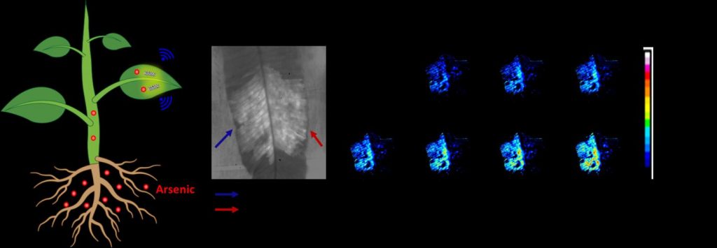
Researchers have developed a living plant-based sensor that can in real-time detect and monitor levels of arsenic, a highly toxic heavy metal, in the soil. Arsenic pollution is a major threat to humans and ecosystems in many Asia Pacific countries.

Caption: Non-destructive plant nanobionic sensor embedded within leaves to report arsenic levels within plants to portable electronics, enabling real-time monitoring of arsenic uptake in living plants. Credit: Dr. Tedrick Thomas Salim Lew

I was not able to find the source for the news item but I did locate something close. From a December 13, 2020 Singapore-Massachusetts Institute of Technology (MIT) Alliance for Research and Technology (SMART), also on EurekAlert,

Scientists from the Disruptive and Sustainable Technologies for Agricultural Precision (DiSTAP) research group at the Singapore-MIT Alliance for Research and Technology (SMART), MIT’s research enterprise in Singapore, have engineered a novel type of plant nanobionic optical sensor that can detect and monitor, in real time, levels of the highly toxic heavy metal arsenic in the underground environment. This development provides significant advantages over conventional methods used to measure arsenic in the environment and will be important for both environmental monitoring and agricultural applications to safeguard food safety, as arsenic is a contaminant in many common agricultural products such as rice, vegetables, and tea leaves.

Arsenic and its compounds are a serious threat to humans and ecosystems. Long-term exposure to arsenic in humans can cause a wide range of detrimental health effects, including cardiovascular disease such as heart attack, diabetes, birth defects, severe skin lesions, and numerous cancers including those of the skin, bladder, and lung. Elevated levels of soil arsenic as a result of anthropogenic activities such as mining and smelting are also harmful to plants, inhibiting growth and resulting in substantial crop losses.

Food crops can absorb arsenic from the soil, leading to contamination of food and produce consumed by humans. Arsenic in underground environments can also contaminate groundwater and other underground water sources, the long-term consumption of which can cause severe health issues. As such, developing accurate, effective, and easy-to-deploy arsenic sensors is important to protect both the agriculture industry and wider environmental safety.

The novel optical nanosensors exhibit changes in their fluorescence intensity upon detecting arsenic. Embedded in plant tissues, with no detrimental effects on the plant, these sensors provide a nondestructive way to monitor the internal dynamics of arsenic taken up by plants from the soil. This integration of optical nanosensors within living plants enables the conversion of plants into self-powered detectors of arsenic from their natural environment, marking a significant upgrade from the time- and equipment-intensive arsenic sampling methods of current conventional methods.

“Our plant-based nanosensor is notable not only for being the first of its kind, but also for the significant advantages it confers over conventional methods of measuring arsenic levels in the below-ground environment, requiring less time, equipment, and manpower,” says Lew. “We envision that this innovation will eventually see wide use in the agriculture industry and beyond. I am grateful to SMART DiSTAP and the Temasek Life Sciences Laboratory (TLL), both of which were instrumental in idea generation and scientific discussion as well as research funding for this work.”

Besides detecting arsenic in rice and spinach, the team also used a species of fern, Pteris cretica, which can hyperaccumulate arsenic. This fern species can absorb and tolerate high levels of arsenic with no detrimental effect — engineering an ultrasensitive plant-based arsenic detector, capable of detecting very low concentrations of arsenic, as low as 0.2 parts per billion. In contrast, the regulatory limit for arsenic detectors is 10 parts per billion. Notably, the novel nanosensors can also be integrated into other species of plants. The researchers say this is the first successful demonstration of living plant-based sensors for arsenic and represents a groundbreaking advancement that could prove highly useful in both agricultural research (e.g., to monitor arsenic taken up by edible crops for food safety) and general environmental monitoring.

Previously, conventional methods of measuring arsenic levels included regular field sampling, plant tissue digestion, extraction, and analysis using mass spectrometry. These methods are time-consuming, require extensive sample treatment, and often involve the use of bulky and expensive instrumentation. The new approach couples nanoparticle sensors with plants’ natural ability to efficiently extract analytes via the roots and transport them. This allows for the detection of arsenic uptake in living plants in real time, with portable, inexpensive electronics such as a portable Raspberry Pi platform equipped with a charge-coupled device camera akin to a smartphone camera.

Co-author, DiSTAP co-lead principal investigator, and MIT Professor Michael Strano adds, “This is a hugely exciting development, as, for the first time, we have developed a nanobionic sensor that can detect arsenic — a serious environmental contaminant and potential public health threat. With its myriad advantages over older methods of arsenic detection, this novel sensor could be a game-changer, as it is not only more time-efficient, but also more accurate and easier to deploy than older methods. It will also help plant scientists in organizations such as TLL to further produce crops that resist uptake of toxic elements. Inspired by TLL’s recent efforts to create rice crops which take up less arsenic, this work is a parallel effort to further support SMART DiSTAP’s efforts in food security research, constantly innovating and developing new technological capabilities to improve Singapore’s food quality and safety.”

The research is carried out by SMART and supported by the National Research Foundation (NRF) Singapore under its Campus for Research Excellence And Technological Enterprise (CREATE) program.

Led by MIT’s Strano and Singapore co-lead principal investigator Professor Chua Nam Hai, DiSTAP is one of the five Interdisciplinary Research Groups (IRGs) in SMART. The DiSTAP program addresses deep problems in food production in Singapore and the world by developing a suite of impactful and novel analytical genetic and biosynthetic technologies. The goal is to fundamentally change how plant biosynthetic pathways are discovered, monitored, engineered, and ultimately translated to meet the global demand for food and nutrients. Scientists from MIT, TTL, Nanyang Technological University, and National University of Singapore are collaboratively developing new tools for the continuous measurement of important plant metabolites and hormones for novel discovery, deeper understanding and control of plant biosynthetic pathways in ways not yet possible, especially in the context of green leafy vegetables; leveraging these new techniques to engineer plants with highly desirable properties for global food security, including high yield density production, drought and pathogen resistance and biosynthesis of high-value commercial products; developing tools for producing hydrophobic food components in industry-relevant microbes; developing novel microbial and enzymatic technologies to produce volatile organic compounds that can protect and/or promote growth of leafy vegetables; and applying these technologies to improve urban farming.

Here’s a link to and a citation for the paper,

Plant Nanobionic Sensors for Arsenic Detection by Tedrick Thomas Salim Lew, Minkyung Park, Jianqiao Cui, Michael S. Strano. Advanced Materials DOI: https://doi.org/10.1002/adma.202005683 First published: 26 November 2020

This paper is behind a paywall.

The devil’s (i.e., luciferase) in the bioluminescent plant

The American Chemical Society (ACS) and the Massachusetts Institute of Technology (MIT) have both issued news releases about the latest in bioluminescence.The researchers tested their work on watercress, a vegetable that was viewed in almost sacred terms in my family; it was not easily available in Vancouver (Canada) when I was child.

My father would hunt down fresh watercress by checking out the Chinese grocery stores. He could spot the fresh stuff from across the street while driving at 30 miles or more per hour. Spotting it entailed an immediate hunt for parking (my father hated to pay so we might have go around the block a few times or more) and a dash out of the car to ensure that he got his watercress before anyone else spotted it. These days it’s much more easily available and, thankfully, my father has passed on so he won’t have to think about glowing watercress.

Getting back to bioluninescent vegetable research, the American Chemical Society’s Dec. 13, 2017 news release on EurekAlert (and as a Dec. 13, 2017 news item on ScienceDaily) makes the announcement,

The 2009 film “Avatar” created a lush imaginary world, illuminated by magical, glowing plants. Now researchers are starting to bring this spellbinding vision to life to help reduce our dependence on artificial lighting. They report in ACS’ journal Nano Letters a way to infuse plants with the luminescence of fireflies.

Nature has produced many bioluminescent organisms, however, plants are not among them. Most attempts so far to create glowing greenery — decorative tobacco plants in particular — have relied on introducing the genes of luminescent bacteria or fireflies through genetic engineering. But getting all the right components to the right locations within the plants has been a challenge. To gain better control over where light-generating ingredients end up, Michael S. Strano and colleagues recently created nanoparticles that travel to specific destinations within plants. Building on this work, the researchers wanted to take the next step and develop a “nanobionic,” glowing plant.

The team infused watercress and other plants with three different nanoparticles in a pressurized bath. The nanoparticles were loaded with light-emitting luciferin; luciferase, which modifies luciferin and makes it glow; and coenzyme A, which boosts luciferase activity. Using size and surface charge to control where the sets of nanoparticles could go within the plant tissues, the researchers could optimize how much light was emitted. Their watercress was half as bright as a commercial 1 microwatt LED and 100,000 times brighter than genetically engineered tobacco plants. Also, the plant could be turned off by adding a compound that blocks luciferase from activating luciferin’s glow.

Here’s a video from MIT detailing their research,

A December 13, 2017 MIT news release (also on EurekAlert) casts more light on the topic (I couldn’t resist the word play),

Imagine that instead of switching on a lamp when it gets dark, you could read by the light of a glowing plant on your desk.

MIT engineers have taken a critical first step toward making that vision a reality. By embedding specialized nanoparticles into the leaves of a watercress plant, they induced the plants to give off dim light for nearly four hours. They believe that, with further optimization, such plants will one day be bright enough to illuminate a workspace.

“The vision is to make a plant that will function as a desk lamp — a lamp that you don’t have to plug in. The light is ultimately powered by the energy metabolism of the plant itself,” says Michael Strano, the Carbon P. Dubbs Professor of Chemical Engineering at MIT and the senior author of the study

This technology could also be used to provide low-intensity indoor lighting, or to transform trees into self-powered streetlights, the researchers say.

MIT postdoc Seon-Yeong Kwak is the lead author of the study, which appears in the journal Nano Letters.

Nanobionic plants

Plant nanobionics, a new research area pioneered by Strano’s lab, aims to give plants novel features by embedding them with different types of nanoparticles. The group’s goal is to engineer plants to take over many of the functions now performed by electrical devices. The researchers have previously designed plants that can detect explosives and communicate that information to a smartphone, as well as plants that can monitor drought conditions.

Lighting, which accounts for about 20 percent of worldwide energy consumption, seemed like a logical next target. “Plants can self-repair, they have their own energy, and they are already adapted to the outdoor environment,” Strano says. “We think this is an idea whose time has come. It’s a perfect problem for plant nanobionics.”

To create their glowing plants, the MIT team turned to luciferase, the enzyme that gives fireflies their glow. Luciferase acts on a molecule called luciferin, causing it to emit light. Another molecule called co-enzyme A helps the process along by removing a reaction byproduct that can inhibit luciferase activity.

The MIT team packaged each of these three components into a different type of nanoparticle carrier. The nanoparticles, which are all made of materials that the U.S. Food and Drug Administration classifies as “generally regarded as safe,” help each component get to the right part of the plant. They also prevent the components from reaching concentrations that could be toxic to the plants.

The researchers used silica nanoparticles about 10 nanometers in diameter to carry luciferase, and they used slightly larger particles of the polymers PLGA and chitosan to carry luciferin and coenzyme A, respectively. To get the particles into plant leaves, the researchers first suspended the particles in a solution. Plants were immersed in the solution and then exposed to high pressure, allowing the particles to enter the leaves through tiny pores called stomata.

Particles releasing luciferin and coenzyme A were designed to accumulate in the extracellular space of the mesophyll, an inner layer of the leaf, while the smaller particles carrying luciferase enter the cells that make up the mesophyll. The PLGA particles gradually release luciferin, which then enters the plant cells, where luciferase performs the chemical reaction that makes luciferin glow.

The researchers’ early efforts at the start of the project yielded plants that could glow for about 45 minutes, which they have since improved to 3.5 hours. The light generated by one 10-centimeter watercress seedling is currently about one-thousandth of the amount needed to read by, but the researchers believe they can boost the light emitted, as well as the duration of light, by further optimizing the concentration and release rates of the components.

Plant transformation

Previous efforts to create light-emitting plants have relied on genetically engineering plants to express the gene for luciferase, but this is a laborious process that yields extremely dim light. Those studies were performed on tobacco plants and Arabidopsis thaliana, which are commonly used for plant genetic studies. However, the method developed by Strano’s lab could be used on any type of plant. So far, they have demonstrated it with arugula, kale, and spinach, in addition to watercress.

For future versions of this technology, the researchers hope to develop a way to paint or spray the nanoparticles onto plant leaves, which could make it possible to transform trees and other large plants into light sources.

“Our target is to perform one treatment when the plant is a seedling or a mature plant, and have it last for the lifetime of the plant,” Strano says. “Our work very seriously opens up the doorway to streetlamps that are nothing but treated trees, and to indirect lighting around homes.”

The researchers have also demonstrated that they can turn the light off by adding nanoparticles carrying a luciferase inhibitor. This could enable them to eventually create plants that shut off their light emission in response to environmental conditions such as sunlight, the researchers say.

Here’s a link to and a citation for the paper,

A Nanobionic Light-Emitting Plant by Seon-Yeong Kwak, Juan Pablo Giraldo, Min Hao Wong, Volodymyr B. Koman, Tedrick Thomas Salim Lew, Jon Ell, Mark C. Weidman, Rosalie M. Sinclair, Markita P. Landry, William A. Tisdale, and Michael S. Strano. Nano Lett., 2017, 17 (12), pp 7951–7961 DOI: 10.1021/acs.nanolett.7b04369 Publication Date (Web): November 17, 2017

Copyright © 2017 American Chemical Society

This paper is behind a paywall.

Cotton that glows ‘naturally’

Interesting, non? This is causing a bit of excitement but before first, here’s more from the Sept. 14, 2017 American Association for the Advancement of Science (AAAS) news release on EurekAlert,

Cotton that’s grown with molecules that endow appealing properties – like fluorescence or magnetism – may one day eliminate the need for applying chemical treatments to fabrics to achieve such qualities, a new study suggests. Applying synthetic polymers to fabrics can result in a range of appealing properties, but anything added to a fabric can get washed or worn away. Furthermore, while many fibers used in fabrics are synthetic (e.g., polyester), some consumers prefer natural fibers to avoid issues related to sensation, skin irritation, smoothness, and weight. Here, Filipe Natalio and colleagues created cotton fibers that incorporate composites with fluorescent and magnetic properties. They synthesized glucose derivatives that deliver the desirable molecules into the growing ovules of the cotton plant, Gossypium hirsutum. Thus, the molecules are embedded into the cotton fibers themselves, rather than added in the form of a chemical treatment. The resulting fibers exhibited fluorescent or magnetic properties, respectively, although they were weaker than raw fibers lacking the embedded composites, the authors report. They propose that similar techniques could be expanded to other biological systems such as bacteria, bamboo, silk, and flax – essentially opening a new era of “material farming.”

Robert Service’s Sept. 14, 2017 article for Science explores the potential of growing cotton with new properties (Note: A link has been removed),

You may have heard about smartphones and smart homes. But scientists are also designing smart clothes, textiles that can harvest energy, light up, detect pollution, and even communicate with the internet. The problem? Even when they work, these often chemically treated fabrics wear out rapidly over time. Now, researchers have figured out a way to “grow” some of these functions directly into cotton fibers. If the work holds, it could lead to stronger, lighter, and brighter textiles that don’t wear out.

Yet, as the new paper went to press today in Science, editors at the journal were made aware of mistakes in a figure in the supplemental material that prompted them to issue an Editorial Expression of Concern, at least until they receive clarification from the authors. Filipe Natalio, lead author and chemist at the Weizmann Institute of Science in Rehovot, Israel, says the mistakes were errors in the names of pigments used in control experiments, which he is working with the editors to fix.

That hasn’t dampened enthusiasm for the work. “I like this paper a lot,” says Michael Strano, a chemical engineer at the Massachusetts Institute of Technology in Cambridge. The study, he says, lays out a new way to add new functions into plants without changing their genes through genetic engineering. Those approaches face steep regulatory hurdles for widespread use. “Assuming the methods claimed are correct, that’s a big advantage,” Strano says.

Sam Lemonick’s Sept. 14, 2017 article for forbes.com describes how the researchers introduced new properties (in this case, glowing colours) into the cotton plants,

His [Filipe Natalio] team of researchers in Israel, Germany, and Austria used sugar molecules to sneak new properties into cotton. Like a Trojan horse, Natalio says. They tested the method by tagging glucose with a fluorescent dye molecule that glows green when hit with the right kind of light.

They bathed cotton ovules—the part of the plant that makes the fibers—in the glucose. And just like flowers suck up dyed water in grade school experiments, the ovules absorbed the sugar solution and piped the tagged glucose molecules to their cells. As the fibers grew, they took on a yellowish tinge—and glowed bright green under ultraviolet light.

Glowing cotton wasn’t enough for Natalio. It took his group about six months to be sure they were actually delivering the fluorescent protein into the cotton cells and not just coating the fibers in it. Once they were certain, they decided to push the envelope with something very unnatural: magnets.

This time, Natalio’s team modified glucose with the rare earth metal dysprosium, making a molecule that acts like a magnet. And just like they did with the dye, the researchers fed it to cotton ovules and ended up with fibers with magnetic properties.

Both Service and Lemonwick note that the editor of the journal Science (where the research paper was published) Jeremy Berg has written an expression of editorial concern as of Sept. 14, 2017,

In the 15 September [2017] issue, Science published the Report “Biological fabrication of cellulose fibers with tailored properties” by F. Natalio et al. (1). After the issue went to press, we became aware of errors in the labeling and/or identification of the pigments used for the control experiments detailed in figs. S1 and S2 of the supplementary materials. Science is publishing this Editorial Expression of Concern to alert our readers to this information as we await full explanation and clarification from the authors.

The problem seems to be one of terminology (from the Lemonwick article),

… Filipe Natalio, lead author and chemist at the Weizmann Institute of Science in Rehovot, Israel, says the mistakes were errors in the names of pigments used in control experiments, which he is working with the editors to fix.

These things happen. Terminology and spelling aren’t always the same from one country to the next and it can result in confusion. I’m glad to see the discussion is being held openly.

Here’s a link to and a citation for the paper,

Biological fabrication of cellulose fibers with tailored properties by Filipe Natalio, Regina Fuchs, Sidney R. Cohen, Gregory Leitus, Gerhard Fritz-Popovski, Oskar Paris, Michael Kappl, Hans-Jürgen Butt. Science 15 Sep 2017: Vol. 357, Issue 6356, pp. 1118-1122 DOI: 10.1126/science.aan5830

This paper is behind a paywall.

Spinach and plant nanobionics

Who knew that spinach leaves could be turned into electronic devices? The answer is: engineers at the Massachusetts Institute of Technology, according to an Oct. 31, 2016 news item on phys.org,

Spinach is no longer just a superfood: By embedding leaves with carbon nanotubes, MIT engineers have transformed spinach plants into sensors that can detect explosives and wirelessly relay that information to a handheld device similar to a smartphone.

This is one of the first demonstrations of engineering electronic systems into plants, an approach that the researchers call “plant nanobionics.”

An Oct. 31, 2016 MIT news release (also on EurekAlert), which originated the news item, describes the research further (Note: Links have been removed),

“The goal of plant nanobionics is to introduce nanoparticles into the plant to give it non-native functions,” says Michael Strano, the Carbon P. Dubbs Professor of Chemical Engineering at MIT and the leader of the research team.

In this case, the plants were designed to detect chemical compounds known as nitroaromatics, which are often used in landmines and other explosives. When one of these chemicals is present in the groundwater sampled naturally by the plant, carbon nanotubes embedded in the plant leaves emit a fluorescent signal that can be read with an infrared camera. The camera can be attached to a small computer similar to a smartphone, which then sends an email to the user.

“This is a novel demonstration of how we have overcome the plant/human communication barrier,” says Strano, who believes plant power could also be harnessed to warn of pollutants and environmental conditions such as drought.

Strano is the senior author of a paper describing the nanobionic plants in the Oct. 31 [2016] issue of Nature Materials. The paper’s lead authors are Min Hao Wong, an MIT graduate student who has started a company called Plantea to further develop this technology, and Juan Pablo Giraldo, a former MIT postdoc who is now an assistant professor at the University of California at Riverside.

Environmental monitoring

Two years ago, in the first demonstration of plant nanobionics, Strano and former MIT postdoc Juan Pablo Giraldo used nanoparticles to enhance plants’ photosynthesis ability and to turn them into sensors for nitric oxide, a pollutant produced by combustion.

Plants are ideally suited for monitoring the environment because they already take in a lot of information from their surroundings, Strano says.

“Plants are very good analytical chemists,” he says. “They have an extensive root network in the soil, are constantly sampling groundwater, and have a way to self-power the transport of that water up into the leaves.”

Strano’s lab has previously developed carbon nanotubes that can be used as sensors to detect a wide range of molecules, including hydrogen peroxide, the explosive TNT, and the nerve gas sarin. When the target molecule binds to a polymer wrapped around the nanotube, it alters the tube’s fluorescence.

In the new study, the researchers embedded sensors for nitroaromatic compounds into the leaves of spinach plants. Using a technique called vascular infusion, which involves applying a solution of nanoparticles to the underside of the leaf, they placed the sensors into a leaf layer known as the mesophyll, which is where most photosynthesis takes place.

They also embedded carbon nanotubes that emit a constant fluorescent signal that serves as a reference. This allows the researchers to compare the two fluorescent signals, making it easier to determine if the explosive sensor has detected anything. If there are any explosive molecules in the groundwater, it takes about 10 minutes for the plant to draw them up into the leaves, where they encounter the detector.

To read the signal, the researchers shine a laser onto the leaf, prompting the nanotubes in the leaf to emit near-infrared fluorescent light. This can be detected with a small infrared camera connected to a Raspberry Pi, a $35 credit-card-sized computer similar to the computer inside a smartphone. The signal could also be detected with a smartphone by removing the infrared filter that most camera phones have, the researchers say.

“This setup could be replaced by a cell phone and the right kind of camera,” Strano says. “It’s just the infrared filter that would stop you from using your cell phone.”

Using this setup, the researchers can pick up a signal from about 1 meter away from the plant, and they are now working on increasing that distance.

Michael McAlpine, an associate professor of mechanical engineering at the University of Minnesota, says this approach holds great potential for engineering not only sensors but many other kinds of bionic plants that might receive radio signals or change color.

“When you have manmade materials infiltrated into a living organism, you can have plants do things that plants don’t ordinarily do,” says McAlpine, who was not involved in the research. “Once you start to think of living organisms like plants as biomaterials that can be combined with electronic materials, this is all possible.”

“A wealth of information”

In the 2014 plant nanobionics study, Strano’s lab worked with a common laboratory plant known as Arabidopsis thaliana. However, the researchers wanted to use common spinach plants for the latest study, to demonstrate the versatility of this technique. “You can apply these techniques with any living plant,” Strano says.

So far, the researchers have also engineered spinach plants that can detect dopamine, which influences plant root growth, and they are now working on additional sensors, including some that track the chemicals plants use to convey information within their own tissues.

“Plants are very environmentally responsive,” Strano says. “They know that there is going to be a drought long before we do. They can detect small changes in the properties of soil and water potential. If we tap into those chemical signaling pathways, there is a wealth of information to access.”

These sensors could also help botanists learn more about the inner workings of plants, monitor plant health, and maximize the yield of rare compounds synthesized by plants such as the Madagascar periwinkle, which produces drugs used to treat cancer.

“These sensors give real-time information from the plant. It is almost like having the plant talk to us about the environment they are in,” Wong says. “In the case of precision agriculture, having such information can directly affect yield and margins.”

Once getting over the excitement, questions spring to mind. How could this be implemented? Is somebody  going to plant a field of spinach and then embed the leaves so they can detect landmines? How will anyone know where to plant the spinach? And on a different track, is this spinach edible? I suspect that if spinach can be successfully used as a sensor, it might not be for explosives but for pollution as the researchers suggest.

Here’s a link to and a citation for the paper,

Nitroaromatic detection and infrared communication from wild-type plants using plant nanobionics by Min Hao Wong, Juan P. Giraldo, Seon-Yeong Kwak, Volodymyr B. Koman, Rosalie Sinclair, Tedrick Thomas Salim Lew, Gili Bisker, Pingwei Liu, & Michael S. Strano. Nature Materials (2016) doi:10.1038/nmat4771 Published online 31 October 2016

This paper is behind a paywall.

The last posting here which featured Strano’s research is in an Aug. 25, 2015 piece about carbon nanotubes and medical sensors.

Carbon nanotubes as sensors in the body

Rachel Ehrenberg has written an Aug. 21, 2015 news item about the latest and greatest carbon nanotube-based biomedical sensors for the journal Nature,

The future of medical sensors may be going down the tubes. Chemists are developing tiny devices made from carbon nanotubes wrapped with polymers to detect biologically important compounds such as insulin, nitric oxide and the blood-clotting protein fibrinogen. The hope is that these sensors could simplify and automate diagnostic tests.

Preliminary experiments in mice, reported by scientists at a meeting of the American Chemical Society in Boston, Massachusetts, this week [Aug. 16 – 20, 2015], suggest that the devices are safe to introduce into the bloodstream or implant under the skin. Researchers also presented data showing that the nanotube–polymer complexes could measure levels of large molecules, a feat that has been difficult for existing technologies.

Ehrenberg focuses on one laboratory in particular (Note: Links have been removed),

“Anything the body makes, it is meant to degrade,” says chemical engineer Michael Strano, whose lab at the Massachusetts Institute of Technology (MIT) in Cambridge is behind much of the latest work1. “Our vision is to make a sensing platform that can monitor a whole range of molecules, and do it in the long term.”

To design one sensor, MIT  researchers coated nanotubes with a mix of polymers and nucleotides and screened for configurations that would bind to the protein fibrinogen. This large molecule is important for building blood clots; its concentration can indicate bleeding disorders, liver disease or impending cardiovascular trouble. The team recently hit on a material that worked — a first for such a large molecule, according to MIT nanotechnology specialist Gili Bisker. Bisker said at the chemistry meeting that the fibrinogen-detecting nanotubes could be used to measure levels of the protein in blood samples, or implanted in body tissue to detect changing fibrinogen levels that might indicate a clot.

The MIT team has also developed2 a sensor that can be inserted beneath the skin to monitor glucose or insulin levels in real time, Bisker reported. The team imagines putting a small patch that contains a wireless device on the skin just above the embedded sensor. The patch would shine light on the sensor and measure its fluorescence, then transmit that data to a mobile phone for real-time monitoring.

Another version of the sensor, developed3 at MIT by biomedical engineer Nicole Iverson and colleagues, detects nitric oxide. This signalling molecule typically indicates inflammation and is associated with many cancer cells. When embedded in a hydrogel matrix, the sensor kept working in mice for more than 400 days and caused no local inflammation, MIT chemical engineer Michael Lee reported. The nitric oxide sensors also performed well when injected into the bloodstreams of mice, successfully passing through small capillaries in the lungs, which are an area of concern for nanotube toxicity. …

There’s at least one corporate laboratory (Google X), working on biosensors although their focus is a little different. From a Jan. 9, 2015 article by Brian Womack and Anna Edney for BloombergBusiness,

Google Inc. sent employees with ties to its secretive X research group to meet with U.S. regulators who oversee medical devices, raising the possibility of a new product that may involve biosensors from the unit that developed computerized glasses.

The meeting included at least four Google workers, some of whom have connections with Google X — and have done research on sensors, including contact lenses that help wearers monitor their biological data. Google staff met with those at the Food and Drug Administration who regulate eye devices and diagnostics for heart conditions, according to the agency’s public calendar. [emphasis mine]

This approach from Google is considered noninvasive,

“There is actually one interface on the surface of the body that can literally provide us with a window of what happens inside, and that’s the surface of the eye,” Parviz [Babak Parviz, … was involved in the Google Glass project and has talked about putting displays on contact lenses, including lenses that monitor wearer’s health]  said in a video posted on YouTube. “It’s a very interesting chemical interface.”

Of course, the assumption is that all this monitoring is going to result in  healthier people but I can’t help thinking about an old saying ‘a little knowledge can be a dangerous thing’. For example, we lived in a world where bacteria roamed free and then we learned how to make them visible, determined they were disease-causing, and began campaigns for killing them off. Now, it turns out that at least some bacteria are good for us and, moreover, we’ve created other, more dangerous bacteria that are drug-resistant. Based on the bacteria example, is it possible that with these biosensors we will observe new phenomena and make similar mistakes?