Tag Archives: luciferase

Use kombucha to produce bacterial cellulose

The combination of the US Army, bacterial cellulose, and kombucha seems a little unusual. However, this January 26, 2021 U.S. Army Research Laboratory news release (also on EurekAlert) provides some clues as to how this combination makes sense,

Kombucha tea, a trendy fermented beverage, inspired researchers to develop a new way to generate tough, functional materials using a mixture of bacteria and yeast similar to the kombucha mother used to ferment tea.

With Army funding, using this mixture, also called a SCOBY, or symbiotic culture of bacteria and yeast, engineers at MIT [Massachusetts Institute of Technology] and Imperial College London produced cellulose embedded with enzymes that can perform a variety of functions, such as sensing environmental pollutants and self-healing materials.

The team also showed that they could incorporate yeast directly into the cellulose, creating living materials that could be used to purify water for Soldiers in the field or make smart packaging materials that can detect damage.

“This work provides insights into how synthetic biology approaches can harness the design of biotic-abiotic interfaces with biological organization over multiple length scales,” said Dr. Dawanne Poree, program manager, Army Research Office, an element of the U.S. Army Combat Capabilities Development Command, now known as DEVCOM, Army Research Laboratory. “This is important to the Army as this can lead to new materials with potential applications in microbial fuel cells, sense and respond systems, and self-reporting and self-repairing materials.”

The research, published in Nature Materials was funded by ARO [Army Research Office] and the Army’s Institute for Soldier Nanotechnologies [ISN] at the Massachusetts Institute of Technology. The U.S. Army established the ISN in 2002 as an interdisciplinary research center devoted to dramatically improving the protection, survivability, and mission capabilities of the Soldier and Soldier-supporting platforms and systems.

“We foresee a future where diverse materials could be grown at home or in local production facilities, using biology rather than resource-intensive centralized manufacturing,” said Timothy Lu, an MIT associate professor of electrical engineering and computer science and of biological engineering.

Researchers produced cellulose embedded with enzymes, creating living materials that could be used to purify water for Soldiers in the field or make smart packaging materials that can detect damage. These fermentation factories, which usually contain one species of bacteria and one or more yeast species, produce ethanol, cellulose, and acetic acid that gives kombucha tea its distinctive flavor.

Most of the wild yeast strains used for fermentation are difficult to genetically modify, so the researchers replaced them with a strain of laboratory yeast called Saccharomyces cerevisiae. They combined the yeast with a type of bacteria called Komagataeibacter rhaeticus that their collaborators at Imperial College London had previously isolated from a kombucha mother. This species can produce large quantities of cellulose.

Because the researchers used a laboratory strain of yeast, they could engineer the cells to do any of the things that lab yeast can do, such as producing enzymes that glow in the dark, or sensing pollutants or pathogens in the environment. The yeast can also be programmed so that they can break down pollutants/pathogens after detecting them, which is highly relevant to Army for chem/bio defense applications.

“Our community believes that living materials could provide the most effective sensing of chem/bio warfare agents, especially those of unknown genetics and chemistry,” said Dr. Jim Burgess ISN program manager for ARO.

The bacteria in the culture produced large-scale quantities of tough cellulose that served as a scaffold. The researchers designed their system so that they can control whether the yeast themselves, or just the enzymes that they produce, are incorporated into the cellulose structure. It takes only a few days to grow the material, and if left long enough, it can thicken to occupy a space as large as a bathtub.

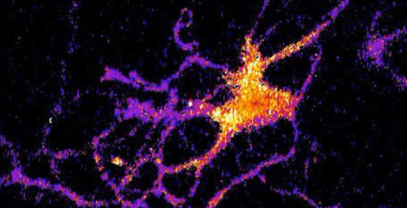
“We think this is a good system that is very cheap and very easy to make in very large quantities,” said MIT graduate student and the paper’s lead author, Tzu-Chieh Tang. To demonstrate the potential of their microbe culture, which they call Syn-SCOBY, the researchers created a material incorporating yeast that senses estradiol, which is sometimes found as an environmental pollutant. In another version, they used a strain of yeast that produces a glowing protein called luciferase when exposed to blue light. These yeasts could be swapped out for other strains that detect other pollutants, metals, or pathogens.

The researchers are now looking into using the Syn-SCOBY system for biomedical or food applications. For example, engineering the yeast cells to produce antimicrobials or proteins that could benefit human health.

Here’s a link to and a citation for the paper,

Living materials with programmable functionalities grown from engineered microbial co-cultures by Charlie Gilbert, Tzu-Chieh Tang, Wolfgang Ott, Brandon A. Dorr, William M. Shaw, George L. Sun, Timothy K. Lu & Tom Ellis. Nature Materials (2021) DOI: https://doi.org/10.1038/s41563-020-00857-5 Published: 11 January 2021

This paper is behind a paywall.

Mystery of North American insect bioluminescent systems unraveled by Brazilian scientists

I’ve always been fond of ‘l’ words and so it is that I’m compelled to post a story about a “luciferin-luciferase system” or, in this case, a story about insect bioluminescence.

Caption: Researchers isolated molecules present in the larvae of the fungus gnat Orfelia fultoni Credit: Vadim Viviani, UFSCar

A September 9, 2020 Fundação de Amparo à Pesquisa do Estado de São Paulo (FAPESP) press release (also on EurekAlert but published Sept. 11, 2020) announces research into ‘blue’ bioluminescence,

Molecules belonging to an almost unknown bioluminescent system found in larvae of the fungus gnat Orfelia fultoni (subfamily Keroplatinae) have been isolated for the first time by researchers at the Federal University of São Carlos (UFSCar) in the state of São Paulo, Brazil. The small fly is one of the few terrestrial organisms that produce blue light. It inhabits riverbanks in the Appalachian Mountains in the eastern United States. A key part of its bioluminescent system is a molecule also present in two recently discovered Brazilian flies.

The study, supported by Paulo Research Foundation – FAPESP, is published in Scientific Reports. Five authors are affiliated with UFSCar and two with universities in the United States.

The bioluminescent systems of glow-worms, fireflies and other insects are normally made up of luciferin (a low molecular weight molecule) and luciferase, an enzyme that catalyzes the oxidation of luciferin by oxygen, producing light. While some bioluminescent systems are well known and even used in biotechnological applications, others are poorly understood, including blue light-emitting systems, such as that of O. fultoni.

“In the published paper, we describe the properties of the insect’s luciferase and luciferin and their anatomical location in its larvae. We also specify several possible proteins that are possible candidates for the luciferase. We don’t yet know what type of protein it is, but it’s likely to be a hexamerin. In insects, hexamerins are storage proteins that provide amino acids, besides having other functions, such as binding low molecular weight compounds, like luciferin,” said Vadim Viviani, a professor in UFSCar’s Sustainability Science and Technology Center (CCTS) in Sorocaba, São Paulo, and principal investigator for the study.

The study was part of the FAPESP-funded project “Arthropod bioluminescence“. The partnership with United States-based researchers dates from a previous project, supported by FAPESP and the United States National Science Foundation (NSF), in partnership with Vanderbilt University (VU), located in Nashville, Tennessee.

In addition to luciferin and luciferase, researchers began characterizing a complex found in insects of the family Keroplatidae, which, in addition to O. fultoni, also includes a Brazilian species in the genus Neoditomyia that produces only luciferin and hence does not emit light.

Because they do not use it to emit light, the luciferin in O. fultoni and the Brazilian Neoditomyia has been named keroplatin. In larvae of this subfamily, keroplatin is associated with “black bodies” – large cells containing dark granules, proteins and probably mitochondria (energy-producing organelles). Researchers are still investigating the biological significance of this association between keroplatin and mitochondria.

“It’s a mystery,” Viviani said. “This luciferin may play a role in the mitochondrial energy metabolism. At night, probably in the presence of a natural chemical reducer, the luciferin is released by these black bodies and reacts with the surrounding luciferase to produce blue light. These are possibilities we plan to study.”

Brazilian cousins

An important factor in the elucidation of the United States insect’s bioluminescent system was the discovery of a larva that lives in Intervales State Park in São Paulo in 2018. It does not emit light but produces luciferin, similar to O. fultoni (read more at: agencia.fapesp.br/29066).

In their latest study, the group injected purified luciferase from the United States species into larvae of the Brazilian species, which then produced blue light. The nonluminescent Brazilian species is more abundant in nature than the United States species, so a larger amount of the material could be obtained for study purposes, especially to characterize the luciferin (keroplatin) present in both species.

In 2019, the group discovered and described Neoceroplatus betaryensis, a new species of fungus gnat, in collaboration with Cassius Stevani, a professor at the University of São Paulo’s Institute of Chemistry (IQ-USP). It was the first blue light-emitting insect found in South America and was detected in a privately held forest reserve near the Upper Ribeira State Tourist Park (PETAR) in the southern portion of the state of São Paulo. A close relative of O. fultoni, N. betaryensis inhabits fallen tree trunks in humid places (read more at: agencia.fapesp.br/31797).

“We show that the bioluminescent system of this Brazilian species is identical to that of O. fultoni. However, the insect is very rare, and so it’s hard to obtain sufficient material for research purposes,” Viviani said.

The researchers are now cloning the insect’s luciferase and characterizing it in molecular terms. They are also analyzing the chemical structure of its luciferin and the morphology of its lanterns.

“Once all this has been determined, we’ll be able to synthesize the luciferin and luciferase in the lab and use these systems in a range of biotech applications, such as studying cells. This will help us understand more about human diseases, among other things,” Viviani said.

Here’s a link to and a citation for the paper,

A new brilliantly blue-emitting luciferin-luciferase system from Orfelia fultoni and Keroplatinae (Diptera) by Vadim R. Viviani, Jaqueline R. Silva, Danilo T. Amaral, Vanessa R. Bevilaqua, Fabio C. Abdalla, Bruce R. Branchini & Carl H. Johnson. Scientific Reports volume 10, Article number: 9608 (2020) DOI: https://doi.org/10.1038/s41598-020-66286-1 Published 15 June 2020

This paper is open access.

The devil’s (i.e., luciferase) in the bioluminescent plant

The American Chemical Society (ACS) and the Massachusetts Institute of Technology (MIT) have both issued news releases about the latest in bioluminescence.The researchers tested their work on watercress, a vegetable that was viewed in almost sacred terms in my family; it was not easily available in Vancouver (Canada) when I was child.

My father would hunt down fresh watercress by checking out the Chinese grocery stores. He could spot the fresh stuff from across the street while driving at 30 miles or more per hour. Spotting it entailed an immediate hunt for parking (my father hated to pay so we might have go around the block a few times or more) and a dash out of the car to ensure that he got his watercress before anyone else spotted it. These days it’s much more easily available and, thankfully, my father has passed on so he won’t have to think about glowing watercress.

Getting back to bioluninescent vegetable research, the American Chemical Society’s Dec. 13, 2017 news release on EurekAlert (and as a Dec. 13, 2017 news item on ScienceDaily) makes the announcement,

The 2009 film “Avatar” created a lush imaginary world, illuminated by magical, glowing plants. Now researchers are starting to bring this spellbinding vision to life to help reduce our dependence on artificial lighting. They report in ACS’ journal Nano Letters a way to infuse plants with the luminescence of fireflies.

Nature has produced many bioluminescent organisms, however, plants are not among them. Most attempts so far to create glowing greenery — decorative tobacco plants in particular — have relied on introducing the genes of luminescent bacteria or fireflies through genetic engineering. But getting all the right components to the right locations within the plants has been a challenge. To gain better control over where light-generating ingredients end up, Michael S. Strano and colleagues recently created nanoparticles that travel to specific destinations within plants. Building on this work, the researchers wanted to take the next step and develop a “nanobionic,” glowing plant.

The team infused watercress and other plants with three different nanoparticles in a pressurized bath. The nanoparticles were loaded with light-emitting luciferin; luciferase, which modifies luciferin and makes it glow; and coenzyme A, which boosts luciferase activity. Using size and surface charge to control where the sets of nanoparticles could go within the plant tissues, the researchers could optimize how much light was emitted. Their watercress was half as bright as a commercial 1 microwatt LED and 100,000 times brighter than genetically engineered tobacco plants. Also, the plant could be turned off by adding a compound that blocks luciferase from activating luciferin’s glow.

Here’s a video from MIT detailing their research,

A December 13, 2017 MIT news release (also on EurekAlert) casts more light on the topic (I couldn’t resist the word play),

Imagine that instead of switching on a lamp when it gets dark, you could read by the light of a glowing plant on your desk.

MIT engineers have taken a critical first step toward making that vision a reality. By embedding specialized nanoparticles into the leaves of a watercress plant, they induced the plants to give off dim light for nearly four hours. They believe that, with further optimization, such plants will one day be bright enough to illuminate a workspace.

“The vision is to make a plant that will function as a desk lamp — a lamp that you don’t have to plug in. The light is ultimately powered by the energy metabolism of the plant itself,” says Michael Strano, the Carbon P. Dubbs Professor of Chemical Engineering at MIT and the senior author of the study

This technology could also be used to provide low-intensity indoor lighting, or to transform trees into self-powered streetlights, the researchers say.

MIT postdoc Seon-Yeong Kwak is the lead author of the study, which appears in the journal Nano Letters.

Nanobionic plants

Plant nanobionics, a new research area pioneered by Strano’s lab, aims to give plants novel features by embedding them with different types of nanoparticles. The group’s goal is to engineer plants to take over many of the functions now performed by electrical devices. The researchers have previously designed plants that can detect explosives and communicate that information to a smartphone, as well as plants that can monitor drought conditions.

Lighting, which accounts for about 20 percent of worldwide energy consumption, seemed like a logical next target. “Plants can self-repair, they have their own energy, and they are already adapted to the outdoor environment,” Strano says. “We think this is an idea whose time has come. It’s a perfect problem for plant nanobionics.”

To create their glowing plants, the MIT team turned to luciferase, the enzyme that gives fireflies their glow. Luciferase acts on a molecule called luciferin, causing it to emit light. Another molecule called co-enzyme A helps the process along by removing a reaction byproduct that can inhibit luciferase activity.

The MIT team packaged each of these three components into a different type of nanoparticle carrier. The nanoparticles, which are all made of materials that the U.S. Food and Drug Administration classifies as “generally regarded as safe,” help each component get to the right part of the plant. They also prevent the components from reaching concentrations that could be toxic to the plants.

The researchers used silica nanoparticles about 10 nanometers in diameter to carry luciferase, and they used slightly larger particles of the polymers PLGA and chitosan to carry luciferin and coenzyme A, respectively. To get the particles into plant leaves, the researchers first suspended the particles in a solution. Plants were immersed in the solution and then exposed to high pressure, allowing the particles to enter the leaves through tiny pores called stomata.

Particles releasing luciferin and coenzyme A were designed to accumulate in the extracellular space of the mesophyll, an inner layer of the leaf, while the smaller particles carrying luciferase enter the cells that make up the mesophyll. The PLGA particles gradually release luciferin, which then enters the plant cells, where luciferase performs the chemical reaction that makes luciferin glow.

The researchers’ early efforts at the start of the project yielded plants that could glow for about 45 minutes, which they have since improved to 3.5 hours. The light generated by one 10-centimeter watercress seedling is currently about one-thousandth of the amount needed to read by, but the researchers believe they can boost the light emitted, as well as the duration of light, by further optimizing the concentration and release rates of the components.

Plant transformation

Previous efforts to create light-emitting plants have relied on genetically engineering plants to express the gene for luciferase, but this is a laborious process that yields extremely dim light. Those studies were performed on tobacco plants and Arabidopsis thaliana, which are commonly used for plant genetic studies. However, the method developed by Strano’s lab could be used on any type of plant. So far, they have demonstrated it with arugula, kale, and spinach, in addition to watercress.

For future versions of this technology, the researchers hope to develop a way to paint or spray the nanoparticles onto plant leaves, which could make it possible to transform trees and other large plants into light sources.

“Our target is to perform one treatment when the plant is a seedling or a mature plant, and have it last for the lifetime of the plant,” Strano says. “Our work very seriously opens up the doorway to streetlamps that are nothing but treated trees, and to indirect lighting around homes.”

The researchers have also demonstrated that they can turn the light off by adding nanoparticles carrying a luciferase inhibitor. This could enable them to eventually create plants that shut off their light emission in response to environmental conditions such as sunlight, the researchers say.

Here’s a link to and a citation for the paper,

A Nanobionic Light-Emitting Plant by Seon-Yeong Kwak, Juan Pablo Giraldo, Min Hao Wong, Volodymyr B. Koman, Tedrick Thomas Salim Lew, Jon Ell, Mark C. Weidman, Rosalie M. Sinclair, Markita P. Landry, William A. Tisdale, and Michael S. Strano. Nano Lett., 2017, 17 (12), pp 7951–7961 DOI: 10.1021/acs.nanolett.7b04369 Publication Date (Web): November 17, 2017

Copyright © 2017 American Chemical Society

This paper is behind a paywall.

Getting your brain cells to glow in the dark

The extraordinary effort to colonize our brains continues apace with a new sensor from Vanderbilt University. From an Oct. 27, 2016 news item on ScienceDaily,

A new kind of bioluminescent sensor causes individual brain cells to imitate fireflies and glow in the dark.

The probe, which was developed by a team of Vanderbilt scientists, is a genetically modified form of luciferase, the enzyme that a number of other species including fireflies use to produce light. …

The scientists created the technique as a new and improved method for tracking the interactions within large neural networks in the brain.

“For a long time neuroscientists relied on electrical techniques for recording the activity of neurons. These are very good at monitoring individual neurons but are limited to small numbers of neurons. The new wave is to use optical techniques to record the activity of hundreds of neurons at the same time,” said Carl Johnson, Stevenson Professor of Biological Sciences, who headed the effort.

Individual neuron glowing with bioluminescent light produced by a new genetically engineered sensor. (Johnson Lab / Vanderbilt University)

Individual neuron glowing with bioluminescent light produced by a new genetically engineered sensor. (Johnson Lab / Vanderbilt University)

An Oct. 27, 2016 Vanderbilt University news release (also on EurekAlert) by David Salisbury, which originated the news item, explains the work in more detail,

“Most of the efforts in optical recording use fluorescence, but this requires a strong external light source which can cause the tissue to heat up and can interfere with some biological processes, particularly those that are light sensitive,” he [Carl Johnson] said.

Based on their research on bioluminescence in “a scummy little organism, the green alga Chlamydomonas, that nobody cares much about” Johnson and his colleagues realized that if they could combine luminescence with optogenetics – a new biological technique that uses light to control cells, particularly neurons, in living tissue – they could create a powerful new tool for studying brain activity.

“There is an inherent conflict between fluorescent techniques and optogenetics. The light required to produce the fluorescence interferes with the light required to control the cells,” said Johnson. “Luminescence, on the other hand, works in the dark!”

Johnson and his collaborators – Associate Professor Donna Webb, Research Assistant Professor Shuqun Shi, post-doctoral student Jie Yang and doctoral student Derrick Cumberbatch in biological sciences and Professor Danny Winder and postdoctoral student Samuel Centanni in molecular physiology and biophysics – genetically modified a type of luciferase obtained from a luminescent species of shrimp so that it would light up when exposed to calcium ions. Then they hijacked a virus that infects neurons and attached it to their sensor molecule so that the sensors are inserted into the cell interior.

The researchers picked calcium ions because they are involved in neuron activation. Although calcium levels are high in the surrounding area, normally they are very low inside the neurons. However, the internal calcium level spikes briefly when a neuron receives an impulse from one of its neighbors.

They tested their new calcium sensor with one of the optogenetic probes (channelrhodopsin) that causes the calcium ion channels in the neuron’s outer membrane to open, flooding the cell with calcium. Using neurons grown in culture they found that the luminescent enzyme reacted visibly to the influx of calcium produced when the probe was stimulated by brief light flashes of visible light.

To determine how well their sensor works with larger numbers of neurons, they inserted it into brain slices from the mouse hippocampus that contain thousands of neurons. In this case they flooded the slices with an increased concentration of potassium ions, which causes the cell’s ion channels to open. Again, they found that the sensor responded to the variations in calcium concentrations by brightening and dimming.

“We’ve shown that the approach works,” Johnson said. “Now we have to determine how sensitive it is. We have some indications that it is sensitive enough to detect the firing of individual neurons, but we have to run more tests to determine if it actually has this capability.”

Here’s a link to and a citation for the paper,

Coupling optogenetic stimulation with NanoLuc-based luminescence (BRET) Ca++ sensing by Jie Yang, Derrick Cumberbatch, Samuel Centanni, Shu-qun Shi, Danny Winder, Donna Webb, & Carl Hirschie Johnson. Nature Communications 7, Article number: 13268 (2016)  doi:10.1038/ncomms13268 Published online: 27 October 2016

This paper is open access.