Tag Archives: luciferin

Mystery of North American insect bioluminescent systems unraveled by Brazilian scientists

I’ve always been fond of ‘l’ words and so it is that I’m compelled to post a story about a “luciferin-luciferase system” or, in this case, a story about insect bioluminescence.

Caption: Researchers isolated molecules present in the larvae of the fungus gnat Orfelia fultoni Credit: Vadim Viviani, UFSCar

A September 9, 2020 Fundação de Amparo à Pesquisa do Estado de São Paulo (FAPESP) press release (also on EurekAlert but published Sept. 11, 2020) announces research into ‘blue’ bioluminescence,

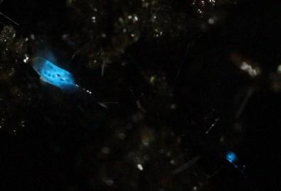
Molecules belonging to an almost unknown bioluminescent system found in larvae of the fungus gnat Orfelia fultoni (subfamily Keroplatinae) have been isolated for the first time by researchers at the Federal University of São Carlos (UFSCar) in the state of São Paulo, Brazil. The small fly is one of the few terrestrial organisms that produce blue light. It inhabits riverbanks in the Appalachian Mountains in the eastern United States. A key part of its bioluminescent system is a molecule also present in two recently discovered Brazilian flies.

The study, supported by Paulo Research Foundation – FAPESP, is published in Scientific Reports. Five authors are affiliated with UFSCar and two with universities in the United States.

The bioluminescent systems of glow-worms, fireflies and other insects are normally made up of luciferin (a low molecular weight molecule) and luciferase, an enzyme that catalyzes the oxidation of luciferin by oxygen, producing light. While some bioluminescent systems are well known and even used in biotechnological applications, others are poorly understood, including blue light-emitting systems, such as that of O. fultoni.

“In the published paper, we describe the properties of the insect’s luciferase and luciferin and their anatomical location in its larvae. We also specify several possible proteins that are possible candidates for the luciferase. We don’t yet know what type of protein it is, but it’s likely to be a hexamerin. In insects, hexamerins are storage proteins that provide amino acids, besides having other functions, such as binding low molecular weight compounds, like luciferin,” said Vadim Viviani, a professor in UFSCar’s Sustainability Science and Technology Center (CCTS) in Sorocaba, São Paulo, and principal investigator for the study.

The study was part of the FAPESP-funded project “Arthropod bioluminescence“. The partnership with United States-based researchers dates from a previous project, supported by FAPESP and the United States National Science Foundation (NSF), in partnership with Vanderbilt University (VU), located in Nashville, Tennessee.

In addition to luciferin and luciferase, researchers began characterizing a complex found in insects of the family Keroplatidae, which, in addition to O. fultoni, also includes a Brazilian species in the genus Neoditomyia that produces only luciferin and hence does not emit light.

Because they do not use it to emit light, the luciferin in O. fultoni and the Brazilian Neoditomyia has been named keroplatin. In larvae of this subfamily, keroplatin is associated with “black bodies” – large cells containing dark granules, proteins and probably mitochondria (energy-producing organelles). Researchers are still investigating the biological significance of this association between keroplatin and mitochondria.

“It’s a mystery,” Viviani said. “This luciferin may play a role in the mitochondrial energy metabolism. At night, probably in the presence of a natural chemical reducer, the luciferin is released by these black bodies and reacts with the surrounding luciferase to produce blue light. These are possibilities we plan to study.”

Brazilian cousins

An important factor in the elucidation of the United States insect’s bioluminescent system was the discovery of a larva that lives in Intervales State Park in São Paulo in 2018. It does not emit light but produces luciferin, similar to O. fultoni (read more at: agencia.fapesp.br/29066).

In their latest study, the group injected purified luciferase from the United States species into larvae of the Brazilian species, which then produced blue light. The nonluminescent Brazilian species is more abundant in nature than the United States species, so a larger amount of the material could be obtained for study purposes, especially to characterize the luciferin (keroplatin) present in both species.

In 2019, the group discovered and described Neoceroplatus betaryensis, a new species of fungus gnat, in collaboration with Cassius Stevani, a professor at the University of São Paulo’s Institute of Chemistry (IQ-USP). It was the first blue light-emitting insect found in South America and was detected in a privately held forest reserve near the Upper Ribeira State Tourist Park (PETAR) in the southern portion of the state of São Paulo. A close relative of O. fultoni, N. betaryensis inhabits fallen tree trunks in humid places (read more at: agencia.fapesp.br/31797).

“We show that the bioluminescent system of this Brazilian species is identical to that of O. fultoni. However, the insect is very rare, and so it’s hard to obtain sufficient material for research purposes,” Viviani said.

The researchers are now cloning the insect’s luciferase and characterizing it in molecular terms. They are also analyzing the chemical structure of its luciferin and the morphology of its lanterns.

“Once all this has been determined, we’ll be able to synthesize the luciferin and luciferase in the lab and use these systems in a range of biotech applications, such as studying cells. This will help us understand more about human diseases, among other things,” Viviani said.

Here’s a link to and a citation for the paper,

A new brilliantly blue-emitting luciferin-luciferase system from Orfelia fultoni and Keroplatinae (Diptera) by Vadim R. Viviani, Jaqueline R. Silva, Danilo T. Amaral, Vanessa R. Bevilaqua, Fabio C. Abdalla, Bruce R. Branchini & Carl H. Johnson. Scientific Reports volume 10, Article number: 9608 (2020) DOI: https://doi.org/10.1038/s41598-020-66286-1 Published 15 June 2020

This paper is open access.

The devil’s (i.e., luciferase) in the bioluminescent plant

The American Chemical Society (ACS) and the Massachusetts Institute of Technology (MIT) have both issued news releases about the latest in bioluminescence.The researchers tested their work on watercress, a vegetable that was viewed in almost sacred terms in my family; it was not easily available in Vancouver (Canada) when I was child.

My father would hunt down fresh watercress by checking out the Chinese grocery stores. He could spot the fresh stuff from across the street while driving at 30 miles or more per hour. Spotting it entailed an immediate hunt for parking (my father hated to pay so we might have go around the block a few times or more) and a dash out of the car to ensure that he got his watercress before anyone else spotted it. These days it’s much more easily available and, thankfully, my father has passed on so he won’t have to think about glowing watercress.

Getting back to bioluninescent vegetable research, the American Chemical Society’s Dec. 13, 2017 news release on EurekAlert (and as a Dec. 13, 2017 news item on ScienceDaily) makes the announcement,

The 2009 film “Avatar” created a lush imaginary world, illuminated by magical, glowing plants. Now researchers are starting to bring this spellbinding vision to life to help reduce our dependence on artificial lighting. They report in ACS’ journal Nano Letters a way to infuse plants with the luminescence of fireflies.

Nature has produced many bioluminescent organisms, however, plants are not among them. Most attempts so far to create glowing greenery — decorative tobacco plants in particular — have relied on introducing the genes of luminescent bacteria or fireflies through genetic engineering. But getting all the right components to the right locations within the plants has been a challenge. To gain better control over where light-generating ingredients end up, Michael S. Strano and colleagues recently created nanoparticles that travel to specific destinations within plants. Building on this work, the researchers wanted to take the next step and develop a “nanobionic,” glowing plant.

The team infused watercress and other plants with three different nanoparticles in a pressurized bath. The nanoparticles were loaded with light-emitting luciferin; luciferase, which modifies luciferin and makes it glow; and coenzyme A, which boosts luciferase activity. Using size and surface charge to control where the sets of nanoparticles could go within the plant tissues, the researchers could optimize how much light was emitted. Their watercress was half as bright as a commercial 1 microwatt LED and 100,000 times brighter than genetically engineered tobacco plants. Also, the plant could be turned off by adding a compound that blocks luciferase from activating luciferin’s glow.

Here’s a video from MIT detailing their research,

A December 13, 2017 MIT news release (also on EurekAlert) casts more light on the topic (I couldn’t resist the word play),

Imagine that instead of switching on a lamp when it gets dark, you could read by the light of a glowing plant on your desk.

MIT engineers have taken a critical first step toward making that vision a reality. By embedding specialized nanoparticles into the leaves of a watercress plant, they induced the plants to give off dim light for nearly four hours. They believe that, with further optimization, such plants will one day be bright enough to illuminate a workspace.

“The vision is to make a plant that will function as a desk lamp — a lamp that you don’t have to plug in. The light is ultimately powered by the energy metabolism of the plant itself,” says Michael Strano, the Carbon P. Dubbs Professor of Chemical Engineering at MIT and the senior author of the study

This technology could also be used to provide low-intensity indoor lighting, or to transform trees into self-powered streetlights, the researchers say.

MIT postdoc Seon-Yeong Kwak is the lead author of the study, which appears in the journal Nano Letters.

Nanobionic plants

Plant nanobionics, a new research area pioneered by Strano’s lab, aims to give plants novel features by embedding them with different types of nanoparticles. The group’s goal is to engineer plants to take over many of the functions now performed by electrical devices. The researchers have previously designed plants that can detect explosives and communicate that information to a smartphone, as well as plants that can monitor drought conditions.

Lighting, which accounts for about 20 percent of worldwide energy consumption, seemed like a logical next target. “Plants can self-repair, they have their own energy, and they are already adapted to the outdoor environment,” Strano says. “We think this is an idea whose time has come. It’s a perfect problem for plant nanobionics.”

To create their glowing plants, the MIT team turned to luciferase, the enzyme that gives fireflies their glow. Luciferase acts on a molecule called luciferin, causing it to emit light. Another molecule called co-enzyme A helps the process along by removing a reaction byproduct that can inhibit luciferase activity.

The MIT team packaged each of these three components into a different type of nanoparticle carrier. The nanoparticles, which are all made of materials that the U.S. Food and Drug Administration classifies as “generally regarded as safe,” help each component get to the right part of the plant. They also prevent the components from reaching concentrations that could be toxic to the plants.

The researchers used silica nanoparticles about 10 nanometers in diameter to carry luciferase, and they used slightly larger particles of the polymers PLGA and chitosan to carry luciferin and coenzyme A, respectively. To get the particles into plant leaves, the researchers first suspended the particles in a solution. Plants were immersed in the solution and then exposed to high pressure, allowing the particles to enter the leaves through tiny pores called stomata.

Particles releasing luciferin and coenzyme A were designed to accumulate in the extracellular space of the mesophyll, an inner layer of the leaf, while the smaller particles carrying luciferase enter the cells that make up the mesophyll. The PLGA particles gradually release luciferin, which then enters the plant cells, where luciferase performs the chemical reaction that makes luciferin glow.

The researchers’ early efforts at the start of the project yielded plants that could glow for about 45 minutes, which they have since improved to 3.5 hours. The light generated by one 10-centimeter watercress seedling is currently about one-thousandth of the amount needed to read by, but the researchers believe they can boost the light emitted, as well as the duration of light, by further optimizing the concentration and release rates of the components.

Plant transformation

Previous efforts to create light-emitting plants have relied on genetically engineering plants to express the gene for luciferase, but this is a laborious process that yields extremely dim light. Those studies were performed on tobacco plants and Arabidopsis thaliana, which are commonly used for plant genetic studies. However, the method developed by Strano’s lab could be used on any type of plant. So far, they have demonstrated it with arugula, kale, and spinach, in addition to watercress.

For future versions of this technology, the researchers hope to develop a way to paint or spray the nanoparticles onto plant leaves, which could make it possible to transform trees and other large plants into light sources.

“Our target is to perform one treatment when the plant is a seedling or a mature plant, and have it last for the lifetime of the plant,” Strano says. “Our work very seriously opens up the doorway to streetlamps that are nothing but treated trees, and to indirect lighting around homes.”

The researchers have also demonstrated that they can turn the light off by adding nanoparticles carrying a luciferase inhibitor. This could enable them to eventually create plants that shut off their light emission in response to environmental conditions such as sunlight, the researchers say.

Here’s a link to and a citation for the paper,

A Nanobionic Light-Emitting Plant by Seon-Yeong Kwak, Juan Pablo Giraldo, Min Hao Wong, Volodymyr B. Koman, Tedrick Thomas Salim Lew, Jon Ell, Mark C. Weidman, Rosalie M. Sinclair, Markita P. Landry, William A. Tisdale, and Michael S. Strano. Nano Lett., 2017, 17 (12), pp 7951–7961 DOI: 10.1021/acs.nanolett.7b04369 Publication Date (Web): November 17, 2017

Copyright © 2017 American Chemical Society

This paper is behind a paywall.

The science behind a firefly’s glow

A Dec. 17, 2014 news item on Nanotechnology Now describes research into the phenomenon of bioluminescence and fireflies,

 Fireflies used rapid light flashes to communicate. This “bioluminescence” is an intriguing phenomenon that has many potential applications, from drug testing and monitoring water contamination, and even lighting up streets using glow-in-dark trees and plants. Fireflies emit light when a compound called luciferin breaks down. We know that this reaction needs oxygen, but what we don’t know is how fireflies actually supply oxygen to their light-emitting cells. Using state-of-the-art imaging techniques, scientists from Switzerland and Taiwan have determined how fireflies control oxygen distribution to light up their cells. The work is published in Physical Review Letters.

A Dec. 17, 2014 EPFL (Ecole Polytechnique Fédérale de Lausanne) news release on EurekAlert provides more description of the work,

The firefly’s light-producing organ is called the “lantern”, and it is located in the insect’s abdomen. It looks like a series of tubes progressing into smaller ones and so one, like a tree’s branches growing into twigs. The function of these tubes, called, is to supply oxygen to the cells of the lantern, which contain luciferase and can produce light. However, the complexity of the firefly’s lantern has made it difficult to study this mechanism in depth, and reproduce it for technological applications.

Giorgio Margaritondo at EPFL, Yeukuang Hwu at the Academia Sinica and their colleagues at the National Tsing Hua University in Taiwan have successfully used two sophisticated imaging techniques to overcome the complexity of the firefly lantern and map out how oxygen is supplied to light-emitting cells. The techniques are called synchrotron phase contrast microtomography and transmission x-ray microscopy. They can scan down to the level of a single cell, even allowing researchers to look inside it.

By applying these techniques on live fireflies, the scientists were able to see the entire structure of the lantern for the first time, and to also make quantitative evaluations of oxygen distribution.

The imaging showed that the firefly diverts oxygen from other cellular functions and puts it into the reaction that breaks up luciferin. Specifically, the researchers found that oxygen consumption in the cell decreased, slowing down energy production. At the same time, oxygen supply switched to light-emission.

The study is the first to ever show the firefly’s lantern in such detail, while also providing clear evidence that it is optimized for light emission thanks to the state-of-the-art techniques used by the scientists. But Margaritondo points out another innovation: “The techniques we used have an advantage over, say, conventional x-ray techniques, which cannot easily distinguish between soft tissues. By using an approach based on changes in light intensity (phase-contrast) as opposed to light absorption (x-rays), we were able to achieve high-resolution imaging of the delicate firefly lantern.”

Here’s an image illustrating the work,

Tomographic Reconstruction of Part of the Firefly Lantern;  This detailed microimage shows larger channels branching into smaller ones, supplying oxygen for the firefly's light emission. The smallest channels are ten thousand times smaller than a millimeter and therefore invisible to other experimental probes: this has prevented scientists so far to unlock the mystery of firefly light flashes. Credit: Giorgio Margaritondo/EPFL

Tomographic Reconstruction of Part of the Firefly Lantern; This detailed microimage shows larger channels branching into smaller ones, supplying oxygen for the firefly’s light emission. The smallest channels are ten thousand times smaller than a millimeter and therefore invisible to other experimental probes: this has prevented scientists so far to unlock the mystery of firefly light flashes. Credit: Giorgio Margaritondo/EPFL

Here’s a link to and a citation for the paper,

Firefly Light Flashing: Oxygen Supply Mechanism by Yueh-Lin Tsai, Chia-Wei Li, Tzay-Ming Hong, Jen-Zon Ho, En-Cheng Yang, Wen-Yen Wu, G. Margaritondo, Su-Ting Hsu, Edwin B. L. Ong, and Y. Hwu. Phys. Rev. Lett. 113, 258103 – Published 17 December 2014 DOI:  http://dx.doi.org/10.1103/PhysRevLett.113.258103

This paper is behind a paywall.