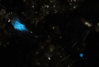
Caption: These soft, living materials glow in response to mechanical stress, such as compression, stretching or twisting. Credit: UC San Diego Jacobs School of Engineering
An October 20, 2023 news item on phys.org describes research into bioluminescent materials, Note: A link has been removed,
A team of researchers led by the University of California San Diego has developed soft yet durable materials that glow in response to mechanical stress, such as compression, stretching or twisting. The materials derive their luminescence from single-celled algae known as dinoflagellates.
The work, inspired by the bioluminescent waves observed during red tide events at San Diego’s beaches, was published Oct. 20 [2023] in Science Advances.
An exciting feature of these materials is their inherent simplicity—they need no electronics, no external power source,” said study senior author Shengqiang Cai, a professor of mechanical and aerospace engineering at the UC San Diego Jacobs School of Engineering. “We demonstrate how we can harness the power of nature to directly convert mechanical stimuli into light emission.”
This study was a multi-disciplinary collaboration involving engineers and materials scientists in Cai’s lab, marine biologist Michael Latz at UC San Diego’s Scripps Institution of Oceanography, and physics professor Maziyar Jalaal at University of Amsterdam.
The primary ingredients of the bioluminescent materials are dinoflagellates and a seaweed-based polymer called alginate. These elements were mixed to form a solution, which was then processed with a 3D printer to create a diverse array of shapes, such as grids, spirals, spiderwebs, balls, blocks and pyramid-like structures. The 3D-printed structures were then cured as a final step.
When the materials are subjected to compression, stretching or twisting, the dinoflagellates within them respond by emitting light. This response mimics what happens in the ocean, when dinoflagellates produce flashes of light as part of a predator defense strategy. In tests, the materials glowed when the researchers pressed on them and traced patterns on their surface. The materials were even sensitive enough to glow under the weight of a foam ball rolling on their surface.
The greater the applied stress, the brighter the glow. The researchers were able to quantify this behavior and developed a mathematical model that can predict the intensity of the glow based on the magnitude of the mechanical stress applied.
The researchers also demonstrated techniques to make these materials resilient in various experimental conditions. To reinforce the materials so that they can bear substantial mechanical loads, a second polymer, poly(ethylene glycol) diacrylate, was added to the original blend. Also, coating the materials with a stretchy rubber-like polymer called Ecoflex provided protection in acidic and basic solutions. With this protective layer, the materials could even be stored in seawater for up to five months without losing their form or bioluminescent properties.
Another beneficial feature of these materials is their minimal maintenance requirements. To keep working, the dinoflagellates within the materials need periodic cycles of light and darkness. During the light phase, they photosynthesize to produce food and energy, which are then used in the dark phase to emit light when mechanical stress is applied. This behavior mirrors the natural processes at play when the dinoflagellates cause bioluminescence in the ocean during red tide events.
“This current work demonstrates a simple method to combine living organisms with non-living components to fabricate novel materials that are self-sustaining and are sensitive to fundamental mechanical stimuli found in nature,” said study first author Chenghai Li, a mechanical and aerospace engineering Ph.D. candidate in Cai’s lab.
The researchers envision that these materials could potentially be used as mechanical sensors to gauge pressure, strain or stress. Other potential applications include soft robotics and biomedical devices that use light signals to perform treatment or controlled drug release.
However, there is much work to be done before these applications can be realized. The researchers are working on further improving and optimizing the materials.
I’ve always been fond of ‘l’ words and so it is that I’m compelled to post a story about a “luciferin-luciferase system” or, in this case, a story about insect bioluminescence.
Caption: Researchers isolated molecules present in the larvae of the fungus gnat Orfelia fultoni Credit: Vadim Viviani, UFSCar
Molecules belonging to an almost unknown bioluminescent system found in larvae of the fungus gnat Orfelia fultoni (subfamily Keroplatinae) have been isolated for the first time by researchers at the Federal University of São Carlos (UFSCar) in the state of São Paulo, Brazil. The small fly is one of the few terrestrial organisms that produce blue light. It inhabits riverbanks in the Appalachian Mountains in the eastern United States. A key part of its bioluminescent system is a molecule also present in two recently discovered Brazilian flies.
The study, supported by Paulo Research Foundation – FAPESP, is published in Scientific Reports. Five authors are affiliated with UFSCar and two with universities in the United States.
The bioluminescent systems of glow-worms, fireflies and other insects are normally made up of luciferin (a low molecular weight molecule) and luciferase, an enzyme that catalyzes the oxidation of luciferin by oxygen, producing light. While some bioluminescent systems are well known and even used in biotechnological applications, others are poorly understood, including blue light-emitting systems, such as that of O. fultoni.
“In the published paper, we describe the properties of the insect’s luciferase and luciferin and their anatomical location in its larvae. We also specify several possible proteins that are possible candidates for the luciferase. We don’t yet know what type of protein it is, but it’s likely to be a hexamerin. In insects, hexamerins are storage proteins that provide amino acids, besides having other functions, such as binding low molecular weight compounds, like luciferin,” said Vadim Viviani, a professor in UFSCar’s Sustainability Science and Technology Center (CCTS) in Sorocaba, São Paulo, and principal investigator for the study.
The study was part of the FAPESP-funded project “Arthropod bioluminescence“. The partnership with United States-based researchers dates from a previous project, supported by FAPESP and the United States National Science Foundation (NSF), in partnership with Vanderbilt University (VU), located in Nashville, Tennessee.
In addition to luciferin and luciferase, researchers began characterizing a complex found in insects of the family Keroplatidae, which, in addition to O. fultoni, also includes a Brazilian species in the genus Neoditomyia that produces only luciferin and hence does not emit light.
Because they do not use it to emit light, the luciferin in O. fultoni and the Brazilian Neoditomyia has been named keroplatin. In larvae of this subfamily, keroplatin is associated with “black bodies” – large cells containing dark granules, proteins and probably mitochondria (energy-producing organelles). Researchers are still investigating the biological significance of this association between keroplatin and mitochondria.
“It’s a mystery,” Viviani said. “This luciferin may play a role in the mitochondrial energy metabolism. At night, probably in the presence of a natural chemical reducer, the luciferin is released by these black bodies and reacts with the surrounding luciferase to produce blue light. These are possibilities we plan to study.”
Brazilian cousins
An important factor in the elucidation of the United States insect’s bioluminescent system was the discovery of a larva that lives in Intervales State Park in São Paulo in 2018. It does not emit light but produces luciferin, similar to O. fultoni (read more at: agencia.fapesp.br/29066).
In their latest study, the group injected purified luciferase from the United States species into larvae of the Brazilian species, which then produced blue light. The nonluminescent Brazilian species is more abundant in nature than the United States species, so a larger amount of the material could be obtained for study purposes, especially to characterize the luciferin (keroplatin) present in both species.
In 2019, the group discovered and described Neoceroplatus betaryensis, a new species of fungus gnat, in collaboration with Cassius Stevani, a professor at the University of São Paulo’s Institute of Chemistry (IQ-USP). It was the first blue light-emitting insect found in South America and was detected in a privately held forest reserve near the Upper Ribeira State Tourist Park (PETAR) in the southern portion of the state of São Paulo. A close relative of O. fultoni, N. betaryensis inhabits fallen tree trunks in humid places (read more at: agencia.fapesp.br/31797).
“We show that the bioluminescent system of this Brazilian species is identical to that of O. fultoni. However, the insect is very rare, and so it’s hard to obtain sufficient material for research purposes,” Viviani said.
The researchers are now cloning the insect’s luciferase and characterizing it in molecular terms. They are also analyzing the chemical structure of its luciferin and the morphology of its lanterns.
“Once all this has been determined, we’ll be able to synthesize the luciferin and luciferase in the lab and use these systems in a range of biotech applications, such as studying cells. This will help us understand more about human diseases, among other things,” Viviani said.
The American Chemical Society (ACS) and the Massachusetts Institute of Technology (MIT) have both issued news releases about the latest in bioluminescence.The researchers tested their work on watercress, a vegetable that was viewed in almost sacred terms in my family; it was not easily available in Vancouver (Canada) when I was child.
My father would hunt down fresh watercress by checking out the Chinese grocery stores. He could spot the fresh stuff from across the street while driving at 30 miles or more per hour. Spotting it entailed an immediate hunt for parking (my father hated to pay so we might have go around the block a few times or more) and a dash out of the car to ensure that he got his watercress before anyone else spotted it. These days it’s much more easily available and, thankfully, my father has passed on so he won’t have to think about glowing watercress.
The 2009 film “Avatar” created a lush imaginary world, illuminated by magical, glowing plants. Now researchers are starting to bring this spellbinding vision to life to help reduce our dependence on artificial lighting. They report in ACS’ journal Nano Letters a way to infuse plants with the luminescence of fireflies.
Nature has produced many bioluminescent organisms, however, plants are not among them. Most attempts so far to create glowing greenery — decorative tobacco plants in particular — have relied on introducing the genes of luminescent bacteria or fireflies through genetic engineering. But getting all the right components to the right locations within the plants has been a challenge. To gain better control over where light-generating ingredients end up, Michael S. Strano and colleagues recently created nanoparticles that travel to specific destinations within plants. Building on this work, the researchers wanted to take the next step and develop a “nanobionic,” glowing plant.
The team infused watercress and other plants with three different nanoparticles in a pressurized bath. The nanoparticles were loaded with light-emitting luciferin; luciferase, which modifies luciferin and makes it glow; and coenzyme A, which boosts luciferase activity. Using size and surface charge to control where the sets of nanoparticles could go within the plant tissues, the researchers could optimize how much light was emitted. Their watercress was half as bright as a commercial 1 microwatt LED and 100,000 times brighter than genetically engineered tobacco plants. Also, the plant could be turned off by adding a compound that blocks luciferase from activating luciferin’s glow.
Imagine that instead of switching on a lamp when it gets dark, you could read by the light of a glowing plant on your desk.
MIT engineers have taken a critical first step toward making that vision a reality. By embedding specialized nanoparticles into the leaves of a watercress plant, they induced the plants to give off dim light for nearly four hours. They believe that, with further optimization, such plants will one day be bright enough to illuminate a workspace.
“The vision is to make a plant that will function as a desk lamp — a lamp that you don’t have to plug in. The light is ultimately powered by the energy metabolism of the plant itself,” says Michael Strano, the Carbon P. Dubbs Professor of Chemical Engineering at MIT and the senior author of the study
This technology could also be used to provide low-intensity indoor lighting, or to transform trees into self-powered streetlights, the researchers say.
MIT postdoc Seon-Yeong Kwak is the lead author of the study, which appears in the journal Nano Letters.
Nanobionic plants
Plant nanobionics, a new research area pioneered by Strano’s lab, aims to give plants novel features by embedding them with different types of nanoparticles. The group’s goal is to engineer plants to take over many of the functions now performed by electrical devices. The researchers have previously designed plants that can detect explosives and communicate that information to a smartphone, as well as plants that can monitor drought conditions.
Lighting, which accounts for about 20 percent of worldwide energy consumption, seemed like a logical next target. “Plants can self-repair, they have their own energy, and they are already adapted to the outdoor environment,” Strano says. “We think this is an idea whose time has come. It’s a perfect problem for plant nanobionics.”
To create their glowing plants, the MIT team turned to luciferase, the enzyme that gives fireflies their glow. Luciferase acts on a molecule called luciferin, causing it to emit light. Another molecule called co-enzyme A helps the process along by removing a reaction byproduct that can inhibit luciferase activity.
The MIT team packaged each of these three components into a different type of nanoparticle carrier. The nanoparticles, which are all made of materials that the U.S. Food and Drug Administration classifies as “generally regarded as safe,” help each component get to the right part of the plant. They also prevent the components from reaching concentrations that could be toxic to the plants.
The researchers used silica nanoparticles about 10 nanometers in diameter to carry luciferase, and they used slightly larger particles of the polymers PLGA and chitosan to carry luciferin and coenzyme A, respectively. To get the particles into plant leaves, the researchers first suspended the particles in a solution. Plants were immersed in the solution and then exposed to high pressure, allowing the particles to enter the leaves through tiny pores called stomata.
Particles releasing luciferin and coenzyme A were designed to accumulate in the extracellular space of the mesophyll, an inner layer of the leaf, while the smaller particles carrying luciferase enter the cells that make up the mesophyll. The PLGA particles gradually release luciferin, which then enters the plant cells, where luciferase performs the chemical reaction that makes luciferin glow.
The researchers’ early efforts at the start of the project yielded plants that could glow for about 45 minutes, which they have since improved to 3.5 hours. The light generated by one 10-centimeter watercress seedling is currently about one-thousandth of the amount needed to read by, but the researchers believe they can boost the light emitted, as well as the duration of light, by further optimizing the concentration and release rates of the components.
Plant transformation
Previous efforts to create light-emitting plants have relied on genetically engineering plants to express the gene for luciferase, but this is a laborious process that yields extremely dim light. Those studies were performed on tobacco plants and Arabidopsis thaliana, which are commonly used for plant genetic studies. However, the method developed by Strano’s lab could be used on any type of plant. So far, they have demonstrated it with arugula, kale, and spinach, in addition to watercress.
For future versions of this technology, the researchers hope to develop a way to paint or spray the nanoparticles onto plant leaves, which could make it possible to transform trees and other large plants into light sources.
“Our target is to perform one treatment when the plant is a seedling or a mature plant, and have it last for the lifetime of the plant,” Strano says. “Our work very seriously opens up the doorway to streetlamps that are nothing but treated trees, and to indirect lighting around homes.”
The researchers have also demonstrated that they can turn the light off by adding nanoparticles carrying a luciferase inhibitor. This could enable them to eventually create plants that shut off their light emission in response to environmental conditions such as sunlight, the researchers say.
Here’s a link to and a citation for the paper,
A Nanobionic Light-Emitting Plant by Seon-Yeong Kwak, Juan Pablo Giraldo, Min Hao Wong, Volodymyr B. Koman, Tedrick Thomas Salim Lew, Jon Ell, Mark C. Weidman, Rosalie M. Sinclair, Markita P. Landry, William A. Tisdale, and Michael S. Strano. Nano Lett., 2017, 17 (12), pp 7951–7961 DOI: 10.1021/acs.nanolett.7b04369 Publication Date (Web): November 17, 2017
I think it was about five years ago thatI wrote a paper on something I called ‘cognitive entanglement’ (mentioned in my July 20,2012 posting) so the latest from Northwestern University (Chicago, Illinois, US) reignited my interest in entanglement. A December 5, 2017 news item on ScienceDaily describes the latest ‘entanglement’ research,
Nearly 75 years ago, Nobel Prize-winning physicist Erwin Schrödinger wondered if the mysterious world of quantum mechanics played a role in biology. A recent finding by Northwestern University’s Prem Kumar adds further evidence that the answer might be yes.
Kumar and his team have, for the first time, created quantum entanglement from a biological system. This finding could advance scientists’ fundamental understanding of biology and potentially open doors to exploit biological tools to enable new functions by harnessing quantum mechanics.
“Can we apply quantum tools to learn about biology?” said Kumar, professor of electrical engineering and computer science in Northwestern’s McCormick School of Engineering and of physics and astronomy in the Weinberg College of Arts and Sciences. “People have asked this question for many, many years — dating back to the dawn of quantum mechanics. The reason we are interested in these new quantum states is because they allow applications that are otherwise impossible.”
Partially supported by the [US] Defense Advanced Research Projects Agency [DARPA], the research was published Dec. 5 [2017] in Nature Communications.
Quantum entanglement is one of quantum mechanics’ most mystifying phenomena. When two particles — such as atoms, photons, or electrons — are entangled, they experience an inexplicable link that is maintained even if the particles are on opposite sides of the universe. While entangled, the particles’ behavior is tied one another. If one particle is found spinning in one direction, for example, then the other particle instantaneously changes its spin in a corresponding manner dictated by the entanglement. Researchers, including Kumar, have been interested in harnessing quantum entanglement for several applications, including quantum communications. Because the particles can communicate without wires or cables, they could be used to send secure messages or help build an extremely fast “quantum Internet.”
“Researchers have been trying to entangle a larger and larger set of atoms or photons to develop substrates on which to design and build a quantum machine,” Kumar said. “My laboratory is asking if we can build these machines on a biological substrate.”
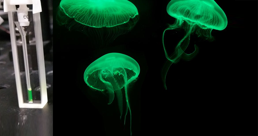
In the study, Kumar’s team used green fluorescent proteins, which are responsible for bioluminescence and commonly used in biomedical research. The team attempted to entangle the photons generated from the fluorescing molecules within the algae’s barrel-shaped protein structure by exposing them to spontaneous four-wave mixing, a process in which multiple wavelengths interact with one another to produce new wavelengths.
Through a series of these experiments, Kumar and his team successfully demonstrated a type of entanglement, called polarization entanglement, between photon pairs. The same feature used to make glasses for viewing 3D movies, polarization is the orientation of oscillations in light waves. A wave can oscillate vertically, horizontally, or at different angles. In Kumar’s entangled pairs, the photons’ polarizations are entangled, meaning that the oscillation directions of light waves are linked. Kumar also noticed that the barrel-shaped structure surrounding the fluorescing molecules protected the entanglement from being disrupted.
“When I measured the vertical polarization of one particle, we knew it would be the same in the other,” he said. “If we measured the horizontal polarization of one particle, we could predict the horizontal polarization in the other particle. We created an entangled state that correlated in all possibilities simultaneously.”
Now that they have demonstrated that it’s possible to create quantum entanglement from biological particles, next Kumar and his team plan to make a biological substrate of entangled particles, which could be used to build a quantum machine. Then, they will seek to understand if a biological substrate works more efficiently than a synthetic one.
Here’s an image accompanying the news release,
Featured in the cuvette on the left, green fluorescent proteins responsible for bioluninescence in jellyfish. Courtesy: Northwestern University
First, animals that flow in the dark and then, updates on other ‘glow in the dark’ projects.
The American Chemical Society (ACS) has produced a video about animals that glow in the dark, here’s more from their August 14, 2017 news release on EurekAlert,
Fireflies, frogs, jellyfish, mushrooms and even parrots have the ability to emit light from their bodies. These creatures use either bioluminescence or fluorescence to put on their light shows.
To learn more about Marc Zimmer’s research at Connecticut College and to find more great info and images involving GFP, visit his website: http://www.conncoll.edu/ccacad/zimmer…
Updates on a previous glowing plants and animals posting
In a May 5, 2013 posting I featured a Kickstarter campaign for a synthetic biology project focused on plants that emit light in the dark. I also mentioned Eduardo Kac (pronounced Katz) and his art project/transgenic bunny called Alba. At the time, I did not realize that Alba had been declared dead in 2002 adding more controversy to an already controversial topice according to Kristen Philipkoski in an Aug. 12, 2002 article (how did I miss this article in 2013?) for Wired magazine (Note: Links have been removed),
Alba, the glowing rabbit that made headlines two years ago for being, well, a glowing rabbit, has met an untimely death, according to the French researcher who genetically engineered her.
Alba the glowing rabbit was 4 years old. Or 2-1/2, depending on who’s talking.
The bunny died about a month ago for reasons that are not clear, said Louis-Marie Houdebine, a genetic researcher at France’s National Institute of Agronomic Research.
“I was informed one day that bunny was dead without any reason,” Houdebine said. “So, rabbits die often. It was about 4 years old, which is a normal lifespan in our facilities.”
Alba was an albino rabbit engineered by splicing the green fluorescent protein (GFP) of a jellyfish into her genome. Houdebine said he did not believe the GFP gene played a role in the animal’s demise.
Eduardo Kac, the artist who created a flurry by making her a work of art, doesn’t buy it, however.
First, Alba’s not 4, she’s 2-1/2, Kac says (a rabbit’s lifespan is up to 12 years), because she was bred by Houdebine specifically for him in January 2000.
Houdebine says he simply picked a rabbit with a gentle disposition that was already in his lab.
Second, he believes Houdebine might be declaring the bunny gone in order to put an end to a two-year, unwelcome barrage of media attention.
If she really is dead, Kac will never realize the final phase of his project, which was to take Alba home and keep her as a pet.
Kac says he and Houdebine originally collaborated on the GFP bunny project, until Houdebine’s director put the kibosh on it.
“My director did not understand,” Houdebine said. “He said I should not give the rabbit (to someone) outside the lab.”
Houdebine said that yes, they spoke about preliminary plans for Kac to use the bunny for his project and take it to an art show in Avignon. But he denies he bred an animal specifically for Kac.
…
Houdebine says he would not have agreed to engineer one animal specifically for any artist.
This disputed point has led fellow artists and critics to question whether Kac can rightly take credit for the Alba project.
But Kac insists that Houdebine did, in fact, agree to make the bunny specifically for him.
Kac found out sometime in mid-2000 that Houdebine’s director had a problem with the project and would not allow the rabbit to be taken from the lab.
Houdebine was initially apologetic, Kac said. But after an article ran on the front page of the Boston Globe on Sept. 17, 2000, their relationship cooled.
…
Houdebine and his director were opposed to the now-famous, brilliantly glowing photograph of Alba. They and other researchers say the rabbit doesn’t actually glow so brightly and uniformly.
“Kac fabricated data for his personal use,” Houdebine said. “This is why we totally stopped any contact with him.”
“The scientific fact is that the rabbit is not green,” he said. “He should have never published that. This was very disagreeable for me.”
Kac believes the scientists were simply afraid of public criticism. Meanwhile, he wanted to do the opposite – to encourage discourse on the transgenic rabbit.
“This director refuses to participate openly in a debate about what is done with public money,” he said. “It’s very easy to fear and reject what you don’t know. As long as they continue to isolate themselves, this mistrust will continue.”
The eyes and ears of the rabbit are green under ultraviolet light, Houdebine said, but the fur does not glow, because it’s dead tissue that doesn’t express the gene. Only if the rabbit were shaved would the body glow, he said.
…
Philipkosk’s article provides some insight into the interface between art and science and is worth reading in its entirety if you have the time.
I’ve also found an update for the glowing plants Kickstarter campaign in an April 20, 2017 article by Sarah Zhang for The Atlantic (Note: Links have been removed),
The latest update came quietly on Tuesday night [April 18, 2017?]. “We’re sorry to say that we have reached a significant transition point,” wrote the Glowing Plant project’s creator, Antony Evans. This “transition point” was more of an endpoint: The project had run out of money. The quest to genetically engineer a glow-in-the-dark plant was no more.
Four years ago, the Glowing Plant project raised nearly half a million dollars on Kickstarter, easily blowing past its initial ask of $65,000. Of course it did. The vision it presented was such potent fantasy. “What if,” Evans asked over swelling music in the pitch video, “we use trees to light our streets instead of street lamps?” What if you could get lighting without electricity? What if the natural world glowed like in Avatar?
This romantic vision so perfectly encapsulated the promises of synthetic biology, a field that treats the natural world as another system to be designed and engineered. In this case, synthetic biology became a possible solution to one of the world’s most pressing energy problems: electricity generation. Plus, it sounded really damn cool.
The Kickstarter campaign only promised a small, potted glowing plant to it backers, and I doubt many backers actually harbored illusions about trees lighting up the night sky soon. But backing the project was a small way to buy into a much grander vision.
…
At a time when “genetically modified organism,” or GMO, is such a poisoned phrase, the project’s crowdfunding success seemed to suggest that a pervasive if vague distrust of genetic modification might be countered by the sense of wonder for a glowing plant. (As the Kickstarter campaign grew, though, environmental groups raised questions and the crowdfunding site later banned giving away genetically modified organisms.)
The team also encountered the hard realities of engineering even a small plant that glows. “We did not anticipate some of the unknown technical challenges that we would get into,” Evans told me. (Plenty of scientists at the time were skeptical of the project’s timeline, though.) Evans is an MBA with a background in mobile apps, though his two original cofounders, who have both since left the project, had backgrounds in synthetic biology.
To get the plant to glow well, the research team had to insert six genes. But they never could get all six in at once. At best, some plants glowed very dimly. (The photo above of the glowing plant is a long exposure, making it appear much brighter than it actually is.) Evans says that he realizes now trying to insert six genes into a complex organism like a plant—rather than single-celled bacteria or yeast—was premature.
…
“I’m really afraid of disappointing that 16-year-old who saw this and imagined a bright wonderful future, of jading and disappointing people,” he says. Despite a few angry backers asking for a refund, most of the comments under the Kickstarter update so far have been supportive. The project had been providing regular, detailed updates on the difficulty of engineering the plants. The latest update was its 67th.
…
Zhang’s article goes on to detail other synthetic biology projects, which are showing some promise.
When you take this work into consideration with CRISPR-CAS9 and the beginnings of genetic germline editing, the question has to be asked: Will public discussion (if there’s any) be considered upstream (early in the process) or downstream (after the work has been done)? Public engagement professionals tend to favour upstream discussions, i.e., before people start demanding fear-based policy.
The extraordinary effort to colonize our brains continues apace with a new sensor from Vanderbilt University. From an Oct. 27, 2016 news item on ScienceDaily,
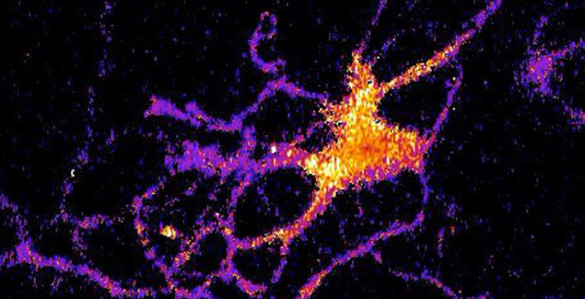
A new kind of bioluminescent sensor causes individual brain cells to imitate fireflies and glow in the dark.
The probe, which was developed by a team of Vanderbilt scientists, is a genetically modified form of luciferase, the enzyme that a number of other species including fireflies use to produce light. …
The scientists created the technique as a new and improved method for tracking the interactions within large neural networks in the brain.
“For a long time neuroscientists relied on electrical techniques for recording the activity of neurons. These are very good at monitoring individual neurons but are limited to small numbers of neurons. The new wave is to use optical techniques to record the activity of hundreds of neurons at the same time,” said Carl Johnson, Stevenson Professor of Biological Sciences, who headed the effort.
Individual neuron glowing with bioluminescent light produced by a new genetically engineered sensor. (Johnson Lab / Vanderbilt University)
“Most of the efforts in optical recording use fluorescence, but this requires a strong external light source which can cause the tissue to heat up and can interfere with some biological processes, particularly those that are light sensitive,” he [Carl Johnson] said.
Based on their research on bioluminescence in “a scummy little organism, the green alga Chlamydomonas, that nobody cares much about” Johnson and his colleagues realized that if they could combine luminescence with optogenetics – a new biological technique that uses light to control cells, particularly neurons, in living tissue – they could create a powerful new tool for studying brain activity.
“There is an inherent conflict between fluorescent techniques and optogenetics. The light required to produce the fluorescence interferes with the light required to control the cells,” said Johnson. “Luminescence, on the other hand, works in the dark!”
Johnson and his collaborators – Associate Professor Donna Webb, Research Assistant Professor Shuqun Shi, post-doctoral student Jie Yang and doctoral student Derrick Cumberbatch in biological sciences and Professor Danny Winder and postdoctoral student Samuel Centanni in molecular physiology and biophysics – genetically modified a type of luciferase obtained from a luminescent species of shrimp so that it would light up when exposed to calcium ions. Then they hijacked a virus that infects neurons and attached it to their sensor molecule so that the sensors are inserted into the cell interior.
The researchers picked calcium ions because they are involved in neuron activation. Although calcium levels are high in the surrounding area, normally they are very low inside the neurons. However, the internal calcium level spikes briefly when a neuron receives an impulse from one of its neighbors.
They tested their new calcium sensor with one of the optogenetic probes (channelrhodopsin) that causes the calcium ion channels in the neuron’s outer membrane to open, flooding the cell with calcium. Using neurons grown in culture they found that the luminescent enzyme reacted visibly to the influx of calcium produced when the probe was stimulated by brief light flashes of visible light.
To determine how well their sensor works with larger numbers of neurons, they inserted it into brain slices from the mouse hippocampus that contain thousands of neurons. In this case they flooded the slices with an increased concentration of potassium ions, which causes the cell’s ion channels to open. Again, they found that the sensor responded to the variations in calcium concentrations by brightening and dimming.
“We’ve shown that the approach works,” Johnson said. “Now we have to determine how sensitive it is. We have some indications that it is sensitive enough to detect the firing of individual neurons, but we have to run more tests to determine if it actually has this capability.”
Here’s a link to and a citation for the paper,
Coupling optogenetic stimulation with NanoLuc-based luminescence (BRET) Ca++ sensing by Jie Yang, Derrick Cumberbatch, Samuel Centanni, Shu-qun Shi, Danny Winder, Donna Webb, & Carl Hirschie Johnson. Nature Communications 7, Article number: 13268 (2016) doi:10.1038/ncomms13268 Published online: 27 October 2016
A Dec. 17, 2014 news item on Nanotechnology Now describes research into the phenomenon of bioluminescence and fireflies,
Fireflies used rapid light flashes to communicate. This “bioluminescence” is an intriguing phenomenon that has many potential applications, from drug testing and monitoring water contamination, and even lighting up streets using glow-in-dark trees and plants. Fireflies emit light when a compound called luciferin breaks down. We know that this reaction needs oxygen, but what we don’t know is how fireflies actually supply oxygen to their light-emitting cells. Using state-of-the-art imaging techniques, scientists from Switzerland and Taiwan have determined how fireflies control oxygen distribution to light up their cells. The work is published in Physical Review Letters.
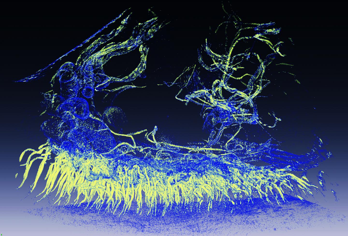
The firefly’s light-producing organ is called the “lantern”, and it is located in the insect’s abdomen. It looks like a series of tubes progressing into smaller ones and so one, like a tree’s branches growing into twigs. The function of these tubes, called, is to supply oxygen to the cells of the lantern, which contain luciferase and can produce light. However, the complexity of the firefly’s lantern has made it difficult to study this mechanism in depth, and reproduce it for technological applications.
Giorgio Margaritondo at EPFL, Yeukuang Hwu at the Academia Sinica and their colleagues at the National Tsing Hua University in Taiwan have successfully used two sophisticated imaging techniques to overcome the complexity of the firefly lantern and map out how oxygen is supplied to light-emitting cells. The techniques are called synchrotron phase contrast microtomography and transmission x-ray microscopy. They can scan down to the level of a single cell, even allowing researchers to look inside it.
By applying these techniques on live fireflies, the scientists were able to see the entire structure of the lantern for the first time, and to also make quantitative evaluations of oxygen distribution.
The imaging showed that the firefly diverts oxygen from other cellular functions and puts it into the reaction that breaks up luciferin. Specifically, the researchers found that oxygen consumption in the cell decreased, slowing down energy production. At the same time, oxygen supply switched to light-emission.
The study is the first to ever show the firefly’s lantern in such detail, while also providing clear evidence that it is optimized for light emission thanks to the state-of-the-art techniques used by the scientists. But Margaritondo points out another innovation: “The techniques we used have an advantage over, say, conventional x-ray techniques, which cannot easily distinguish between soft tissues. By using an approach based on changes in light intensity (phase-contrast) as opposed to light absorption (x-rays), we were able to achieve high-resolution imaging of the delicate firefly lantern.”
Here’s an image illustrating the work,
Tomographic Reconstruction of Part of the Firefly Lantern; This detailed microimage shows larger channels branching into smaller ones, supplying oxygen for the firefly’s light emission. The smallest channels are ten thousand times smaller than a millimeter and therefore invisible to other experimental probes: this has prevented scientists so far to unlock the mystery of firefly light flashes. Credit: Giorgio Margaritondo/EPFL
Here’s a link to and a citation for the paper,
Firefly Light Flashing: Oxygen Supply Mechanism by Yueh-Lin Tsai, Chia-Wei Li, Tzay-Ming Hong, Jen-Zon Ho, En-Cheng Yang, Wen-Yen Wu, G. Margaritondo, Su-Ting Hsu, Edwin B. L. Ong, and Y. Hwu. Phys. Rev. Lett. 113, 258103 – Published 17 December 2014 DOI: http://dx.doi.org/10.1103/PhysRevLett.113.258103
Mark Schrope has written a wonderful piece on the search for the giant squid in his Jan. 25, 2013 posting on Slate.com. It’s a story about adventure, myth, scientific pursuits, and, very cunningly, environmental issues.
I will excerpt a few bits from the piece but I encourage you to read it in its entirety,
Deep-sea biologist Edith Widder was working on a ship positioned off Japan’s Ogasawara Islands when Wen-Sung Chung asked her to step into the lab to see something. Cameras followed her as she got up. This was not unusual, since the Japan Broadcasting Commission (NHK) and the Discovery Channel were funding the expedition, which was being conducted from a research yacht named Alucia leased from a billionaire hedge fund owner. Chung was nonchalant, so it didn’t occur to Widder that she was about to see the culmination of a quest that has driven ocean explorers for more than a century. She thought maybe it was going to be video of a cool shark.
The purpose of the expedition was to capture footage of the enigmatic giant squid in its natural habitat. The animal can grow to 35 feet or longer, and its eye is as big as your head. But it lives about 1,000 feet below the surface and deeper, and it had only been glimpsed a few times at the surface and photographed alive once.
Widder is a world expert on bioluminescence, the light that countless marine animals use to communicate, especially in the dark world of the deep sea.
Schopes introduces a mystery, ‘What is Widder about to see?’, and then doesn’t answer it for several paragraphs while he explains who she is, her area of research, and the legend of the giant squid. Note: A link has been removed.
The giant squid has been the stuff of legend for about as long as people have sailed across oceans. Aristotle and Pliny the Elder described what may have been giant squid, which occasionally wash ashore or end up in fishermen’s nets, and the species is thought to be the origin of the Norwegian kraken myth.
Countless groups in past decades have tried to manufacture giant squid encounters, investing millions, getting all the best advice from the experts, only to come back as failed crusaders. One of the other scientists aboard the Alucia, Tsunemi Kubodera of Japan’s National Museum of Nature and Science, has been hunting giant squid in these waters for years. He managed to capture some still images of one giant squid and video of another after it was caught and brought to the surface. But none of that could compare to video of the animal alive in the deep, a view that would finally allow scientists to begin to understand the mysterious animal.
The expedition has not released expense figures, but it must have cost millions. When Chung, a graduate student at the University of Queensland, brought Widder into the lab and started fast-forwarding through the video, the scientists were already a week into a six-week expedition with nothing significant to show. Producer-types were growing tense.
Apparently, giant squid have a good sense of drama,
Now Widder is the first person to capture footage of a giant squid in its natural habitat. But even she admits that the grainy black-and-white footage, by itself, would have been a little unsatisfying. Some high-def footage would be the ultimate satisfaction. The drama-savvy squid would come through again.
…
Seven days after the first Medusa footage of a giant squid, Kubodera was in the clear sphere of a Triton submersible with pilot Jim Harris and NHK cameraman Tatsuhiko “Magic Man” Sugita when it happened. Kubodera was exploiting a different hypothesis: that the elusive squid find their prey by looking up with those huge eyes to see the faint silhouette of prey.
On Kubodera’s dives, the team tied a smaller, diamondback squid to the front of the sub and wrapped the bait around foam so that it would sink slower. Up and down, up and down the sub had gone for hours, using another low-light camera.
A giant squid latched on at 2,000 feet. As it drifted down, Harris matched the descent to keep the squid in full camera view. After the first few minutes they had flipped on the big lights, thinking the squid would flee, but it was committed to the bait. The sub’s maximum safe depth is 3,300 feet. Had the squid held on that far, Harris would have had to hit the brakes and the squid would have dropped out of view. But instead, at the last minute—3,000 feet—the squid swam off, so they got the entire encounter on film.
…
“I’ll never forget how beautiful it was,” says Harris. “It looked like it was covered in gold leaf.” That was a surprise to everyone because the dead ones certainly hadn’t looked like that. They were pasty. Kubodera says it was like seeing an entirely different animal.
Once Schrope has established the adventure aspect and revealed a giant squid covered in gold while, incidentally, establishing Widder’s credentials as a scientist and lover of marine life, there’s this,
For Widder, deep exploration remains a delight, but it’s no longer the primary focus of her career. In 2005, she left her longtime research post at the Harbor Branch Oceanographic Institution to found the Ocean Research and Conservation Association [ORCA], headquartered in a scenic old Coast Guard station on the Fort Pierce inlet. She wanted to take a step away from academia, where scientists are expected to stay relatively quiet in public and avoid anything that smacks of activism.
Widder had been growing increasingly overwhelmed by the environmental decline she was seeing, particularly pollution in coastal waters and estuaries, which are plagued by the polluted runoff of a Florida lifestyle dependent on constant growth and lots of fertilizer.
It gets better,
… She wants to wipe away the fallacy that pollution is an amorphous, intractable problem by gathering the information needed to pinpoint key problems. [emphasis mine] The group wants to create the aquatic equivalent of weather maps. Red shows polluted waters, blue the areas in the best shape. If people know the spot their kids swim in is in the red, they’ll take much more notice, she reasons. Perhaps more importantly, tourists would gravitate to cleaner waters if they could, creating a strong motivation for improvements.
Already the project has had success. [emphasis mine] Mapping the pollution in a stretch of Indian River Lagoon—Widder’s home and her office are both on the lagoon—she was surprised to find that two canals came up blue in a field of red. After some checking, the team learned that the golf course on those canals had switched to better environmental practices. They were preventing mowed grass clippings and runoff from the course from making it into the water. It was the perfect example for the local government, and in short order, a new fertilizer ordinance was passed.
The pièce de résistance,
They seem a world apart, but to Widder, the deep-sea exploration for fantastic creatures and the coastal environmental work guided by microbes are intimately tied. Not just because it’s all one big sea. Attention from the higher profile deep-sea work gives her a bully pulpit for focusing attention on things people don’t want to hear about, like water pollution. “I don’t want to hear about that stuff either,” she says. “But we’ve got to deal with it.” …
Too often in environmental stories writers and activists, in an attempt to communicate the seriousness of the issues, project a sense of doom. Necessary in the early days, the time has come to change the tone otherwise there’s a risk of inculcating hopelessness (some might say it’s already happening), which is the last thing we need. As Widder says, ” … we’ve got to deal with it.”
Very nicely done Mr. Schrope and Dr. Widder!
You can find more about ORCA here, by the way, the story has videos of the giant squid, and Discovery Channel (which broadcast the documentary on Jan. 27, 2013) also has information about the giant squid. Canadians are not allowed to view the video on the US website, we are required to visit the .ca website.
ETA Mar. 20, 2013: Danish scientists have determined that all giant squid no matter where they are found are related as per a Mar. 19, 2013 news item on ScienceDaily,
The giant squid is one of the most enigmatic animals on the planet. It is extremely rarely seen, except as the remains of animals that have been washed ashore, and placed in the formalin or ethanol collections of museums. But now, researchers at the University of Copenhagen leading an international team, have discovered that no matter where in the world they are found, the fabled animals are so closely related at the genetic level that they represent a single, global population, and thus despite previous statements to the contrary, a single species worldwide.