Tag Archives: photoluminescence

New system for imaging rare-earth doped nanoparticles

The Institut national de la recherche scientifique (INRS; Québec, Canada) has issued a January 30,2024 news release (also on EurekAlert) announcing new work in the field of imaging, Note: Links have been removed,

Teams led by professors Jinyang Liang and Fiorenzo Vetrone from the Énergie Matériaux Télécommunications Research Centre at the Institut national de la recherche scientifique (INRS) have developed a new system for imaging nanoparticles. It consists of a high-precision, short-wave infrared imaging technique capable of capturing the photoluminescence lifetimes of rare-earth doped nanoparticles in the micro- to millisecond range.

This groundbreaking discovery, which was published in the journal Advanced Science, paves the way for promising applications, particularly in the biomedical and information security fields.

Rare-earth elements are strategic metals that possess unique light-emitting properties that make them very attractive research tools in cutting-edge science. What’s more, the photoluminescence lifetime of nanoparticles doped with these ions has the advantage of being minimally affected by external conditions. As a result, measuring it through imaging provides data from which accurate and highly reliable information can be derived.

Although this field is seeing remarkable progress, existing optical systems for this type of measurement were less than ideal.

“Until now, existing optical systems have offered limited possibilities due to inefficient photon detection, limited imaging speed, and low sensitivity,” explains Professor Jinyang Liang, a specialist in ultrafast imaging and biophotonics.

To date, the most common technique for measuring the photoluminescence lifetime of rare-earth doped nanoparticles has involved counting time-correlated single photons.

“This method requires a large number of repeated excitations at the same location because the detector can only process a limited number of photons for each excitation,” says the study’s first author Miao Liu, a Ph.D. student in energy and materials science supervised by Profs. Liang and Vetrone.

However, the long photoluminescence lifetimes of rare-earth doped nanoparticles in the infrared spectrum, from hundreds of microseconds to several milliseconds, restrict the excitation’s repetition rate. As a result, the pixel dwelling time needed to build the photoluminescence intensity decay curve is much longer.

Pushing the limits

To overcome this challenge, Liang and Vetrone’s teams have combined streak optics with a high-sensitivity camera. The resulting device is called SWIR-PLIMASC (SWIR for short-wave infrared and PLIMASC for photoluminescence lifetime imaging microscopy using an all-optical streak camera). It vastly improves mapping of the optical properties of short-wave infrared photoluminescence lifetimes. It is the first high-sensitivity, high-speed SWIR imaging system in the optics field.

“It has several advantages,” says Miao Liu. “For instance, it responds to a wide spectral range, from 900 nm to 1700 nm, allowing photoluminescence to be detected at different wavelengths and/or spectral bands.”

The Ph.D. student adds that with the help of this device, photoluminescence lifetimes in the infrared spectrum, from microseconds to milliseconds, can be directly captured in one snapshot with a 1D imaging speed that can be tuned from 10.3 kHz to 138.9 kHz.

Finally, the operation that allocates the temporal information of photoluminescence to different spatial positions ensures that the entire process of 1D photoluminescence intensity decay can be recorded in a single snapshot, without repeated excitation. “You save time, but still get high sensitivity,” sums up Miao Liu.

Biomedical and security applications

The work carried out as part of this research will have a very tangible impact. In the biomedical field, the advances made possible by SWIR-PLIMASC could be used to fight cancer, believes Professor Fiorenzo Vetrone, whose expertise lies in nanomedicine.

“As our system applies to the temperature-based photoluminescence lifetime imaging of rare-earth ions, we believe that the data obtained could, for example, help to detect cancer cells even earlier and more accurately. The metabolism of those cells raises the temperature of the surrounding tissues,” says Professor Vetrone.

The innovative system can also be used to store information at enhanced security levels, more specifically to prevent documents and data from being falsified. Finally, in fundamental science, these unprecedented results will allow scientists to synthesize rare-earth nanoparticles with even more interesting optical properties.

Here’s a link to and a citation for the paper,

Short-wave Infrared Photoluminescence Lifetime Mapping of Rare-Earth Doped Nanoparticles Using All-Optical Streak Imaging by Miao Liu, Yingming Lai, Miguel Marquez, Fiorenzo Vetrone, Jinyang Liang. Advanced Science DOI: https://doi.org/10.1002/advs.202305284 First published: 06 January 2024

This paper is open access.
Using natural proteins to grow gold nanoclusters for hybrid bionanomaterials

While there’s a January 10, 2022 news item on Nanowerk, the research being announced was made available online in the Fall of 2021 and is now available in print,

Gold nanoclusters are groups of a few gold atoms with interesting photoluminescent properties. The features of gold nanoclusters depend not only on their structure, but their size and also by the ligands coordinated to them. These inorganic nanomaterials have been used in sensing, biomedicine and optics and their coordination with biomolecules can endow multiple capabilities in biological media.

A research collaboration between the groups of Dr. Juan Cabanillas, Research Professor at IMDEA Nanociencia and Dr. Aitziber L. Cortajarena, Ikerbasque Professor and Principal Investigator at CIC biomaGUNE have explored the use of natural proteins to grow gold nanoclusters, resulting in hybrid bionanomaterials with tunable photoluminescent properties and with a plethora of potential applications.

A January 10, 2022 IMDEA Nanociencia press release, which originated the news item, provides more technical detail about the research,

The nanoclusters –with less than 2 nm in size- differentiate from larger nanoparticles (plasmonic) since they present discrete energy levels coupled optically. The groups of amino acids within the proteins coordinate the gold atoms and allow the groups to be arranged around the gold nanocluster, facilitating the stabilization and adding an extra level of tailoring. These nanoclusters have interesting energy harvesting features. Since the discrete energy levels are optically coupled, the absorption of a photon leads to promotion of an electron to higher levels, which can trigger a photophysical process or a photochemical reaction.  

The results by Cabanillas and Cortajarena groups, published in Advanced Optical Materials and Nano Letters, explore the origin of the photoluminescence in protein-designed gold nanoclusters and shed light into the strong influence of environmental conditions on the nature of luminescence. Nanocluster capping by two types of amino acids (histidine and cysteine) allow for changing the emission spectral range from blue to red, paving the way to tune the optical properties by an appropriate ligand choice. The nature of emission is also changed with capping, from fluorescence to phosphorescence, respectively. The synergistic protein-nanocluster effects on emission are still not clear, and the groups at IMDEA Nanociencia and CIC biomaGUNE are working to elucidate the mechanisms behind. There are potential applications for the aforementioned nanoclusters, in solid state as active medium in laser cavities. Optical gain properties from these nanoclusters are yet to be demonstrated, which could pave the way to a new generation of potentially interesting laser devices. As the combination of gold plus proteins is potentially biocompatible, many potential applications in biomedicine can also be envisaged.

A related publication of the groups in Nano Letters demonstrates that the insertion of tryptophans, amino acids with high electron density, in the vicinity of the nanocluster boosts its photoluminescence quantum efficiency up to 40% in some cases, values relevant for solid state light emission applications. Researchers also observed an antenna effect: the tryptophans can absorb light in a discrete manner and transfer the energy to the cluster. This effect has interest for energy harvesting and for sensing purposes as well.

The proteins through the biocapping enable the synthesis of the nanoclusters and largely improve their quantum efficiency. “The photoluminescence quantum efficiency is largely improved when using the biocapping” Dr. Cabanillas says. He believes this research work means “a new field opening for the tuning of optical properties of nanoclusters through protein engineering, and much work is ahead for the understanding of the amplification mechanism”. Dr. Cortajarena emphasizes “we have already demonstrated the great potential of engineered photoluminescent protein-nanocluster in biomedical and technological fields, and understanding the fundamental emission mechanisms is pivotal for future applications“. A variety of further applications include biosensors, as the protein admits functionalization with recognition molecules, energy harvesting, imaging and photodynamic therapies. Further work is ahead this opening avenue for photophysics research.

This research is a collaboration led by Dr. Juan Cabanillas and Dr. Aitziber L. Cortajarena research groups at IMDEA Nanociencia and CIC biomaGUNE, with contributions from researchers at the Diamond Light Source Ltd. [synchrotron] and DIPC. It has been cofounded by the projects AMAPOLA, NMAT2D, FULMATEN, Atracción de Talento from Comunidad de Madrid and the Severo Ochoa Centre of Excellence award to IMDEA Nanociencia. CIC biomaGUNE acknowledges support by the projects ERC-ProNANO, ERC-NIMM, ProTOOLs and the Maria de Maeztu Units of Excellence Programme.

Here are links to and citations for the papers,

Tuning the Optical Properties of Au Nanoclusters by Designed Proteins by Elena Lopez-Martinez, Diego Gianolio, Saül Garcia-Orrit, Victor Vega-Mayoral, Juan Cabanillas-Gonzalez, Carlos Sanchez-Cano, Aitziber L. Cortajarena. Advanced Optical Materials Volume 10, Issue 1 January 4, 2022 2101332 DOI: https://doi.org/10.1002/adom.202101332 First published: 31 October 2021

This paper is open access.

Boosting the Photoluminescent Properties of Protein-Stabilized Gold Nanoclusters through Protein Engineering by Antonio Aires, Ahmad Sousaraei, Marco Möller, Juan Cabanillas-Gonzalez, and Aitziber L. Cortajarena. Nano Lett. 2021, 21, 21, 9347–9353 DOI: https://doi.org/10.1021/acs.nanolett.1c03768 Publication Date: November 1, 2021 Copyright © 2021 American Chemical Society

This paper is behind a paywall.

Not being familiar with either of the two research institutions mentioned in the press release, I did a little digging.

Here’s a little information about IMDEA Nanociencia (IMDEA Nanoscience Institute), from its Wikipedia entry, Note: All links have been removed,

IMDEA Nanoscience Institute is a private non-profit foundation within the IMDEA Institutes network, created in 2006-2007 as a result of collaboration agreement between the Community of Madrid and Spanish Ministry of Education and Science. The foundation manages IMDEA-Nanoscience Institute,[1] a scientific centre dedicated to front-line research in nanoscience, nanotechnology and molecular design and aiming at transferable innovations and close contact with industries. IMDEA Nanoscience is a member of the Campus of International excellence, a consortium of research institutes promoted by the Autonomous University of Madrid and Spanish National Research Council (UAM/CSIC).[2]

As for CIC biomaGUNE, here’s more from its institutional profile on the science.eus website,

The Centre for Cooperative Research in Biomaterials-CIC biomaGUNE, located in San Sebastian (Spain), was officially opened in December 2006. CIC biomaGUNE is a non-profit research organization created to promote scientific research and technological innovation at the highest levels in the Basque Country following the BioBasque policy in order to create a new business sector based on biosciences. Established by the Department of Industry, Technology & Innovation of the Government of the Autonomous Community of the Basque Country, CIC biomaGUNE constitutes one of the Centres of the CIC network, the largest Basque Country research network on specific strategic areas, having the mission to contribute to the economical and social development of the country through the generation of knowledge and speeding up the process that leads to technological innovation.

Surgery on nanoparticles?

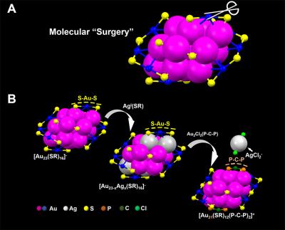
Chemists performed “surgery” on a 23-gold-atom nanoparticle according to a June 12, 2017 news item on Nanowerk (Note: A link has been removed),

A team of chemists led by Carnegie Mellon University’s [CMU] Rongchao Jin has for the first time conducted site-specific surgery on a nanoparticle. The procedure, which allows for the precise tailoring of nanoparticles, stands to advance the field of nanochemistry.

The surgical technique developed by Qi Li, the study’s lead author and a 3rd year graduate student in the Jin group, will allow researchers to enhance nanoparticles’ functional properties, such as catalytic activity and photoluminescence, increasing their usefulness in a wide variety of fields including health care, electronics and manufacturing. The findings were published in Science Advances (“Molecular “surgery” on a 23-gold-atom nanoparticle”).

Here’s an image the researchers have provided,

Caption: Carnegie Mellon chemists used a two-step metal exchange method to remove two S-Au-S staples from the surface of a nanoparticle. Credit: Carnegie Mellon University

A June 12, 2017 CMU press release (also on EurekAlert), which originated the news item, provides more details about the research,

“Nanochemistry is a relatively new field, it’s only about 20 years old. We’ve been racing to catch up to fields like organic chemistry that are more than 100 years old,” said Jin, a chemistry professor in the Mellon College of Science. “Organic chemists have been able to tailor the functional groups of molecules for quite some time, like tailoring penicillin for better medical functions, for example. We dreamed that we could do something similar in nanoscience. Developing atomically precise nanoparticles has allowed us to make this dream come true.”

In order to make this “nano-surgery” a reality, researchers needed to begin with atomically precise nanoparticles that could be reliably produced time after time. Jin’s lab has been at the forefront of this research. Working with gold nanoparticles, he and his team have developed methods to precisely control the number of atoms in each nanoparticle, resulting in uniformly-sized nanoparticles with every batch. With reliably precise particles, Jin and colleagues were able to identify the particles’ structures, and begin to tease out how that structure impacted the particles’ properties and functionality.

With these well-defined nanoparticles in hand, Jin’s next step was to find a way to surgically tailor the particles in order to learn more about­ – and hopefully enhance – their functionality.

In their recent study, Jin and colleagues performed nano-surgery on a gold nanoparticle made up of 23 gold atoms surrounded by a protective surface of ligands in staple-like motifs. Using a two-step metal exchange method, they removed two S-Au-S staples from the particle’s surface. In doing this they revealed the structural factors that determine the particle’s optical properties and established the role that the surface plays in photoluminescence. Significantly, the surgery increased the particle’s photoluminescence by about 10-fold. Photoluminescence plays a critical role in biological imaging, cancer diagnosis and LED technology, among other applications.

Jin and coworkers are now trying to generalize this site-specific surgery method to other nanoparticles.

Here’s a link to and a citation for the paper,

Molecular “surgery” on a 23-gold-atom nanoparticle by Qi Li, Tian-Yi Luo, Michael G. Taylor, Shuxin Wang, Xiaofan Zhu, Yongbo Song, Giannis Mpourmpakis, Nathaniel L. Rosi, and Rongchao Jin. Science Advances 19 May 2017: Vol. 3, no. 5, e1603193 DOI: 10.1126/sciadv.1603193

This paper is open access.

Namdiatream; a European multimodal diagnostics project

I’ve written about lab-on-a-chip projects, point-of-care diagnostics, and other such initiatives on several occasions, most recently in a Mar. 1, 2013 posting about a technique where powder is used to make the diagnostic device more portable. This time it was a Europe-wide project described in a Mar. 4, 2013 news item on Nanowerk,which caught my attention (Note: A link has been removed),

The plan of the EU-funded consortium Nanotechnological toolkits for multi-modal disease diagnostics and treatment monitoring (Namdiatream) is not to cure cancer, per se, but to boost the sensitivity of diagnostics and the ability to monitor progress during treatment. They focused on three types – breast, prostate and lung cancer.

… The prototype devices being developed during the four-year project will detect common cancer cells much earlier and, with timely treatment, improve the chances of recovery.

According to the project leader, Professor Yuri Volkov of Trinity College Dublin’s School of Medicine, the portable nanodevices are based on innovative lab-on-a-chip, -bead and -wire technologies applicable in different settings – clinical, research, or point of care (i.e. hospitals). These lab-on-x technologies exploit the photo-luminescent (‘glow-in-the-dark’ light emitting), plasmonic (‘light-on-a-wire’), magnetic and unique optical properties of nanomaterials.

Volkov offers some insight into how the project started and its current state of evolution (from the news item),

This is ground-breaking work made possible thanks to advanced technology but also to EU funding for cross-border investigations. Teams across Europe were doing related but fragmented research, suggests Prof. Volkov. This risked leaving a team dangling if their approach failed or lacked funding.

“So we integrated our research and identified joint strengths to help one another develop the best technological approaches in case something didn’t work in one, or synergies were identified, thereby increasing the chances of wider success.”

At its half-way stage, notes Prof. Volkov, Namdiatream underwent a natural evolution when it became clear that by merging and refocusing work in some areas – i.e. in fluorescent nanomaterial technology and magnetic nanowire barcodes – it would speed up industrial implementation efforts.

“Now, work on the preclinical prototype devices is well under way,” he confirms. But one of the many remaining challenges is to calibrate their sensitivity, so that they do not give false readings, for instance.

The Namdiatream (Nanotechnological Toolkits for Multi-Modal Disease Diagnostics and Treatment Monitoring) home page offers more detail about the project,

Namdiatream is a truly interdisciplinary and Pan-european consortium that builds around 7 High-Tech SMEs [small to medium enterprises], 2 Multinational industries and 13 academic institutions. NAMDIATREAM will develop nanotechnology-based toolkit to enable early detection and imaging of molecular biomarkers of the most common cancer types and of cancer metastases, as well as permitting the identification of cells indicative of early-stage disease onset. The project is built on the innovative technology concepts of super-sensitive “lab-on-a-bead”, “lab-on-a-chip” and “lab-on-a-wire” nano-devices.

Interestingly, this too was on the home page,

The ETP Nanomedicine documents point out that nanotechnology has yet to deliver practical solutions for the patients and clinicians in their struggle against common, socially and economically important diseases such as cancer. Therefore NAMDIATREAM results will firstly aim to deliver to the diagnostic and medical imaging device companies involved in the consortium, and the clinical and academic partners. This could further provide the basis for cancer therapeutics as it will be possible to accurately assess the kinetics of cancer cell destruction during the course of appropriate therapy.