Tag Archives: bionics

Nanoscale tattoos for individual cells

It’s fascinating to read about a technique for applying ‘tattoos’ to living cells and I have two news items and news releases with different perspectives about this same research.

First out the door was the August 7, 2023 news item on ScienceDaily,

Engineers have developed nanoscale tattoos — dots and wires that adhere to live cells — in a breakthrough that puts researchers one step closer to tracking the health of individual cells.

The new technology allows for the first time the placement of optical elements or electronics on live cells with tattoo-like arrays that stick on cells while flexing and conforming to the cells’ wet and fluid outer structure.

“If you imagine where this is all going in the future, we would like to have sensors to remotely monitor and control the state of individual cells and the environment surrounding those cells in real time,” said David Gracias, a professor of chemical and biomolecular engineering at Johns Hopkins University who led the development of the technology. “If we had technologies to track the health of isolated cells, we could maybe diagnose and treat diseases much earlier and not wait until the entire organ is damaged.”

An August 7, 2023 Johns Hopkins University news release by (also on EurekAlert), which originated the news item, describes the research in an accessible fashion before delving into technical details,

Gracias, who works on developing  biosensor technologies that are nontoxic and noninvasive for the body, said the tattoos bridge the gap between living cells or tissue and conventional sensors and electronic materials. They’re essentially like barcodes or QR codes, he said.

“We’re talking about putting something like an electronic tattoo on a living object tens of times smaller than the head of a pin,” Gracias said. “It’s the first step towards attaching sensors and electronics on live cells.”

The structures were able to stick to soft cells for 16 hours even as the cells moved.

The researchers built the tattoos in the form of arrays with gold, a material known for its ability to prevent signal loss or distortion in electronic wiring. They attached the arrays to cells that make and sustain tissue in the human body, called fibroblasts. The arrays were then treated with  molecular glues and transferred onto the cells using an alginate hydrogel film, a gel-like laminate that can be dissolved after the gold adheres to the cell. The molecular glue on the array bonds to a film secreted by the cells called the extracellular matrix.

Previous research has demonstrated how to use hydrogels to stick nanotechnology onto human skin and internal animal organs. By showing how to adhere nanowires and nanodots onto single cells, Gracias’ team is addressing the long-standing challenge of making optical sensors and electronics compatible with biological matter at the single cell level. 

“We’ve shown we can attach complex nanopatterns to living cells, while ensuring that the cell doesn’t die,” Gracias said. “It’s a very important result that the cells can live and move with the tattoos because there’s often a significant incompatibility between living cells and the methods engineers use to fabricate electronics.”

The team’s ability to attach the dots and wires in an array form is also crucial. To use this technology to track bioinformation, researchers must be able to arrange sensors and wiring into specific patterns not unlike how they are arranged in electronic chips. 

“This is an array with specific spacing,” Gracias explained, “not a haphazard bunch of dots.”

The team plans to try to attach more complex nanocircuits that can stay in place for longer periods. They also want to experiment with different types of cells.

Other Johns Hopkins authors are Kam Sang Kwok, Yi Zuo, Soo Jin Choi, Gayatri J. Pahapale, and Luo Gu.

This looks more like a sea creature to me but it’s not,
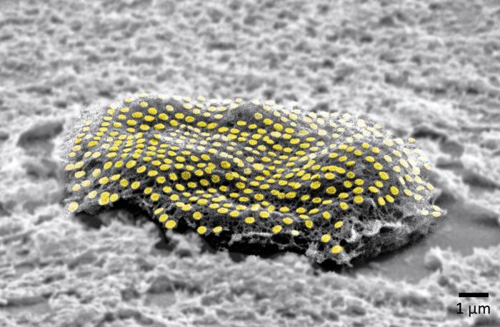
Caption: False-colored gold nanodot array on a fibroblast cell. Credit: Kam Sang Kwok and Soo Jin Choi, Gracias Lab/Johns Hopkins University.[The measurement, i.e., what looks like a ‘u’ with a preceding tail, in the lower right corner of the image is one micron/one millionth add that to the ‘m’ and you have what’s commonly described as one micrometre.]

An August 10, 2023 news item on ScienceDaily offers a different perspective from the American Chemical Society (ACS) on this research,

For now, cyborgs exist only in fiction, but the concept is becoming more plausible as science progresses. And now, researchers are reporting in ACS’ Nano Letters that they have developed a proof-of-concept technique to “tattoo” living cells and tissues with flexible arrays of gold nanodots and nanowires. With further refinement, this method could eventually be used to integrate smart devices with living tissue for biomedical applications, such as bionics and biosensing.

An August 10, 2023 ACS news release (also on EurekAlert), which originated the news item, explains some of the issues with attaching electronics to living tissue,

Advances in electronics have enabled manufacturers to make integrated circuits and sensors with nanoscale resolution. More recently, laser printing and other techniques have made it possible to assemble flexible devices that can mold to curved surfaces. But these processes often use harsh chemicals, high temperatures or pressure extremes that are incompatible with living cells. Other methods are too slow or have poor spatial resolution. To avoid these drawbacks, David Gracias, Luo Gu and colleagues wanted to develop a nontoxic, high-resolution, lithographic method to attach nanomaterials to living tissue and cells.

The team used nanoimprint lithography to print a pattern of nanoscale gold lines or dots on a polymer-coated silicon wafer. The polymer was then dissolved to free the gold nanoarray so it could be transferred to a thin piece of glass. Next, the gold was functionalized with cysteamine and covered with a hydrogel layer, which, when peeled away, removed the array from the glass. The patterned side of this flexible array/hydrogel layer was coated with gelatin and attached to individual live fibroblast cells. In the final step, the hydrogel was degraded to expose the gold pattern on the surface of the cells. The researchers used similar techniques to apply gold nanoarrays to sheets of fibroblasts or to rat brains. Experiments showed that the arrays were biocompatible and could guide cell orientation and migration.

The researchers say their cost-effective approach could be used to attach other nanoscale components, such as electrodes, antennas and circuits, to hydrogels or living organisms, thereby opening up opportunities for the development of biohybrid materials, bionic devices and biosensors.

The authors acknowledge funding from the Air Force Office of Scientific Research, the National Institute on Aging, the National Science Foundation and the Johns Hopkins University Surpass Program.

Here’s a link to and a citation for the paper,

Toward Single Cell Tattoos: Biotransfer Printing of Lithographic Gold Nanopatterns on Live Cells by Kam Sang Kwok, Yi Zuo, Soo Jin Choi, Gayatri J. Pahapale, Luo Gu, and David H. Gracias. Nano Lett. 2023, 23, 16, 7477–7484 DOI: https://doi.org/10.1021/acs.nanolett.3c01960 Publication Date:August 1, 2023 Copyright © 2023 American Chemical Society

This paper is behind a paywall.
Neural and technological inequalities

I’m always happy to see discussions about the social implications of new and emerging technologies. In this case, the discussion was held at the Fast Company (magazine) European Innovation Festival. KC Ifeanyi wrote a July 10, 2019 article for Fast Company highlighting a session between two scientists focusing on what I’ve termed ‘machine/flesh’ or is, sometimes, called a cyborg but not with these two scientists (Note: A link has been removed),

At the Fast Company European Innovation Festival today, scientists Moran Cerf and Riccardo Sabatini had a wide-ranging discussion on the implications of technology that can hack humanity. From ethical questions to looking toward human biology for solutions, here are some of the highlights:

The ethics of ‘neural inequality’

There are already chips that can be implanted in the brain to help recover bodily functions after a stroke or brain injury. However, what happens if (more likely when) a chip in your brain can be hacked or even gain internet access, essentially making it possible for some people (more likely wealthy people) to process information much more quickly than others?

“It’s what some call neural inequality,” says Cerf, a neuroscientist and business professor at the Kellogg School of Management and at the neuroscience program at Northwestern University. …

Opening new pathways to thought through bionics

Cerf mentioned a colleague who was born without his left hand. He engineered a bionic one that he can control with an app and that has the functionality of doing things no human hand can do, like rotating 360 degrees. As fun of a party trick as that is, Cerf brings up a good point in that his colleague’s brain is processing something we can’t, thereby possibly opening new pathways of thought.

“The interesting thing, and this is up to us to investigate, is his brain can think thoughts that you cannot think [emphasis mine] because he has a function you don’t have,” Cerf says. …

The innovation of your human body

As people look to advanced bionics to amplify their senses or abilities, Sabatini, chief data scientist at Orionis Biosciences, makes the argument that our biological bodies are far more advanced than we give them credit for. …

Democratizing tech’s edges

Early innovation so often comes with a high price tag. The cost of experimenting with nascent technology or running clinical trials can be exorbitant. And Sabatini believes democratizing that part of the process is where the true innovation will be. …

Earlier technology that changed our thinking and thoughts

This isn’t the first time that technology has altered our thinking and the kinds of thoughts we have as per ” brain can think thoughts that you cannot think.” According to Walter J. Ong’s 1982 book, ‘Orality and Literacy’,that’s what writing did to us; it changed our thinking and the kinds of thoughts we have.

It took me quite a while to understand ‘writing’ as a technology, largely due to how much I took it for granted. Once I made that leap, it changed how I understood the word technology. Then, the idea that ‘writing’ could change your brain didn’t require as dramatic a leap although it fundamentally altered my concept of the relationship between technology and humans. Up to that time, I had viewed technology as an instrument that allowed me to accomplish goals (e.g., driving a car from point a to point b) but it had very little impact on me as a person.

You can find out more about Walter J. Ong and his work in his Wikipedia entry. Pay special attention to the section about, Orality and Literacy.

Who’s talking about technology and our thinking?

The article about the scientists (Cerf and Sabatini) at the Fast Company European Innovation Festival (held July 9 -10, 2019 in Milan, Italy) never mentions cyborgs. Presumably, neither did Sabatini or Cerf. It seems odd. Two thinkers were discussing ‘neural inequality’ and there was no mention of a cyborg (human and machine joined together).

Interestingly, the lead sponsor for this innovation festival was Gucci. That company would not have been my first guess or any other guess for that matter as having an interest in neural inequality.

So, Gucci sponsored a festival that is not not cheap. A two-day pass was $1600. (early birds got a discount of $457) and a ‘super’ pass was $2,229 (with an early bird discount of $629). So, you didn’t get into the room unless you had a fair chunk of change and time.

The tension, talking about inequality at a festival or other venue that most people can’t afford to attend, is discussed at more length in Anand Giridharadas’s 2018 book, ‘Winners Take All; The Elite Charade of Changing the World’.

It’s not just who gets to discuss ‘neural inequality’, it’s when you get to discuss it, which affects how the discussion is framed.

There aren’t an easy answers to these questions but I find the easy assumption that the wealthy and the science and technology communities get first dibs at the discussion a little disconcerting while being perfectly predictable.

On the plus side, there are artists and others who have jumped in and started the discussion by turning themselves into cyborgs. This August 14, 2015 article (Body-hackers: the people who turn themselves into cyborgs) by Oliver Wainwright for the Guardian is very informative and not for the faint of heart.

For the curious, I’ve been covering these kinds of stories here since 2009. The category ‘human enhancement’ and the search term ‘machine/flesh’ should provide you with an assortment of stories on the topic.

Singapore’s* new chip could make low-powered wireless neural implants a possibility and Australians develop their own neural implant

Singapore

This research from Singapore could make neuroprosthetics and exoskeletons a little easier to manage as long as you don’t mind having a neural implant. From a Feb. 11, 2016 news item on ScienceDaily,

A versatile chip offers multiple applications in various electronic devices, report researchers, suggested that there is now hope that a low-powered, wireless neural implant may soon be a reality. Neural implants when embedded in the brain can alleviate the debilitating symptoms of Parkinson’s disease or give paraplegic people the ability to move their prosthetic limbs.

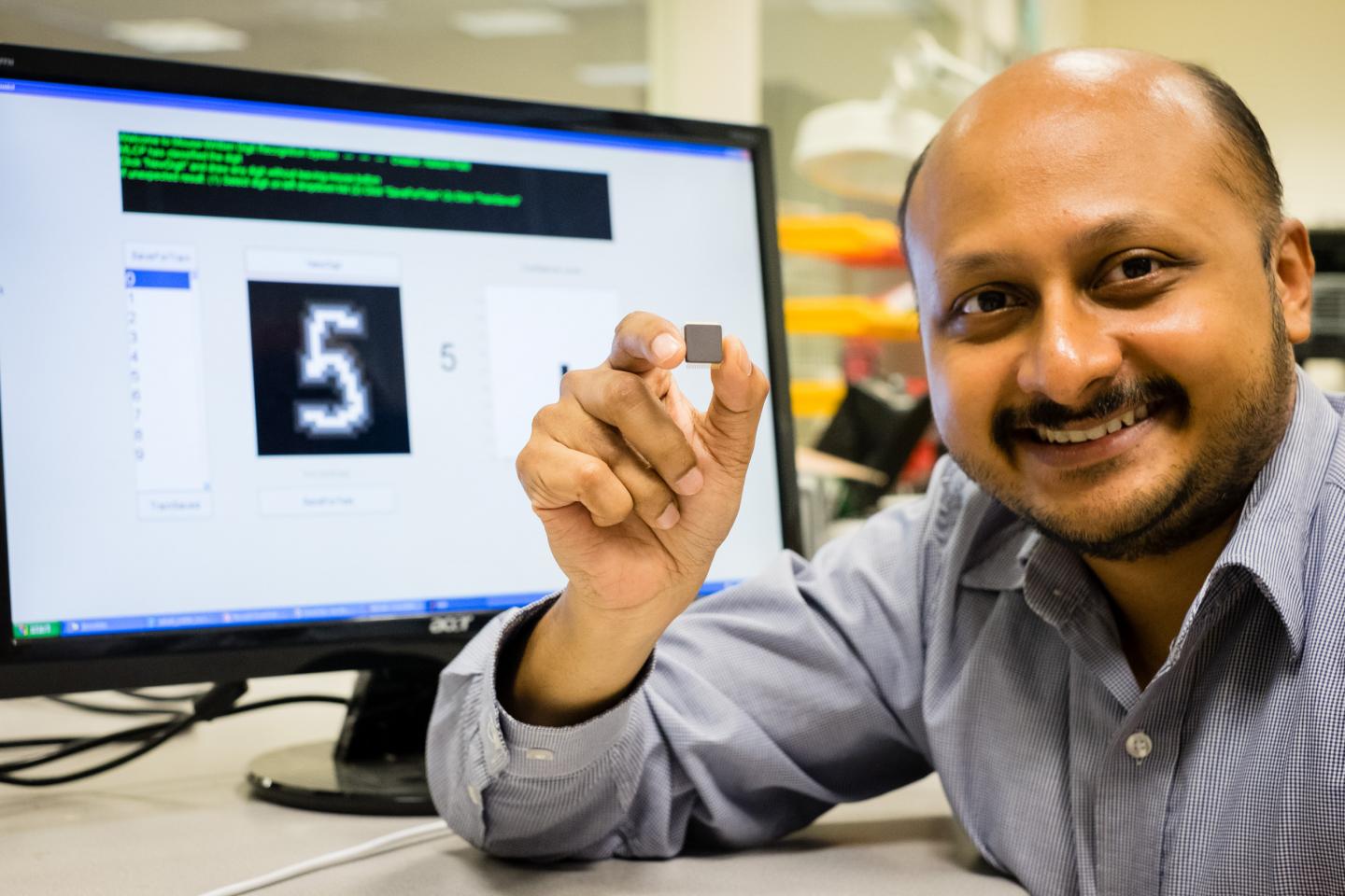
Caption: NTU Asst Prof Arindam Basu is holding his low-powered smart chip. Credit: NTU Singapore

Caption: NTU Asst Prof Arindam Basu is holding his low-powered smart chip. Credit: NTU Singapore

A Feb. 11, 2016 Nanyang Technological University (NTU) press release (also on EurekAlert), which originated the news item, provides more detail,

Scientists at Nanyang Technological University, Singapore (NTU Singapore) have developed a small smart chip that can be paired with neural implants for efficient wireless transmission of brain signals.

Neural implants when embedded in the brain can alleviate the debilitating symptoms of Parkinson’s disease or give paraplegic people the ability to move their prosthetic limbs.

However, they need to be connected by wires to an external device outside the body. For a prosthetic patient, the neural implant is connected to a computer that decodes the brain signals so the artificial limb can move.

These external wires are not only cumbersome but the permanent openings which allow the wires into the brain increases the risk of infections.

The new chip by NTU scientists can allow the transmission of brain data wirelessly and with high accuracy.

Assistant Professor Arindam Basu from NTU’s School of Electrical and Electronic Engineering said the research team have tested the chip on data recorded from animal models, which showed that it could decode the brain’s signal to the hand and fingers with 95 per cent accuracy.

“What we have developed is a very versatile smart chip that can process data, analyse patterns and spot the difference,” explained Prof Basu.

“It is about a hundred times more efficient than current processing chips on the market. It will lead to more compact medical wearable devices, such as portable ECG monitoring devices and neural implants, since we no longer need large batteries to power them.”

Different from other wireless implants

To achieve high accuracy in decoding brain signals, implants require thousands of channels of raw data. To wirelessly transmit this large amount of data, more power is also needed which means either bigger batteries or more frequent recharging.

This is not feasible as there is limited space in the brain for implants while frequent recharging means the implants cannot be used for long-term recording of signals.

Current wireless implant prototypes thus suffer from a lack of accuracy as they lack the bandwidth to send out thousands of channels of raw data.

Instead of enlarging the power source to support the transmission of raw data, Asst Prof Basu tried to reduce the amount of data that needs to be transmitted.

Designed to be extremely power-efficient, NTU’s patented smart chip will analyse and decode the thousands of signals from the neural implants in the brain, before compressing the results and sending it wirelessly to a small external receiver.

This invention and its findings were published last month [December 2015] in the prestigious journal, IEEE Transactions on Biomedical Circuits & Systems, by the Institute of Electrical and Electronics Engineers, the world’s largest professional association for the advancement of technology.

Its underlying science was also featured in three international engineering conferences (two in Atlanta, USA and one in China) over the last three months.

Versatile smart chip with multiple uses

This new smart chip is designed to analyse data patterns and spot any abnormal or unusual patterns.

For example, in a remote video camera, the chip can be programmed to send a video back to the servers only when a specific type of car or something out of the ordinary is detected, such as an intruder.

This would be extremely beneficial for the Internet of Things (IOT), where every electrical and electronic device is connected to the Internet through a smart chip.

With a report by marketing research firm Gartner Inc predicting that 6.4 billion smart devices and appliances will be connected to the Internet by 2016, and will rise to 20.8 billion devices by 2020, reducing network traffic will be a priority for most companies.

Using NTU’s new chip, the devices can process and analyse the data on site, before sending back important details in a compressed package, instead of sending the whole data stream. This will reduce data usage by over a thousand times.

Asst Prof Basu is now in talks with Singapore Technologies Electronics Limited to adapt his smart chip that can significantly reduce power consumption and the amount of data transmitted by battery-operated remote sensors, such as video cameras.

The team is also looking to expand the applications of the chip into commercial products, such as to customise it for smart home sensor networks, in collaboration with a local electronics company.

The chip, measuring 5mm by 5mm can now be licensed by companies from NTU’s commercialisation arm, NTUitive.

Here’s a link to and a citation for the paper,

A 128-Channel Extreme Learning Machine-Based Neural Decoder for Brain Machine Interfaces by Yi Chen, Enyi Yao, Arindam Basu. IEEE Transactions on Biomedical Circuits and Systems, 2015; 1 DOI: 10.1109/TBCAS.2015.2483618

This paper is behind a paywall.

Australia

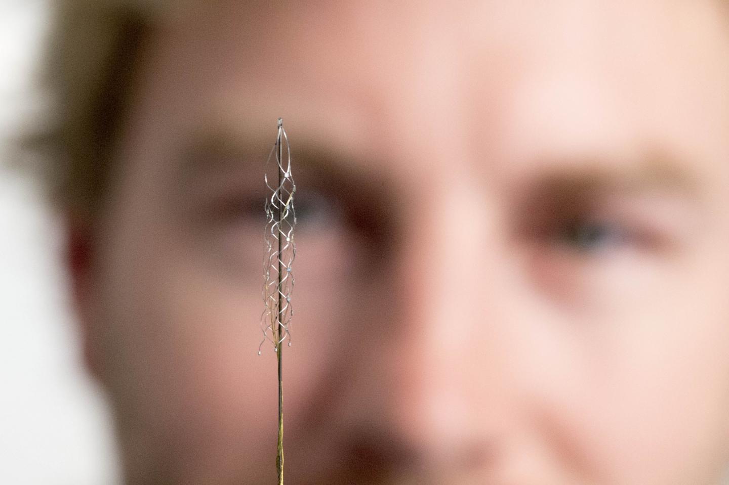
Earlier this month there was a Feb. 9, 2016 announcement about a planned human clinical trial in Australia for a new brain-machine interface (neural implant). Before proceeding with the news, here’s what this implant looks like,

Caption: This tiny device, the size of a small paperclip, is implanted in to a blood vessel next to the brain and can read electrical signals from the motor cortex, the brain's control centre. These signals can then be transmitted to an exoskeleton or wheelchair to give paraplegic patients greater mobility. Users will need to learn how to communicate with their machinery, but over time, it is thought it will become second nature, like driving or playing the piano. The first human trials are slated for 2017 in Melbourne, Australia. Credit: The University of Melbourne.

Caption: This tiny device, the size of a small paperclip, is implanted in to a blood vessel next to the brain and can read electrical signals from the motor cortex, the brain’s control centre. These signals can then be transmitted to an exoskeleton or wheelchair to give paraplegic patients greater mobility. Users will need to learn how to communicate with their machinery, but over time, it is thought it will become second nature, like driving or playing the piano. The first human trials are slated for 2017 in Melbourne, Australia. Credit: The University of Melbourne.

A Feb. 9, 2016 University of Melbourne press release (also on EurekAlert), which originated the news item, provides more detail,

Melbourne medical researchers have created a new minimally invasive brain-machine interface, giving people with spinal cord injuries new hope to walk again with the power of thought.

The brain machine interface consists of a stent-based electrode (stentrode), which is implanted within a blood vessel next to the brain, and records the type of neural activity that has been shown in pre-clinical trials to move limbs through an exoskeleton or to control bionic limbs.

The new device is the size of a small paperclip and will be implanted in the first in-human trial at The Royal Melbourne Hospital in 2017.

The results published today in Nature Biotechnology show the device is capable of recording high-quality signals emitted from the brain’s motor cortex, without the need for open brain surgery.

Principal author and Neurologist at The Royal Melbourne Hospital and Research Fellow at The Florey Institute of Neurosciences and the University of Melbourne, Dr Thomas Oxley, said the stentrode was revolutionary.

“The development of the stentrode has brought together leaders in medical research from The Royal Melbourne Hospital, The University of Melbourne and the Florey Institute of Neuroscience and Mental Health. In total 39 academic scientists from 16 departments were involved in its development,” Dr Oxley said.

“We have been able to create the world’s only minimally invasive device that is implanted into a blood vessel in the brain via a simple day procedure, avoiding the need for high risk open brain surgery.

“Our vision, through this device, is to return function and mobility to patients with complete paralysis by recording brain activity and converting the acquired signals into electrical commands, which in turn would lead to movement of the limbs through a mobility assist device like an exoskeleton. In essence this a bionic spinal cord.”

Stroke and spinal cord injuries are leading causes of disability, affecting 1 in 50 people. There are 20,000 Australians with spinal cord injuries, with the typical patient a 19-year old male, and about 150,000 Australians left severely disabled after stroke.

Co-principal investigator and biomedical engineer at the University of Melbourne, Dr Nicholas Opie, said the concept was similar to an implantable cardiac pacemaker – electrical interaction with tissue using sensors inserted into a vein, but inside the brain.

“Utilising stent technology, our electrode array self-expands to stick to the inside wall of a vein, enabling us to record local brain activity. By extracting the recorded neural signals, we can use these as commands to control wheelchairs, exoskeletons, prosthetic limbs or computers,” Dr Opie said.

“In our first-in-human trial, that we anticipate will begin within two years, we are hoping to achieve direct brain control of an exoskeleton for three people with paralysis.”

“Currently, exoskeletons are controlled by manual manipulation of a joystick to switch between the various elements of walking – stand, start, stop, turn. The stentrode will be the first device that enables direct thought control of these devices”

Neurophysiologist at The Florey, Professor Clive May, said the data from the pre-clinical study highlighted that the implantation of the device was safe for long-term use.

“Through our pre-clinical study we were able to successfully record brain activity over many months. The quality of recording improved as the device was incorporated into tissue,” Professor May said.

“Our study also showed that it was safe and effective to implant the device via angiography, which is minimally invasive compared with the high risks associated with open brain surgery.

“The brain-computer interface is a revolutionary device that holds the potential to overcome paralysis, by returning mobility and independence to patients affected by various conditions.”

Professor Terry O’Brien, Head of Medicine at Departments of Medicine and Neurology, The Royal Melbourne Hospital and University of Melbourne said the development of the stentrode has been the “holy grail” for research in bionics.

“To be able to create a device that can record brainwave activity over long periods of time, without damaging the brain is an amazing development in modern medicine,” Professor O’Brien said.

“It can also be potentially used in people with a range of diseases aside from spinal cord injury, including epilepsy, Parkinsons and other neurological disorders.”

The development of the minimally invasive stentrode and the subsequent pre-clinical trials to prove its effectiveness could not have been possible without the support from the major funding partners – US Defense Department DARPA [Defense Advanced Research Projects Agency] and Australia’s National Health and Medical Research Council.

So, DARPA is helping fund this, eh? Interesting but not a surprise given the agency’s previous investments in brain research and neuroprosthetics.

For those who like to get their news via video,

Here’s a link to and a citation for the paper,

Minimally invasive endovascular stent-electrode array for high-fidelity, chronic recordings of cortical neural activity by Thomas J Oxley, Nicholas L Opie, Sam E John, Gil S Rind, Stephen M Ronayne, Tracey L Wheeler, Jack W Judy, Alan J McDonald, Anthony Dornom, Timothy J H Lovell, Christopher Steward, David J Garrett, Bradford A Moffat, Elaine H Lui, Nawaf Yassi, Bruce C V Campbell, Yan T Wong, Kate E Fox, Ewan S Nurse, Iwan E Bennett, Sébastien H Bauquier, Kishan A Liyanage, Nicole R van der Nagel, Piero Perucca, Arman Ahnood et al. Nature Biotechnology (2016)  doi:10.1038/nbt.3428 Published online 08 February 2016

This paper is behind a paywall.

I wish the researchers in Singapore, Australia, and elsewhere, good luck!

*’Sinagpore’ in head changed to ‘Singapore’ on May 14, 2019.