Tag Archives: A. John Hart

Detangling carbon nanotubes (CNTs)

An April 27, 2022 news item on ScienceDaily announces research into a solution to a vexing problem associated with the production of carbon nanotubes (CNTs),

Carbon nanotubes that are prone to tangle like spaghetti can use a little special sauce to realize their full potential.

Rice University scientists have come up with just the sauce, an acid-based solvent that simplifies carbon nanotube processing in a way that’s easier to scale up for industrial applications.

The Rice lab of Matteo Pasquali reported in Science Advances on its discovery of a unique combination of acids that helps separate nanotubes in a solution and turn them into films, fibers or other materials with excellent electrical and mechanical properties.

The study co-led by graduate alumnus Robert Headrick and graduate student Steven Williams reports the solvent is compatible with conventional manufacturing processes. That should help it find a place in the production of advanced materials for many applications.

An April 22, 2022 Rice University news release (received via email and also published on April 27, 2022 on EurekAlert), which originated the news item, delves further into how the research has environmental benefits and into its technical aspects (Note Links have been removed),

“There’s a growing realization that it’s probably not a good idea to increase the mining of copper and aluminum and nickel,” said Pasquali, Rice’s A.J. Hartsook Professor and a professor of chemical and biomolecular engineering, chemistry and materials science and nanoengineering. He is also director of the Rice-based Carbon Hub, which promotes the development of advanced carbon materials to benefit the environment.

“But there is this giant opportunity to use hydrocarbons as our ore,” he said. “In that light, we need to broaden as much as possible the range in which we can use carbon materials, especially where it can displace metals with a product that can be manufactured sustainably from a feedstock like hydrocarbons.” Pasquali noted these manufacturing processes produce clean hydrogen as well.

“Carbon is plentiful, we control the supply chains and we know how to get it out in an environmentally responsible way,” he said.

A better way to process carbon will help. The solvent is based on methanesulfonic (MSA), p-toluenesulfonic (pToS)and oleum acids that, when combined, are less corrosive than those currently used to process nanotubes in a solution. Separating nanotubes (which researchers refer to as dissolving) is a necessary step before they can be extruded through a needle or other device where shear forces help turn them into familiar fibers or sheets. 

Oleum and chlorosulfonic acids have long been used to dissolve nanotubes without modifying their structures, but both are highly corrosive. By combining oleum with two weaker acids, the team developed a broadly applicable process that enables new manufacturing for nanotubes products.

“The oleum surrounds each individual nanotube and gives it a very localized positive charge,” said Headrick, now a research scientist at Shell. “That charge makes them repel each other.”

After detangling, the milder acids further separate the nanotubes. They found MSA is best for fiber spinning and roll-to-roll film production, while pToS, a solid that melts at 40 degrees Celsius (104 degrees Fahrenheit), is particularly useful for 3D printing applications because it allows nanotube solutions to be processed at a moderate temperature and then solidified by cooling.

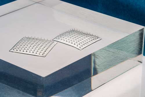
The researchers used these stable liquid crystalline solutions to make things in both modern and traditional ways, 3D printing carbon nanotube aerogels and silk screen printing patterns onto a variety of surfaces, including glass. 

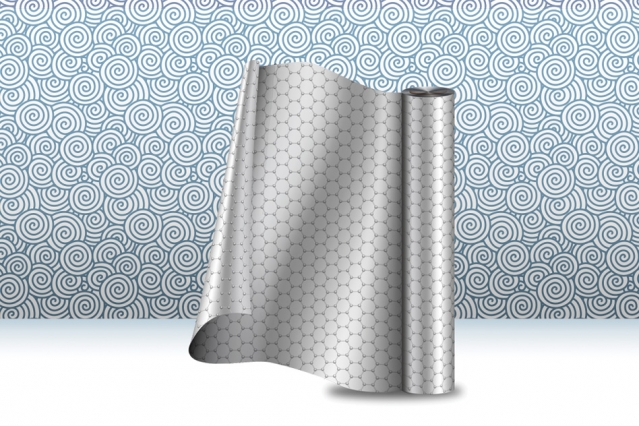
The solutions also enabled roll-to-roll production of transparent films that can be used as electrodes. “Honestly, it was a little surprising how well that worked,” Headrick said. “It came out pretty flawless on the very first try.”

The researchers noted oleum still requires careful handling, but once diluted with the other acids, the solution is much less aggressive to other materials. 

“The acids we’re using are so much gentler that you can use them with common plastics,” Headrick said. “That opens the door to a lot of materials processing and printing techniques that are already in place in manufacturing facilities. 

“It’s also really important for integrating carbon nanotubes into other devices, depositing them as one step in a device-manufacturing process,” he said.

They reported the less-corrosive solutions did not give off harmful fumes and were easier to clean up after production. MSA and pToS can also be recycled after processing nanotubes, lowering their environmental impact and energy and processing costs.

Williams said the next step is to fine-tune the solvent for applications, and to determine how factors like chirality and size affect nanotube processing. “It’s really important that we have high-quality, clean, large diameter tubes,” he said.

Co-authors of the paper are alumna Lauren Taylor and graduate students Oliver Dewey and Cedric Ginestra of Rice; graduate student Crystal Owens and professors Gareth McKinley and A. John Hart at the Massachusetts Institute of Technology; alumna Lucy Liberman, graduate student Asia Matatyaho Ya’akobi and Yeshayahu Talmon, a professor emeritus of chemical engineering, at the Technion-Israel Institute of Technology, Haifa, Israel; and Benji Maruyama, autonomous materials lead in the Materials and Manufacturing Directorate, Air Force Research Laboratory.

Here’s a link to and a citation for the paper,

Versatile acid solvents for pristine carbon nanotube assembly by Robert J. Headrick, Steven M. Williams, Crystal E. Owens, Lauren W. Taylor, Oliver S. Dewey, Cedric J. Ginestra, Lucy Liberman, Asia Matatyaho Ya’akobi, Yeshayahu Talmon, Benji Maruyama, Gareth H. McKinley, A. John Hart, Matteo Pasquali. Science Advances • 27 Apr 2022 • Vol 8, Issue 17 • DOI: 10.1126/sciadv.abm3285

This paper is open access.

Massachusetts Institute of Technology (MIT) researchers achieve highest fraction of CNCs in a composite to date

Cellulose nanocrystals (CNCs), always of interest to me, are featured in research announced in a February 11, 2022 news item on Nanowerk,

The strongest part of a tree lies not in its trunk or its sprawling roots, but in the walls of its microscopic cells.

A single wood cell wall is constructed from fibers of cellulose — nature’s most abundant polymer, and the main structural component of all plants and algae. Within each fiber are reinforcing cellulose nanocrystals, or CNCs, which are chains of organic polymers arranged in nearly perfect crystal patterns. At the nanoscale, CNCs are stronger and stiffer than Kevlar. If the crystals could be worked into materials in significant fractions, CNCs could be a route to stronger, more sustainable, naturally derived plastics.

Now, an MIT team has engineered a composite made mostly from cellulose nanocrystals mixed with a bit of synthetic polymer. The organic crystals take up about 60 to 90 percent of the material — the highest fraction of CNCs achieved in a composite to date.

A February 10, 2022 MIT news release (also on EurekAlert), which originated the news item, delves further into the research (Note: A link has been removed),

The researchers found the cellulose-based composite is stronger and tougher than some types of bone, and harder than typical aluminum alloys. The material has a brick-and-mortar microstructure that resembles nacre, the hard inner shell lining of some molluscs.

The team hit on a recipe for the CNC-based composite that they could fabricate using both 3D printing and conventional casting. They printed and cast the composite into penny-sized pieces of film that they used to test the material’s strength and hardness. They also machined the composite into the shape of a tooth to show that the material might one day be used to make cellulose-based dental implants — and for that matter, any plastic products — that are stronger, tougher, and more sustainable.

“By creating composites with CNCs at high loading, we can give polymer-based materials mechanical properties they never had before,” says A. John Hart, professor of mechanical engineering. “If we can replace some petroleum-based plastic with naturally-derived cellulose, that’s arguably better for the planet as well.”

Hart and his team, including Abhinav Rao PhD ’18, Thibaut Divoux, and Crystal Owens SM ’17, have published their results today in the journal Cellulose.

Gel bonds

Each year, more than 10 billion tons of cellulose is synthesized from the bark, wood, or leaves of plants. Most of this cellulose is used to manufacture paper and textiles, while a portion of it is processed into powder for use in food thickeners and cosmetics.

In recent years, scientists have explored uses for cellulose nanocrystals, which can be extracted from cellulose fibers via acid hydrolysis. The exceptionally strong crystals could be used as natural reinforcements in polymer-based materials. But researchers have only been able to incorporate low fractions of CNCs, as the crystals have tended to clump and only weakly bond with polymer molecules.

Hart and his colleagues looked to develop a composite with a high fraction of CNCs, that they could shape into strong, durable forms. They started by mixing a solution of synthetic polymer with commercially available CNC powder. The team determined the ratio of CNC and polymer that would turn the solution into a gel, with a consistency that could either be fed through the nozzle of a 3-D printer or poured into a mold to be cast. They used an ultrasonic probe to break up any clumps of cellulose in the gel, making it more likely for the dispersed cellulose to form strong bonds with polymer molecules.

They fed some of the gel through a 3-D printer and poured the rest into a mold to be cast. They then let the printed samples dry. In the process, the material shrank, leaving behind a solid composite composed mainly of cellulose nanocrystals.

“We basically deconstructed wood, and reconstructed it,” Rao says. “We took the best components of wood, which is cellulose nanocrystals, and reconstructed them to achieve a new composite material.”

Tough cracks

Interestingly, when the team examined the composite’s structure under a microscope, they observed that grains of cellulose settled into a brick-and-mortar pattern, similar to the architecture of nacre. In nacre, this zig-zagging microstructure stops a crack from running straight through the material. The researchers found this to also be the case with their new cellulose composite.

They tested the material’s resistance to cracks, using tools to initiate first nano- and then micro-scale cracks. They found that, across multiple scales, the composite’s arrangement of cellulose grains prevented the cracks from splitting the material. This resistance to plastic deformation gives the composite a hardness and stiffness at the boundary between conventional plastics and metals.

Going forward, the team is looking for ways to minimize the shrinkage of gels as they dry. While shrinkage isn’t much of a problem when printing small objects, anything bigger could buckle or crack as the composite dries.

“If you could avoid shrinkage, you could keep scaling up, maybe to the meter scale,” Rao says. “Then, if we were to dream big, we could replace a significant fraction of plastics,with cellulose composites.”

This research was supported, in part, by the Proctor and Gamble Corporation, and by the National Defense Science and Engineering Graduate Fellowship.

Here’s a link to and a citation for the paper,

Printable, castable, nanocrystalline cellulose-epoxy composites exhibiting hierarchical nacre-like toughening by Abhinav Rao, Thibaut Divoux, Crystal E. Owens & A. John Hart. Cellulose (2022) DOI: https://doi.org/10.1007/s10570-021-04384-7 Published: 10 February 2022

This paper is behind a paywall.

Food sensor made from of silk microneedles looks like velco

These sensors really do look like velcro,

The Velcro-like food sensor, made from an array of silk microneedles, can pierce through plastic packaging to sample food for signs of spoilage and bacterial contamination. Image: Felice Frankel

A September 9, 2020 news item on Nanowerk announces some research from the Massachusetts Institute (MIT),

MIT engineers have designed a Velcro-like food sensor, made from an array of silk microneedles, that pierces through plastic packaging to sample food for signs of spoilage and bacterial contamination.

The sensor’s microneedles are molded from a solution of edible proteins found in silk cocoons, and are designed to draw fluid into the back of the sensor, which is printed with two types of specialized ink. One of these “bioinks” changes color when in contact with fluid of a certain pH range, indicating that the food has spoiled; the other turns color when it senses contaminating bacteria such as pathogenic E. coli.

A Sept. 9, 2020 MIT news release (also on EurekAlert), which originated the news item, delves further into the research,

The researchers attached the sensor to a fillet of raw fish that they had injected with a solution contaminated with E. coli. After less than a day, they found that the part of the sensor that was printed with bacteria-sensing bioink turned from blue to red — a clear sign that the fish was contaminated. After a few more hours, the pH-sensitive bioink also changed color, signaling that the fish had also spoiled.

The results, published today in the journal Advanced Functional Materials, are a first step toward developing a new colorimetric sensor that can detect signs of food spoilage and contamination.

Such smart food sensors might help head off outbreaks such as the recent salmonella contamination in onions and peaches. They could also prevent consumers from throwing out food that may be past a printed expiration date, but is in fact still consumable.

“There is a lot of food that’s wasted due to lack of proper labeling, and we’re throwing food away without even knowing if it’s spoiled or not,” says Benedetto Marelli, the Paul M. Cook Career Development Assistant Professor in MIT’s Department of Civil and Environmental Engineering. “People also waste a lot of food after outbreaks, because they’re not sure if the food is actually contaminated or not. A technology like this would give confidence to the end user to not waste food.”

Marelli’s co-authors on the paper are Doyoon Kim, Yunteng Cao, Dhanushkodi Mariappan, Michael S. Bono Jr., and A. John Hart.

Silk and printing

The new food sensor is the product of a collaboration between Marelli, whose lab harnesses the properties of silk to develop new technologies, and Hart, whose group develops new manufacturing processes.

Hart recently developed a high-resolution floxography technique, realizing microscopic patterns that can enable low-cost printed electronics and sensors. Meanwhile, Marelli had developed a silk-based microneedle stamp that penetrates and delivers nutrients to plants. In conversation, the researchers wondered whether their technologies could be paired to produce a printed food sensor that monitors food safety.

“Assessing the health of food by just measuring its surface is often not good enough. At some point, Benedetto mentioned his group’s microneedle work with plants, and we realized that we could combine our expertise to make a more effective sensor,” Hart recalls.

The team looked to create a sensor that could pierce through the surface of many types of food. The design they came up with consisted of an array of microneedles made from silk.

“Silk is completely edible, nontoxic, and can be used as a food ingredient, and it’s mechanically robust enough to penetrate through a large spectrum of tissue types, like meat, peaches, and lettuce,” Marelli says.

A deeper detection

To make the new sensor, Kim first made a solution of silk fibroin, a protein extracted from moth cocoons, and poured the solution into a silicone microneedle mold. After drying, he peeled away the resulting array of microneedles, each measuring about 1.6 millimeters long and 600 microns wide — about one-third the diameter of a spaghetti strand.

The team then developed solutions for two kinds of bioink — color-changing printable polymers that can be mixed with other sensing ingredients. In this case, the researchers mixed into one bioink an antibody that is sensitive to a molecule in E. coli. When the antibody comes in contact with that molecule, it changes shape and physically pushes on the surrounding polymer, which in turn changes the way the bioink absorbs light. In this way, the bioink can change color when it senses contaminating bacteria.

The researchers made a bioink containing antibodies sensitive to E. coli, and a second bioink sensitive to pH levels that are associated with spoilage. They printed the bacteria-sensing bioink on the surface of the microneedle array, in the pattern of the letter “E,” next to which they printed the pH-sensitive bioink, as a “C.” Both letters initially appeared blue in color.

Kim then embedded pores within each microneedle to increase the array’s ability to draw up fluid via capillary action. To test the new sensor, he bought several fillets of raw fish from a local grocery store and injected each fillet with a fluid containing either E. coli, Salmonella, or the fluid without any contaminants. He stuck a sensor into each fillet. Then, he waited.

After about 16 hours, the team observed that the “E” turned from blue to red, only in the fillet contaminated with E. coli, indicating that the sensor accurately detected the bacterial antigens. After several more hours, both the “C” and “E” in all samples turned red, indicating that every fillet had spoiled.

The researchers also found their new sensor indicates contamination and spoilage faster than existing sensors that only detect pathogens on the surface of foods.

“There are many cavities and holes in food where pathogens are embedded, and surface sensors cannot detect these,” Kim says. “So we have to plug in a bit deeper to improve the reliability of the detection. Using this piercing technique, we also don’t have to open a package to inspect food quality.”

The team is looking for ways to speed up the microneedles’ absorption of fluid, as well as the bioinks’ sensing of contaminants. Once the design is optimized, they envision the sensor could be used at various stages along the supply chain, from operators in processing plants, who can use the sensors to monitor products before they are shipped out, to consumers who may choose to apply the sensors on certain foods to make sure they are safe to eat.

Here’s a link to and a citation for the paper,

A Microneedle Technology for Sampling and Sensing Bacteria in the Food Supply Chain by Doyoon Kim, Yunteng Cao, Dhanushkodi Mariappan, Michael S. Bono Jr., A. John Hart, Benedetto Marelli. DOI: https://doi.org/10.1002/adfm.202005370 First published: 09 September 2020

This paper is behind a paywall.

Spooling strips of graphene

An April 18, 2018 news item on phys.org highlights an exciting graphene development at the Massachusetts Institute of Technology (MIT),

MIT engineers have developed a continuous manufacturing process that produces long strips of high-quality graphene.

The team’s results are the first demonstration of an industrial, scalable method for manufacturing high-quality graphene that is tailored for use in membranes that filter a variety of molecules, including salts, larger ions, proteins, or nanoparticles. Such membranes should be useful for desalination, biological separation, and other applications.

A new manufacturing process produces strips of graphene, at large scale, for use in membrane technologies and other applications. Image: Christine Daniloff, MIT

An April 17, 2018 MIT news release (also on EurekAlert) by Jennifer Chu, which originated the news item,. provides more detail,

“For several years, researchers have thought of graphene as a potential route to ultrathin membranes,” says John Hart, associate professor of mechanical engineering and director of the Laboratory for Manufacturing and Productivity at MIT. “We believe this is the first study that has tailored the manufacturing of graphene toward membrane applications, which require the graphene to be seamless, cover the substrate fully, and be of high quality.”

Hart is the senior author on the paper, which appears online in the journal Applied Materials and Interfaces. The study includes first author Piran Kidambi, a former MIT postdoc who is now an assistant professor at Vanderbilt University; MIT graduate students Dhanushkodi Mariappan and Nicholas Dee; Sui Zhang of the National University of Singapore; Andrey Vyatskikh, a former student at the Skolkovo Institute of Science and Technology who is now at Caltech; and Rohit Karnik, an associate professor of mechanical engineering at MIT.

Growing graphene

For many researchers, graphene is ideal for use in filtration membranes. A single sheet of graphene resembles atomically thin chicken wire and is composed of carbon atoms joined in a pattern that makes the material extremely tough and impervious to even the smallest atom, helium.

Researchers, including Karnik’s group, have developed techniques to fabricate graphene membranes and precisely riddle them with tiny holes, or nanopores, the size of which can be tailored to filter out specific molecules. For the most part, scientists synthesize graphene through a process called chemical vapor deposition, in which they first heat a sample of copper foil and then deposit onto it a combination of carbon and other gases.

Graphene-based membranes have mostly been made in small batches in the laboratory, where researchers can carefully control the material’s growth conditions. However, Hart and his colleagues believe that if graphene membranes are ever to be used commercially they will have to be produced in large quantities, at high rates, and with reliable performance.

“We know that for industrialization, it would need to be a continuous process,” Hart says. “You would never be able to make enough by making just pieces. And membranes that are used commercially need to be fairly big – some so big that you would have to send a poster-wide sheet of foil into a furnace to make a membrane.”

A factory roll-out

The researchers set out to build an end-to-end, start-to-finish manufacturing process to make membrane-quality graphene.

The team’s setup combines a roll-to-roll approach – a common industrial approach for continuous processing of thin foils – with the common graphene-fabrication technique of chemical vapor deposition, to manufacture high-quality graphene in large quantities and at a high rate. The system consists of two spools, connected by a conveyor belt that runs through a small furnace. The first spool unfurls a long strip of copper foil, less than 1 centimeter wide. When it enters the furnace, the foil is fed through first one tube and then another, in a “split-zone” design.

While the foil rolls through the first tube, it heats up to a certain ideal temperature, at which point it is ready to roll through the second tube, where the scientists pump in a specified ratio of methane and hydrogen gas, which are deposited onto the heated foil to produce graphene.

“Graphene starts forming in little islands, and then those islands grow together to form a continuous sheet,” Hart says. “By the time it’s out of the oven, the graphene should be fully covering the foil in one layer, kind of like a continuous bed of pizza.”

As the graphene exits the furnace, it’s rolled onto the second spool. The researchers found that they were able to feed the foil continuously through the system, producing high-quality graphene at a rate of 5 centimers per minute. Their longest run lasted almost four hours, during which they produced about 10 meters of continuous graphene.

“If this were in a factory, it would be running 24-7,” Hart says. “You would have big spools of foil feeding through, like a printing press.”

Flexible design

Once the researchers produced graphene using their roll-to-roll method, they unwound the foil from the second spool and cut small samples out. They cast the samples with a polymer mesh, or support, using a method developed by scientists at Harvard University, and subsequently etched away the underlying copper.

“If you don’t support graphene adequately, it will just curl up on itself,” Kidambi says. “So you etch copper out from underneath and have graphene directly supported by a porous polymer – which is basically a membrane.”

The polymer covering contains holes that are larger than graphene’s pores, which Hart says act as microscopic “drumheads,” keeping the graphene sturdy and its tiny pores open.

The researchers performed diffusion tests with the graphene membranes, flowing a solution of water, salts, and other molecules across each membrane. They found that overall, the membranes were able to withstand the flow while filtering out molecules. Their performance was comparable to graphene membranes made using conventional, small-batch approaches.

The team also ran the process at different speeds, with different ratios of methane and hydrogen gas, and characterized the quality of the resulting graphene after each run. They drew up plots to show the relationship between graphene’s quality and the speed and gas ratios of the manufacturing process. Kidambi says that if other designers can build similar setups, they can use the team’s plots to identify the settings they would need to produce a certain quality of graphene.

“The system gives you a great degree of flexibility in terms of what you’d like to tune graphene for, all the way from electronic to membrane applications,” Kidambi says.

Looking forward, Hart says he would like to find ways to include polymer casting and other steps that currently are performed by hand, in the roll-to-roll system.

“In the end-to-end process, we would need to integrate more operations into the manufacturing line,” Hart says. “For now, we’ve demonstrated that this process can be scaled up, and we hope this increases confidence and interest in graphene-based membrane technologies, and provides a pathway to commercialization.”

Here’s a link to and a citation for the paper,

A Scalable Route to Nanoporous Large-Area Atomically Thin Graphene Membranes by Roll-to-Roll Chemical Vapor Deposition and Polymer Support Casting by Piran R. Kidambi, Dhanushkodi D. Mariappan, Nicholas T. Dee, Andrey Vyatskikh, Sui Zhang, Rohit Karnik, and A. John Hart. ACS Appl. Mater. Interfaces, 2018, 10 (12), pp 10369–10378 DOI: 10.1021/acsami.8b00846 Publication Date (Web): March 19, 2018

Copyright © 2018 American Chemical Society

This paper is behind a paywall.

Finally, there is a video of the ‘graphene spooling out’ process,

3D printing with cellulose

The scientists seem quite excited about their work with 3D printing and cellulose. From a March 3, 2017 MIT (Massachusetts Institute of Technology) news release (also on EurekAlert),

For centuries, cellulose has formed the basis of the world’s most abundantly printed-on material: paper. Now, thanks to new research at MIT, it may also become an abundant material to print with — potentially providing a renewable, biodegradable alternative to the polymers currently used in 3-D printing materials.

“Cellulose is the most abundant organic polymer in the world,” says MIT postdoc Sebastian Pattinson, lead author of a paper describing the new system in the journal Advanced Materials Technologies. The paper is co-authored by associate professor of mechanical engineering A. John Hart, the Mitsui Career Development Professor in Contemporary Technology.

Cellulose, Pattinson explains, is “the most important component in giving wood its mechanical properties. And because it’s so inexpensive, it’s biorenewable, biodegradable, and also very chemically versatile, it’s used in a lot of products. Cellulose and its derivatives are used in pharmaceuticals, medical devices, as food additives, building materials, clothing — all sorts of different areas. And a lot of these kinds of products would benefit from the kind of customization that additive manufacturing [3-D printing] enables.”

Meanwhile, 3-D printing technology is rapidly growing. Among other benefits, it “allows you to individually customize each product you make,” Pattinson says.

Using cellulose as a material for additive manufacturing is not a new idea, and many researchers have attempted this but faced major obstacles. When heated, cellulose thermally decomposes before it becomes flowable, partly because of the hydrogen bonds that exist between the cellulose molecules. The intermolecular bonding also makes high-concentration cellulose solutions too viscous to easily extrude.

Instead, the MIT team chose to work with cellulose acetate — a material that is easily made from cellulose and is already widely produced and readily available. Essentially, the number of hydrogen bonds in this material has been reduced by the acetate groups. Cellulose acetate can be dissolved in acetone and extruded through a nozzle. As the acetone quickly evaporates, the cellulose acetate solidifies in place. A subsequent optional treatment replaces the acetate groups and increases the strength of the printed parts.

“After we 3-D print, we restore the hydrogen bonding network through a sodium hydroxide treatment,” Pattinson says. “We find that the strength and toughness of the parts we get … are greater than many commonly used materials” for 3-D printing, including acrylonitrile butadiene styrene (ABS) and polylactic acid (PLA).

To demonstrate the chemical versatility of the production process, Pattinson and Hart added an extra dimension to the innovation. By adding a small amount of antimicrobial dye to the cellulose acetate ink, they 3-D-printed a pair of surgical tweezers with antimicrobial functionality.

“We demonstrated that the parts kill bacteria when you shine fluorescent light on them,” Pattinson says. Such custom-made tools “could be useful for remote medical settings where there’s a need for surgical tools but it’s difficult to deliver new tools as they break, or where there’s a need for customized tools. And with the antimicrobial properties, if the sterility of the operating room is not ideal the antimicrobial function could be essential,” he says.

Because most existing extrusion-based 3-D printers rely on heating polymer to make it flow, their production speed is limited by the amount of heat that can be delivered to the polymer without damaging it. This room-temperature cellulose process, which simply relies on evaporation of the acetone to solidify the part, could potentially be faster, Pattinson says. And various methods could speed it up even further, such as laying down thin ribbons of material to maximize surface area, or blowing hot air over it to speed evaporation. A production system would also seek to recover the evaporated acetone to make the process more cost effective and environmentally friendly.

Cellulose acetate is already widely available as a commodity product. In bulk, the material is comparable in price to that of thermoplastics used for injection molding, and it’s much less expensive than the typical filament materials used for 3-D printing, the researchers say. This, combined with the room-temperature conditions of the process and the ability to functionalize cellulose in a variety of ways, could make it commercially attractive.

Here’s a link to and a citation for the paper,

Additive Manufacturing of Cellulosic Materials with Robust Mechanics and Antimicrobial Functionality by Sebastian W. Pattinson and A. John Hart. Advanced Materials Technologies DOI: 10.1002/admt.201600084 Version of Record online: 30 JAN 2017

© 2017 WILEY-VCH Verlag GmbH & Co. KGaA, Weinheim

This paper is behind a paywall.

Understanding how carbon nanotubes grow and self-organize is key to better production

This research may help to commercialize use of carbon nanotubes (CNTs), a  ‘magical’ nanoscale material with great promise and great difficulties (standardizing production being one of the main difficulties). A Feb. 10, 2017 news item on phys.org describes how researchers at the Lawrence Livermore National Laboratory (LLNL) and other collaborators have recorded carbon nanotubes self-organizing,

For the first time, Lawrence Livermore National Laboratory scientists and collaborators have captured a movie of how large populations of carbon nanotubes grow and align themselves.

Understanding how carbon nanotubes (CNT) nucleate, grow and self-organize to form macroscale materials is critical for application-oriented design of next-generation supercapacitors, electronic interconnects, separation membranes and advanced yarns and fabrics.

A Feb. 9, 2017 LLNL news release, which originated the news item, provides more information about the research (Note: Links have been removed),

New research by LLNL scientist Eric Meshot and colleagues from Brookhaven National Laboratory (link is external) (BNL) and Massachusetts Institute of Technology (link is external) (MIT) has demonstrated direct visualization of collective nucleation and self-organization of aligned carbon nanotube films inside of an environmental transmission electron microscope (ETEM).

In a pair of studies reported in recent issues of Chemistry of Materials (link is external) and ACS Nano (link is external), the researchers leveraged a state-of-the-art kilohertz camera in an aberration-correction ETEM at BNL to capture the inherently rapid processes that govern the growth of these exciting nanostructures.

Among other phenomena discovered, the researchers are the first to provide direct proof of how mechanical competition among neighboring carbon nanotubes can simultaneously promote self-alignment while also frustrating and limiting growth.

“This knowledge may enable new pathways toward mitigating self-termination and promoting growth of ultra-dense and aligned carbon nanotube materials, which would directly impact several application spaces, some of which are being pursued here at the Laboratory,” Meshot said.

Meshot has led the CNT synthesis development at LLNL for several projects, including those supported by the Laboratory Directed Research and Development (LDRD) program and the Defense Threat Reduction Agency (link is external) (DTRA) that use CNTs as fluidic nanochannels for applications ranging from single-molecule detection to macroscale membranes for breathable and protective garments.

Here’s a link to and a citation for the both of the papers mentioned in the news release,

Measurement of the Dewetting, Nucleation, and Deactivation Kinetics of Carbon Nanotube Population Growth by Environmental Transmission Electron Microscopy by Mostafa Bedewy, B. Viswanath, Eric R. Meshot, Dmitri N. Zakharov, Eric A. Stach, and A. John Hart. Chem. Mater., 2016, 28 (11), pp 3804–3813 DOI: 10.1021/acs.chemmater.6b00798 Publication Date (Web): May 23, 2016

Copyright © 2016 American Chemical Society

Real-Time Imaging of Self-Organization and Mechanical Competition in Carbon Nanotube Forest Growth by Viswanath Balakrishnan, Mostafa Bedewy, Eric R. Meshot, Sebastian W. Pattinson, Erik S. Polsen, Fabrice Laye, Dmitri N. Zakharov, Eric A. Stach, and A. John Hart. ACS Nano, 2016, 10 (12), pp 11496–11504 DOI: 10.1021/acsnano.6b07251 Publication Date (Web): November 23, 2016

Copyright © 2016 American Chemical Society

Both papers are behind a paywall.

The researchers have also provided this image which allows you to appreciate the difference between a ‘scientific’ version of the work and an artistic version,

This transmission electron microscope image shows growth of a dense carbon nanotube population. Courtesy: LLNL