This environmental monitoring story focused on the roly-poly was announced in an April 18, 2023 news item on Statnano,
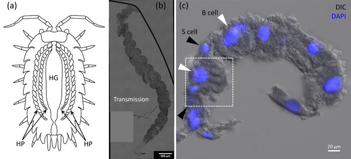
The woodlouse goes by many names: roly-poly, pill bug, potato bug, tomato bug, butchy boy, cheesy bob, and chiggy pig, to name just a few. It is best known for contracting into a ball when agitated. This crustacean (yes, it’s a crustacean, not an insect) thrives in heavily metal-contaminated areas due to its specialized digestive organ, called a hepatopancreas, that stores and expels unwanted metals.
Metal nanoparticles are common in industrial and research plants. However, they can leach into the surrounding environment. Currently, little is known about the toxicity of metal nanoparticles for nearby organisms because detecting metal nanoparticles, particularly gold, requires microscopic, 3D imaging that cannot be done in the field
….
An April 11, 2023 American Institute of Physics (AIP) news release (also on EurekAlert), which originated the news item, describes a new approach to detecting gold nanoparticles in roly-polys,
In Applied Physics Letters, by AIP Publishing, researchers from Cardiff University in the U.K. introduce a novel imaging method to detect gold nanoparticles in woodlice. With information about the quantity, location, and impact of gold nanoparticles within the organism, scientists can better understand the potential harm other metals may have on nature.
“Gold nanoparticles are used extensively for biological research applications owing to their biocompatibility and photostability and are available in a large range of shapes and sizes,” said author Wolfgang Langbein. “By using gold nanoparticles, which would not normally be present in the woodlice diet, we can study the journey of nanoparticles inside complex biological systems.”
The researchers developed an imaging method known as four-wave mixing microscopy, which flashes light that the gold nanoparticles absorb. The light flashes again and the subsequent scattering reveals the nanoparticles’ locations. Using this state-of-the-art technique, they locate the individual gold nanoparticles in the 3D cellular environment.
“By precisely pinpointing the fate of individual gold nanoparticles in the hepatopancreas of woodlice, we can gain a better understanding of how these organisms sequester and respond to metals ingested from the environment,” said Langbein. “Tracking this metal within these organisms is the first step enabling further study to determine, for example, if gold is collected within specific cells, or if it can interfere with the metabolisms in high doses.”
The use of gold nanoparticles in medical devices is increasing and with it, their abundance in the environment. This imaging technique will provide clarity into the little-understood mechanisms in the woodlice hepatopancreas and will also provide helpful environmental monitoring.
In the future, background-free four-wave mixing microscopy could be used to detect other metal nanoparticles and may be applied to organisms like fish larvae and even human cell cultures.
Here’s a link to and a citation for the paper,
Background-free four-wave mixing microscopy of small gold nanoparticles inside a multi-cellular organ by Iestyn Pope, Nuno G.C. Ferreira, Peter Kille, Wolfgang Langbein, and Paola Borri. Appl. Phys. Lett. 122, 153701 (2023) DOI: https://doi.org/10.1063/5.0140651Published online April 11, 2023
This paper is open access.