Tag Archives: Jonathan Rakar

“Skin in a syringe” offers a new way to heal burns

An August 14, 2025 news item on ScienceDaily announced research into burn care from Sweden’s Linköping University,

Researchers have created what could be called “skin in a syringe.” The gel containing live cells can be 3D printed into a skin transplant, as shown in a study conducted on mice. This technology may lead to new ways to treat burns and severe wounds. The study was led from the Center for Disaster Medicine and Traumatology and Linköping University in Sweden, and has been published in Advanced Healthcare Materials.

An August 12, 2025 Linköping University press release (also on EurekAlert), which originated the news item, delves further into the research,

As long as we have a healthy skin, we do not give it much thought. However, if we get major wounds or other injuries, it becomes clear that the skin is the body’s protection from the outside world. Helping the body restore the skin barrier after a serious burn can therefore be a matter of life and death.

Large burns are often treated by transplanting a thin layer of the top part of the skin, the epidermis. This is basically composed of a single cell type. Transplanting only this part of the skin leads to severe scarring.

Under the epidermis there is a thicker and more advanced layer of skin called the dermis. It has blood vessels, nerves, hair follicles and other structures necessary for skin function and elasticity. However, transplanting also the dermis is rarely an option, as the procedure leaves a wound as large as the wound to be healed.

The trick is to create new skin that does not become scar tissue but a functioning dermis.

“The dermis is so complicated that we can’t grow it in a lab. We don’t even know what all its components are. That’s why we, and many others, think that we could possibly transplant the building blocks and then let the body make the dermis itself,” says Johan Junker, researcher at the Swedish Center for Disaster Medicine and Traumatology and docent in plastic surgery at Linköping University, who led the study published in Advanced Healthcare Materials.

The most common cell type in the dermis, the connective tissue cell or fibroblast, is easy to remove from the body and grow in a lab. The connective tissue cell also has the advantage of being able to develop into more specialised cell types depending on what is needed. The researchers behind the study provide a scaffold by having the cells grow on tiny, porous beads of gelatine, a substance similar to skin collagen. But a liquid containing these beads poured on a wound will not stay there.

The researchers’ solution to the problem is mixing the gelatine beads with a gel consisting of another body-specific substance, hyaluronic acid. When the beads and gel are mixed, they are connected using what is known as click chemistry. The result is a gel that, somewhat simplified, can be called skin in a syringe.

“The gel has a special feature that means that it becomes liquid when exposed to light pressure. You can use a syringe to apply it to a wound, for example, and once applied it becomes gel-like again. This also makes it possible to 3D print the gel with the cells in it,” says Daniel Aili, professor of molecular physics at Linköping University, who led the study together with Johan Junker.

In the current study, the researchers 3D-printed small pucks that were placed under the skin of mice. The results point to the potential of this technology to be used to grow the patient’s own cells from a minimal skin biopsy, which are then 3D-printed into a graft and applied to the wound.

“We see that the cells survive and it’s clear that they produce different substances that are needed to create new dermis. In addition, blood vessels are formed in the grafts, which is important for the tissue to survive in the body. We find this material very promising,” says Johan Junker.

Blood vessels are key to a variety of applications for engineered tissue-like materials. Scientists can grow cells in three-dimensional materials that can be used to build organoids, i.e. mini versions of organs. But there is a bottleneck as concerns these tissue models; they lack blood vessels to transport oxygen and nutrients to the cells. This means that there is a limit to how large the structures can get before the cells at the centre die from oxygen and nutrient deficiency.

The LiU researchers may be one step closer to solving the problem of blood vessel supply. In another article, also published in Advanced Healthcare Materials, the researchers describe a method for making threads from materials consisting of 98 per cent water, known as hydrogels.

“The hydrogel threads become quite elastic, so we can tie knots on them. We also show that they can be formed into mini-tubes, which we can pump fluid through or have blood vessel cells grow in,” says Daniel Aili.

The mini-tubes, or the perfusable channels as the researchers also call them, open up new possibilities for the development of blood vessels for e.g. organoids.

Lars Kölby, professor of plastic surgery at Sahlgrenska University Hospital in Gothenburg, also participated in the project. The research has received funding from, among others, the Erling-Persson Foundation, the European Research Council (ERC), the Swedish Research Council and the Knut and Alice Wallenberg Foundation.

Caption: The researchers 3D-printed small pucks of the gel with cells in it. Credit: Magnus Johansson/Linköping University

Here are links to and citations for both papers in the order in which they are mentioned in the press release,

Biphasic Granular Bioinks for Biofabrication of High Cell Density Constructs for Dermal Regeneration by Rozalin Shamasha, Sneha Kollenchery Ramanathan, Kristin Oskarsdotter, Fatemeh Rasti Boroojeni, Aleksandra Zielińska, Sajjad Naeimipour, Philip Lifwergren, Nina Reustle, Lauren Roberts, Annika Starkenberg, Gunnar Kratz, Peter Apelgren, Karin Säljö, Jonathan Rakar, Lars Kölby, Daniel Aili, Johan Junker. Advanced Healthcare Materials Volume 14, Issue 21 August 19, 2025 2501430 DOI:
https://doi.org/10.1002/adhm.202501430 First published online: 12 June 2025

This paper is open access.

Printing and Rerouting of Elastic and Protease Responsive Shape Memory Hydrogel Filaments by Philip Lifwergren, Viktoria Schoen, Sajjad Naeimipour, Lalit Khare, Anna Wunder, Hanna Blom, Jose G. Martinez, Pierfrancesco Pagella, Anders Fridberger, Johan Junker, Daniel Aili. Advanced Healthcare Materials Volume 14, Issue 22 August 28, 2025 2502262 DOI: https://doi.org/10.1002/adhm.202502262 First published online: 20 June 2025

This paper is open access.

Nanocellulose wound dressing reveals early signs of infection?

The wound dressing changes colour from yellow to blue when the wound is infected. Credit: Olov Planthaber Courtesy: Linköping University

An April 18, 2023 news item on Nanowerk announces a new nanocellulose-based wound dressing that can monitor infections, Note: A link has been removed,

A nanocellulose wound dressing that can reveal early signs of infection without interfering with the healing process has been developed by researchers at Linköping University, Sweden. Their study, published in Materials Today Bio (“Nanocellulose composite wound dressings for real-time pH wound monitoring”), is one further step on the road to a new type of wound care.

The wound dressing is made of tight mesh nanocellulose, preventing bacteria and other microbes from getting in. At the same time, the material lets gases and liquid through. Credit: Olov Planthaber Courtesy: Linköping University

An April 19, 2023 Linköping University press release (also on EurekAlert but published April 18, 2023), which originated the news item, provides context for the research and more technical details about it,

The skin is the largest organ of the human body. A wound disrupts the normal function of the skin and can take a long time to heal, be very painful for the patient and may, in a worst case scenario, lead to death if not treated correctly. Also, hard-to-heal wounds pose a great burden on society, representing about half of all costs in out-patient care.

In traditional wound care, dressings are changed regularly, about every two days. To check whether the wound is infected, care staff have to lift the dressing and make an assessment based on appearance and tests. This is a painful procedure that disturbs wound healing as the scab breaks repeatedly. The risk of infection also increases every time the wound is exposed.

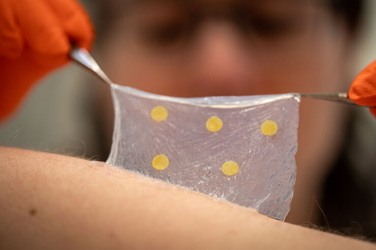
Researchers at Linköping University, in collaboration with colleagues from Örebro and Luleå Universities [Örebro University and Luleå University of Technology in Sweden], have now developed a wound dressing made of nanocellulose that can reveal early signs of infection without interfering with the healing process.

“Being able to see instantly whether a wound has become infected, without having to lift the dressing, opens up for a new type of wound care that can lead to more efficient care and improve life for patients with hard-to-heal wounds. It can also reduce unnecessary use of antibiotics,” says Daniel Aili, professor in the Division of Biophysics and Bioengineering at Linköping University.

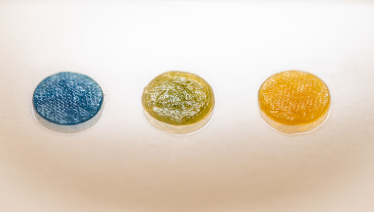
The dressing is made of tight mesh nanocellulose, preventing bacteria and other microbes from getting in. At the same time, the material lets gases and liquid through, something that is important to wound healing. The idea is that once applied, the dressing will stay on during the entire healing process. Should the wound become infected, the dressing will show a colour shift.

Non-infected wounds have a pH value of about 5.5. When an infection occurs, the wound becomes increasingly basic and may have a pH value of 8, or even higher. This is because bacteria in the wound change their surroundings to fit their optimal growth environment. An elevated pH value in the wound can be detected long before any pus, soreness or redness, which are the most common signs of infection.

To make the wound dressing show the elevated pH value, the researchers used bromthymol blue, BTB, a dye that changes colour from yellow to blue when the pH value exceeds 7. For BTB to be used in the dressing without being compromised, it was loaded onto a silica material with pores only a few nanometres in size. The silica material could then be combined with the dressing material without compromising the nanocellulose. The result is a wound dressing that turns blue when there is an infection.

Wound infections are often treated with antibiotics that spread throughout the body. But if the infection is detected at an early stage, local treatment of the wound may suffice. This is why Daniel Aili and his colleagues at Örebro University are also developing anti-microbial substances based on so-called lipopeptides [emphasis mine] that kill off all types of bacteria.

“The use of antibiotics makes infections increasingly problematic, as multi-resistant bacteria are becoming more common. If we can combine the anti-microbial substance with the dressing, we minimise the risk of infection and reduce the overuse of antibiotics,” says Daniel Aili.

Daniel Aili says that the new wound dressing and the anti-microbial substance are part of developing a new type of wound treatment in out-patient care. But as all products to be used in medical care settings have to pass rigorous and expensive testing, he thinks that it will be five to ten years before it will be available there.

Both studies are part of the HEALiX research project financed by the Swedish Foundation for Strategic Research with the objective of developing a new type of wound treatment. Funding was also received from, among others, the Swedish Government Strategic Research Area in Materials Science on Functional Materials (AFM) at Linköping University, Vinnova, the Knut and Alice Wallenberg Foundation and the Swedish Research Council.

For the curious, the HEALiX research project is here.

As noted in the press release, there are two studies. First, here’s a link and citation for the work on antimicrobial lipopeptides,

Development of novel broad-spectrum antimicrobial lipopeptides derived from plantaricin NC8 β by Emanuel Wiman, Elisa Zattarin, Daniel Aili, Torbjörn Bengtsson, Robert Selegård & Hazem Khalaf. Scientific Reports volume 13, Article number: 4104 (2023) DOI: https://doi.org/10.1038/s41598-023-31185-8
Published: 13 March 2023

This paper is open access.

Now, here’s a link to and a citation for the paper about nanocellulose-based wound dressings,

Nanocellulose composite wound dressings for real-time pH wound monitoring by Olof Eskilson, Elisa Zattarin, Linn Berglund, Kristiina Oksman, Kristina Hanna, Jonathan Rakar, Petter Sivlér, Mårten Skog, Ivana Rinklake, Rozalin Shamasha, Zeljana Sotra, Annika Starkenberg, Magnus Odén, Emanuel Wiman, Hazem Khalaf, Torbjörn Bengtsson, Johan P.E. Junker, Robert Selegård, Emma M. Björk, Daniel Aili. Materials Today Bio, Volume 19, April 2023, 100574 DOI: 10.1016/j.mtbio.2023.100574 Published online on 6 February 2023

This paper too is open access.