Tag Archives: adeno-associated viruses (AAV)

Lifesaving moths and nanomagnets

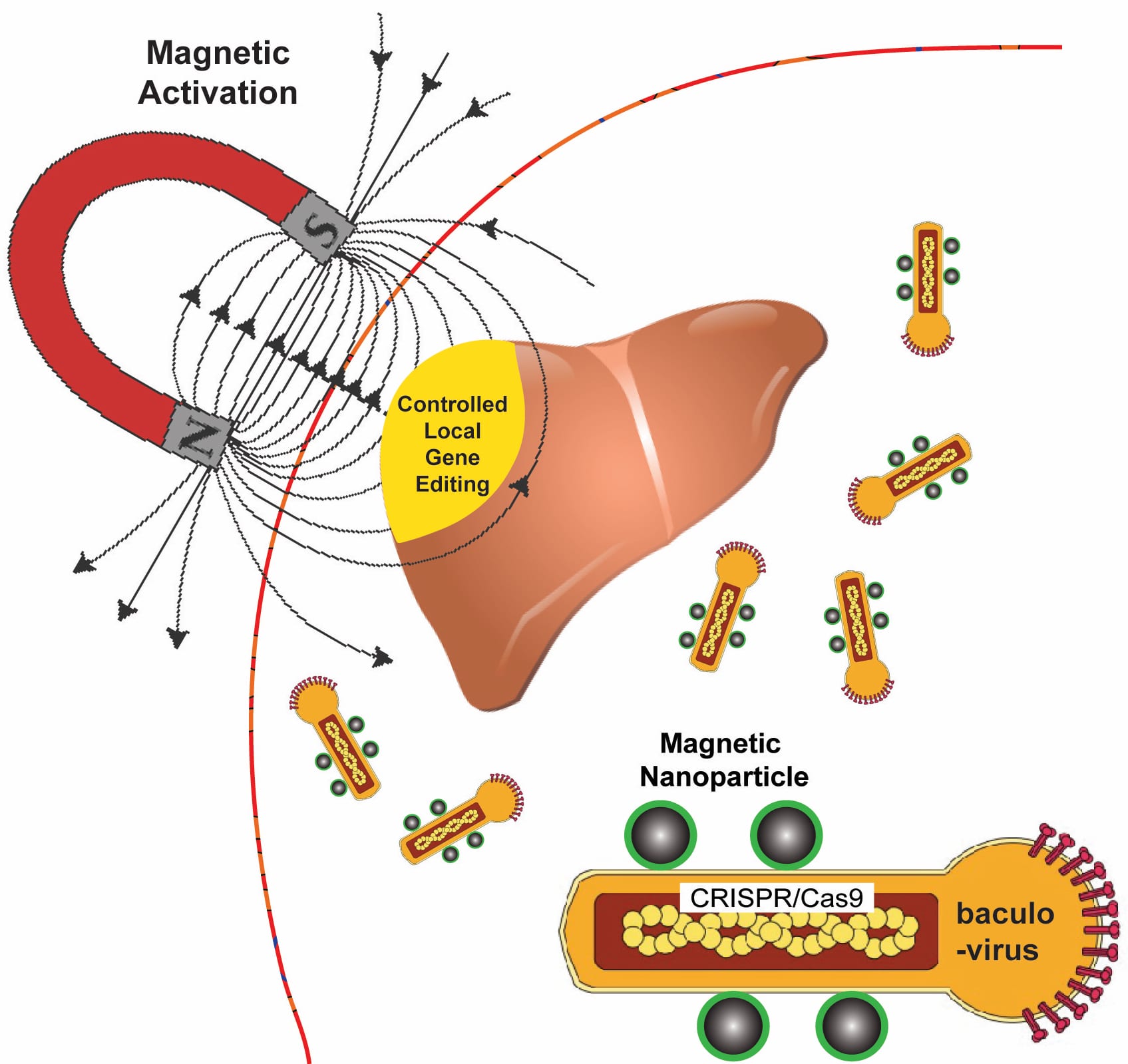
Rice University bioengineers use a magnetic field to activate nanoparticle-attached baculoviruses in a tissue. The viruses, which normally infect alfalfa looper moths, are modified to deliver gene-editing DNA code only to cells that are targeted with magnetic field-induced local transduction. Courtesy of the Laboratory of Biomolecular Engineering and Nanomedicine

Kudos to whomever put that diagram together! That’s a lot of well conveyed information.

Now for the details about how this technology might save lives. From a November 13, 2018 news item on Nanowerk,

A new technology that relies on a moth-infecting virus and nanomagnets could be used to edit defective genes that give rise to diseases like sickle cell, muscular dystrophy and cystic fibrosis.

Rice University bioengineer Gang Bao has combined magnetic nanoparticles with a viral container drawn from a particular species of moth to deliver CRISPR/Cas9 payloads that modify genes in a specific tissue or organ with spatial control.

A November 12, 2018 Rice University news release (also on EurekAlert published on November 13, 2018), which originated the news item, provides detail,

Because magnetic fields are simple to manipulate and, unlike light, pass easily through tissue, Bao and his colleagues want to use them to control the expression of viral payloads in target tissues by activating the virus that is otherwise inactivated in blood.

The research appears in Nature Biomedical Engineering. In nature, CRISPR/Cas9 bolsters microbes’ immune systems by recording the DNA of invaders. That gives microbes the ability to recognize and attack returning invaders, but scientists have been racing to adapt CRISPR/Cas9 to repair mutations that cause genetic diseases and to manipulate DNA in laboratory experiments.

CRISPR/Cas9 has the potential to halt hereditary disease – if scientists can get the genome-editing machinery to the right cells inside the body. But roadblocks remain, especially in delivering the gene-editing payloads with high efficiency.

Bao said it will be necessary to edit cells in the body to treat many diseases. “But efficiently delivering genome-editing machinery into target tissue in the body with spatial control remains a major challenge,” Bao said. “Even if you inject the viral vector locally, it can leak to other tissues and organs, and that could be dangerous.”

The delivery vehicle developed by Bao’s group is based on a virus that infects Autographa californica, aka the alfalfa looper, a moth native to North America. The cylindrical baculovirus vector (BV), the payload-carrying part of the virus, is considered large at up to 60 nanometers in diameter and 200-300 nanometers in length. That’s big enough to transport more than 38,000 base pairs of DNA, which is enough to supply multiple gene-editing units to a target cell, Bao said.

He said the inspiration to combine BV and magnetic nanoparticles came from discussions with Rice postdoctoral researcher and co-lead author Haibao Zhu, who learned about the virus during a postdoctoral stint in Singapore but knew nothing about magnetic nanoparticles until he joined the Bao lab. The Rice team had previous experience using iron oxide nanoparticles and an applied magnetic field to open blood vessel walls just enough to let large-molecule drugs pass through.

“We really didn’t know if this would work for gene editing or not, but we thought, ‘worth a shot,'” Bao said.

The researchers use the magnetic nanoparticles to activate BV and deliver gene-editing payloads only where they’re needed. To do this, they take advantage of an immune-system protein called C3 that normally inactivates baculoviruses.

“If we combine BV with magnetic nanoparticles, we can overcome this deactivation by applying the magnetic field,” Bao said. “The beauty is that when we deliver it, gene editing occurs only at the tissue, or the part of the tissue, where we apply the magnetic field.”

Application of the magnetic field allows BV transduction, the payload-delivery process that introduces gene-editing cargo into the target cell. The payload is also DNA, which encodes both a reporter gene and the CRISPR/Cas9 system.

In tests, the BV was loaded with green fluorescent proteins or firefly luciferase. Cells with the protein glowed brightly under a microscope, and experiments showed the magnets were highly effective at targeted delivery of BV cargoes in both cell cultures and lab animals.

Bao noted his and other labs are working on the delivery of CRISPR/Cas9 with adeno-associated viruses (AAV), but he said BV’s capacity for therapeutic cargo is roughly eight times larger. “However, it is necessary to make BV transduction into target cells more efficient,” he said.

Here’s a link to and a citation for the paper,

Spatial control of in vivo CRISPR–Cas9 genome editing via nanomagnets by Haibao Zhu, Linlin Zhang, Sheng Tong, Ciaran M. Lee, Harshavardhan Deshmukh, & Gang Bao. Nature Biomedical Engineering (2018) DOI: https://doi.org/10.1038/s41551-018-0318-7 Published: 12 November 2018

This paper is behind a paywall.
Resizing viral peptides for nanoscale drug delivery system

A January 9, 2018 news item on Nanowerk sheds some light on the research (Note: A link has been removed),

By chipping away at a viral protein, Rice University scientists have discovered a path toward virus-like, nanoscale devices that may be able to deliver drugs to cells.

The protein is one of three that make up the protective shell, called the capsid, of natural adeno-associated viruses (AAV). By making progressively smaller versions of the protein, the researchers made capsids with unique abilities and learned a great deal about AAV’s mechanisms.

The research appears in the American Chemical Society journal ACS Nano (“Reprogramming the Activatable Peptide Display Function of Adeno-Associated Virus Nanoparticles”).
programmable adeno-associated viruses

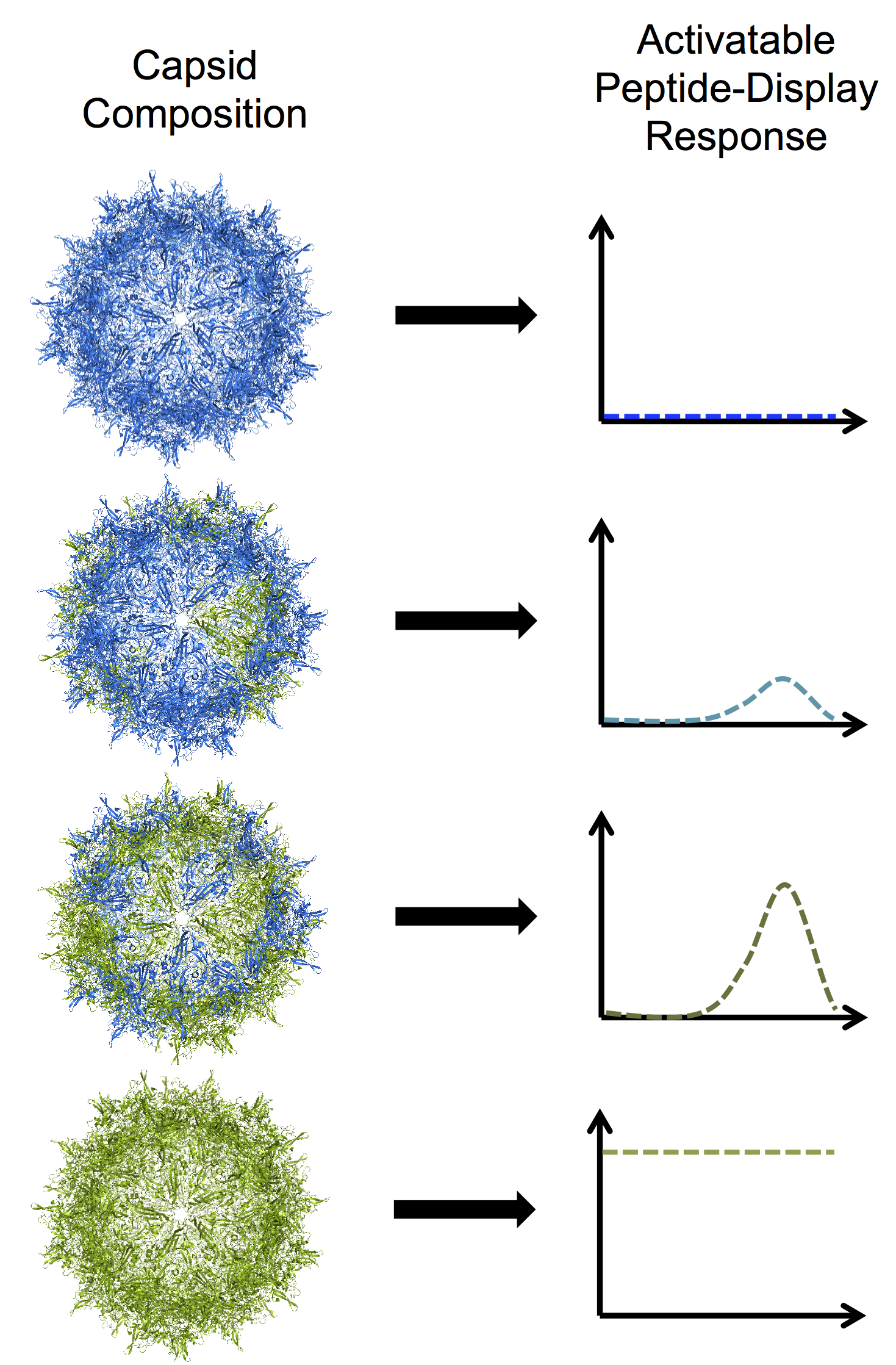
Rice University bioengineers have developed programmable adeno-associated viruses by modifying one of three proteins that assemble into a tough shell called a capsid. In this illustration, blue subunits in the capsid represent the protein VP3 and green subunits represent a truncated mutant of VP2.

Here’s an image illustrating the work,

Rice University bioengineers have developed programmable adeno-associated viruses by modifying one of three proteins that assemble into a tough shell called a capsid. In this illustration, blue subunits in the capsid represent the protein VP3 and green subunits represent a truncated mutant of VP2. From top to bottom: a VP3-only capsid that does not display any peptides; a mosaic capsid with a majority of VP3 and small amount of the VP2 mutant that shows a low level of activable peptide display; a mosaic capsid with equal amounts of VP3 and VP2 mutant that shows a high level of activable peptide display; and a homomeric VP2 mutant capsid with a high level of constant, brush-like peptide display. For a larger version, click on the image. Illustration by Nicole Thadani Courtesy: Rice University

A January 8, 2018 Rice University news release (also on EurekAlert), which originated the news item, expands on the story,

Rice bioengineer Junghae Suh studies the manipulation of nondisease-causing AAVs to deliver helpful cargoes like chemotherapy drugs. Her research has led to the development of viruses that can be triggered by light or by extracellular proteases associated with certain diseases.

AAVs are small — about 25 nanometers — and contain a single strand of DNA inside tough capsids that consist of a mosaic of proteins known as VP1, VP2 and VP3. AAVs have been used to deliver gene-therapy payloads, but nobody has figured out how AAV capsids physically reconfigure themselves when triggered by external stimuli, Suh said. That was the starting point for her lab.

“This virus has intrinsic peptide (small protein) domains hidden inside the capsid,” she said. “When the virus infects a cell, it senses the low pH and other endosomal factors, and these peptide domains pop out onto the surface of the virus capsid.

“This conformational change, which we termed an ‘activatable peptide display,’ is important for the virus because the externalized domains break down the endosomal membrane and allow the virus to escape into the cytoplasm,” Suh said. “In addition, nuclear localization sequences in those domains allow the virus to transit into the nucleus. We believed we could replace that functionality with something else.”

Suh and lead author and Rice graduate student Nicole Thadani think their mutant AAVs can become “biocomputing nanoparticles” that detect and process environmental inputs and produce controllable outputs. Modifying the capsid is the first step.

Of the three natural capsid proteins, only VP1 and VP2 can be triggered to expose their functional peptides, but neither can make a capsid on its own. Shorter VP3s can form capsids by themselves, but do not display peptides. In natural AAVs, VP3 proteins outnumber each of their compadres 10-to-1.

That limits the number of peptides that can be exposed, so Suh, Thadani and their co-authors set out to change the ratio. That led them to truncate VP2 and synthesize mosaic capsids with VP3, resulting in successful alteration of the number of exposed peptides. Based on previous research, they inserted a common hexahistidine tag that made it easy to monitor the surface display of the peptide region.

“We wanted to boost the protein’s activable property beyond what occurs in the native virus capsid,” Thadani said. “Rather than displaying just five copies of the peptide per capsid, now we may be able to display 20 or 30 and get more of the bioactivity that we want.”

They then made a truncated VP2 able to form a capsid on its own. “The results were quite surprising, and not obvious to us,” Suh said. “We chopped down that VP2 component enough to form what we call a homomeric capsid, where the entire capsid is made up of just that mutant subunit. That gave us viruses that appear to have peptide ‘brushes’ that are always on the surface.

“A viral structure like that has never been seen in nature,” she said. “We got a particle with this peptide brush, with loose ends everywhere. Now we want to know if we can use these loose ends to attach other things or carry out other functions.”

Homomeric AAVs display as many as 60 peptides, while mosaic AAVs could be programmed to respond to stimuli specific to particular cells or tissues and display a smaller desired number of peptides, the researchers said.

“Viruses have evolved to invade cells very effectively,” Suh said. “We want to use our virus as a nanoparticle platform to deliver protein- or peptide-based therapeutics more efficiently into cells. We want to harness what nature has already created, tweak it a little bit and use it for our purposes.”

Here’s a link to and a citation for the paper,

Reprogramming the Activatable Peptide Display Function of Adeno-Associated Virus Nanoparticles by Nicole N. Thadani, Christopher Dempsey, Julia Zhao, Sonya M. Vasquez, and Junghae Suh. ACS Nano, Article ASAP DOI: 10.1021/acsnano.7b07804 Publication Date (Web): December 26, 2017

Copyright © 2017 American Chemical Society

This paper is behind a paywall.