Tag Archives: virus

COVID-19: caution and concern not panic

There’s a lot of information being pumped out about COVID-19 and not all of it is as helpful as it might be. In fact, the sheer volume can seem overwhelming despite one’s best efforts to be calm.

Here are a few things I’ve used to help relieve some fo the pressure as numbers in Canada keep rising.

Inspiration from the Italians

I was thrilled to find Emily Rumball’s March 18 ,2020 article titled, “Italians making the most of quarantine is just what the world needs right now (VIDEOS),” on the Daily Hive website. The couple dancing on the balcony while Ginger Rogers and Fred Astaire are shown dancing on the wall above is my favourite.

As the Italians practice social distancing and exercise caution, they are also demonstrating that “life goes on” even while struggling as one of the countries hit hardest by COVID-19.

Investigating viruses and the 1918/19 pandemic vs. COVID-19

There has been some mention of and comparison to the 1918/19 pandemic (also known as the Spanish flu) in articles by people who don’t seem to be particularly well informed about that earlier pandemic. Susan Baxter offers a concise and scathing explanation for why the 1918/19 situation deteriorated as much as it did in her February 8, 2010 posting. As for this latest pandemic (COVID-19), she explains what a virus actually is and suggests we all calm down in her March 17, 2020 posting. BTW, she has an interdisciplinary PhD for work largely focused on health economics. She is also a lecturer in the health sciences programme at Simon Fraser University (Vancouver, Canada). Full disclosure: She and I have a longstanding friendship.

Marilyn J. Roossinck, a professor of Plant Pathology and Environmental Microbiology at Pennsylvania State University, wrote a February 20, 2020 essay for The Conversation titled, “What are viruses anyway, and why do they make us so sick? 5 questions answered,”

4. SARS was a formidable foe, and then seemed to disappear. Why?

Measures to contain SARS started early, and they were very successful. The key is to stop the chain of transmission by isolating infected individuals. SARS had a short incubation period; people generally showed symptoms in two to seven days. There were no documented cases of anyone being a source of SARS without showing symptoms.

Stopping the chain of transmission is much more difficult when the incubation time is much longer, or when some people don’t get symptoms at all. This may be the case with the virus causing CoVID-19, so stopping it may take more time.

1918/19 pandemic vs. COVID-19

Angela Betsaida B. Laguipo, with a Bachelor of Nursing degree from the University of Baguio, Philippine is currently completing her Master’s Degree, has written a March 9, 2020 article for News Medical comparing the two pandemics,

The COVID-19 is fast spreading because traveling is an everyday necessity today, with flights from one country to another accessible to most.

Some places did manage to keep the virus at bay in 1918 with traditional and effective methods, such as closing schools, banning public gatherings, and locking down villages, which has been performed in Wuhan City, in Hubei province, China, where the coronavirus outbreak started. The same method is now being implemented in Northern Italy, where COVID-19 had killed more than 400 people.

The 1918 Spanish flu has a higher mortality rate of an estimated 10 to 20 percent, compared to 2 to 3 percent in COVID-19. The global mortality rate of the Spanish flu is unknown since many cases were not reported back then. About 500 million people or one-third of the world’s population contracted the disease, while the number of deaths was estimated to be up to 50 million.

During that time, public funds are mostly diverted to military efforts, and a public health system was still a budding priority in most countries. In most places, only the middle class or the wealthy could afford to visit a doctor. Hence, the virus has [sic] killed many people in poor urban areas where there are poor nutrition and sanitation. Many people during that time had underlying health conditions, and they can’t afford to receive health services.

I recommend reading Laguipo’s article in its entirety right down to the sources she cites at the end of her article.

Ed Yong’s March 20, 2020 article for The Atlantic, “Why the Coronavirus Has Been So Successful; We’ve known about SARS-CoV-2 for only three months, but scientists can make some educated guesses about where it came from and why it’s behaving in such an extreme way,” provides more information about what is currently know about the coronavirus, SATS-CoV-2,

One of the few mercies during this crisis is that, by their nature, individual coronaviruses are easily destroyed. Each virus particle consists of a small set of genes, enclosed by a sphere of fatty lipid molecules, and because lipid shells are easily torn apart by soap, 20 seconds of thorough hand-washing can take one down. Lipid shells are also vulnerable to the elements; a recent study shows that the new coronavirus, SARS-CoV-2, survives for no more than a day on cardboard, and about two to three days on steel and plastic. These viruses don’t endure in the world. They need bodies.

But why do some people with COVID-19 get incredibly sick, while others escape with mild or nonexistent symptoms? Age is a factor. Elderly people are at risk of more severe infections possibly because their immune system can’t mount an effective initial defense, while children are less affected because their immune system is less likely to progress to a cytokine storm. But other factors—a person’s genes, the vagaries of their immune system, the amount of virus they’re exposed to, the other microbes in their bodies—might play a role too. In general, “it’s a mystery why some people have mild disease, even within the same age group,” Iwasaki [Akiko Iwasaki of the Yale School of Medicine] says.

We still have a lot to learn about this.

Going nuts and finding balance with numbers

Generally speaking,. I find numbers help me to put this situation into perspective. It seems I’m not alone; Dr. Daniel Gillis’ (Guelph University in Ontario, Canada) March 18, 2020 blog post is titled, Statistics In A Time of Crisis.

Hearkening back in history, the Wikipedia entry for Spanish flu offers a low of 17M deaths in a 2018 estimate to a high of !00M deaths in a 2005 estimate. At this writing (Friday, March 20, 2020 at 3 pm PT), the number of coronovirus cases worldwide is 272,820 with 11, 313 deaths.

Articles like Michael Schulman’s March 16, 2020 article for the New Yorker might not be as helpful as one hope (Note: Links have been removed),

Last Wednesday night [March 11, 2020], not long after President Trump’s Oval Office address, I called my mother to check in about the, you know, unprecedented global health crisis [emphasis mine] that’s happening. She told me that she and my father were in a cab on the way home from a fun dinner at the Polo Bar, in midtown Manhattan, with another couple who were old friends.

“You went to a restaurant?!” I shrieked. This was several days after she had told me, through sniffles, that she was recovering from a cold but didn’t see any reason that she shouldn’t go to the school where she works. Also, she was still hoping to make a trip to Florida at the end of the month. My dad, a lawyer, was planning to go into the office on Thursday, but thought that he might work from home on Friday, if he could figure out how to link up his personal computer. …

… I’m thirty-eight, and my mother and father are sixty-eight and seventy-four, respectively. Neither is retired, and both are in good shape. But people sixty-five and older—more than half of the baby-boomer population—are more susceptible to COVID-19 and have a higher mortality rate, and my parents’ blithe behavior was as unsettling as the frantic warnings coming from hospitals in Italy.

Clearly, Schulman is concerned about his parents’ health and well being but the tone of near hysteria is a bit off-putting. We’re not in a crisis (exception: the Italians and, possibly, the Spanish and the French)—yet.

Tyler Dawson’s March 20, 2020 article in The Province newspaper (in Vancouver, British Columbia) offers dire consequences from COVID-19 before pivoting,

COVID-19 will leave no Canadian untouched.

Travel plans halted. First dates postponed. School semesters interrupted. Jobs lost. Retirement savings decimated. Some of us will know someone who has gotten sick, or tragically, died from the virus.

By now we know the terminology: social distancing, flatten the curve. Across the country, each province is taking measures to prepare, to plan for care, and the federal government has introduced financial measures amounting to more than three per cent of the country’s GDP to float the economy onward.

The response, says Steven Taylor, a University of British Columbia psychiatry professor and author of The Psychology of Pandemics, is a “balancing act.” [emphasis mine] Keep people alert, but neither panicked nor tuned out.

“You need to generate some degree of anxiety that gets people’s attention,” says Taylor. “If you overstate the message it could backfire.”

Prepare for uncertainty

In the same way experts still cannot come up with a definitive death rate for the 1918/19 pandemic, they are having trouble with this one too although, now, they’re trying to model the future rather than trying to establish what happened in the past. David Adam’s March 12, 2020 article forThe Scientist, provides some insight into the difficulties (Note: Links have been removed)

Like any other models, the projections of how the outbreak will unfold, how many people will become infected, and how many will die, are only as reliable as the scientific information they rest on. And most modelers’ efforts so far have focused on improving these data, rather than making premature predictions.

“Most of the work that modelers have done recently or in the first part of the epidemic hasn’t really been coming up with models and predictions, which is I think how most people think of it,” says John Edmunds, who works in the Centre for the Mathematical Modelling of Infectious Diseases at the London School of Hygiene & Tropical Medicine. “Most of the work has really been around characterizing the epidemiology, trying to estimate key parameters. I don’t really class that as modeling but it tends to be the modelers that do it.”

These variables include key numbers such as the disease incubation period, how quickly the virus spreads through the population, and, perhaps most contentiously, the case-fatality ratio. This sounds simple: it’s the proportion of infected people who die. But working it out is much trickier than it looks. “The non-specialists do this all the time and they always get it wrong,” Edmunds says. “If you just divide the total numbers of deaths by the total numbers of cases, you’re going to get the wrong answer.”

Earlier this month, Tedros Adhanom Ghebreyesus, the head of the World Health Organization, dismayed disease modelers when he said COVID-19 (the disease caused by the SARS-CoV-2 coronavirus) had killed 3.4 percent of reported cases, and that this was more severe than seasonal flu, which has a death rate of around 0.1 percent. Such a simple calculation does not account for the two to three weeks it usually takes someone who catches the virus to die, for example. And it assumes that reported cases are an accurate reflection of how many people are infected, when the true number will be much higher and the true mortality rate much lower.

Edmunds calls this kind of work “outbreak analytics” rather than true modeling, and he says the results of various specialist groups around the world are starting to converge on COVID-19’s true case-fatality ratio, which seems to be about 1 percent.[emphasis mine]

The 1% estimate in Adam’s article accords with Jeremy Samuel Faust’s (an emergency medicine physician at Brigham and Women’s Hospital in Boston, faculty in its division of health policy and public health, and an instructor at Harvard Medical School) estimates in a March 4, 2020 article (COVID-19 Isn’t As Deadly As We Think featured in my March 9, 2020 posting).

In a March 17, 2020 article by Steven Lewis (a health policy consultant formerly based in Saskatchewan, Canada; now living in Australia) for the Canadian Broadcasting Corporation’s (CBC) news online website, he covers some of the same ground and offers a somewhat higher projected death rate while refusing to commit,

Imagine you’re a chief public health officer and you’re asked the question on everyone’s mind: how deadly is the COVID-19 outbreak?

With the number of cases worldwide approaching 200,000, and 1,000 or more cases in 15 countries, you’d think there would be an answer. But the more data we see, the tougher it is to come up with a hard number.

Overall, the death rate is around four per cent — of reported cases. That’s also the death rate in China, which to date accounts for just under half the total number of global cases.

China is the only country where a) the outcome of almost all cases is known (85 per cent have recovered), and b) the spread has been stopped (numbers plateaued about a month ago). 

A four per cent death rate is pretty high — about 40 times more deadly than seasonal flu — but no experts believe that is the death rate. The latest estimate is that it is around 1.5 per cent. [emphasis mine] Other models suggest that it may be somewhat lower. 

The true rate can be known only if every case is known and confirmed by testing — including the asymptomatic or relatively benign cases, which comprise 80 per cent or more of the total — and all cases have run their course (people have either recovered or died). Aside from those in China, almost all cases identified are still active. 

Unless a jurisdiction systematically tests a large random sample of its population, we may never know the true rate of infection or the real death rate. 

Yet for all this unavoidable uncertainty, it is still odd that the rates vary so widely by country.

His description of the situation in Europe is quite interesting and worthwhile if you have the time to read it.

In the last article I’m including here, Murray Brewster offers some encouraging words in his March 20, 2020 piece about the preparations being made by the Canadian Armed Forces (CAF),

The Canadian military is preparing to respond to multiple waves of the COVID-19 pandemic which could stretch out over a year or more, the country’s top military commander said in his latest planning directive.

Gen. Jonathan Vance, chief of the defence staff, warned in a memo issued Thursday that requests for assistance can be expected “from all echelons of government and the private sector and they will likely come to the Department [of National Defence] through multiple points of entry.”

The directive notes the federal government has not yet directed the military to move into response mode, but if or when it does, a single government panel — likely a deputy-minister level inter-departmental task force — will “triage requests and co-ordinate federal responses.”

It also warns that members of the military will contract the novel coronavirus, “potentially threatening the integrity” of some units.

The notion that the virus caseload could recede and then return is a feature of federal government planning.

The Public Health Agency of Canada has put out a notice looking for people to staff its Centre for Emergency Preparedness and Response during the crisis and the secondment is expected to last between 12 and 24 months.

The Canadian military, unlike those in some other nations, has high-readiness units available. Vance said they are already set to reach out into communities to help when called.

Planners are also looking in more detail at possible missions — such as aiding remote communities in the Arctic where an outbreak could cripple critical infrastructure.

Defence analyst Dave Perry said this kind of military planning exercise is enormously challenging and complicated in normal times, let alone when most of the federal civil service has been sent home.

“The idea that they’re planning to be at this for year is absolutely bang on,” said Perry, a vice-president at the Canadian Global Affairs Institute.

In other words, concern and caution are called for not panic. I realize this post has a strongly Canada-centric focus but I’m hopeful others elsewhere will find this helpful.
Altered virus spins gold into beads

They’re not calling this synthetic biology but I’ m pretty sure that altering a virus gene so the virus can spin gold (Rumpelstiltskin anyone?) qualifies. From an August 24, 2018 news item on ScienceDaily,

The race is on to find manufacturing techniques capable of arranging molecular and nanoscale objects with precision.

Engineers at the University of California, Riverside, have altered a virus to arrange gold atoms into spheroids measuring a few nanometers in diameter. The finding could make production of some electronic components cheaper, easier, and faster.

An August 23, 2018 University of California at Riverside (UCR) news release (also on EurekAlett) by Holly Ober, which originated the news item, adds detail,

“Nature has been assembling complex, highly organized nanostructures for millennia with precision and specificity far superior to the most advanced technological approaches,” said Elaine Haberer, a professor of electrical and computer engineering in UCR’s Marlan and Rosemary Bourns College of Engineering and senior author of the paper describing the breakthrough. “By understanding and harnessing these capabilities, this extraordinary nanoscale precision can be used to tailor and build highly advanced materials with previously unattainable performance.”

Viruses exist in a multitude of shapes and contain a wide range of receptors that bind to molecules. Genetically modifying the receptors to bind to ions of metals used in electronics causes these ions to “stick” to the virus, creating an object of the same size and shape. This procedure has been used to produce nanostructures used in battery electrodes, supercapacitors, sensors, biomedical tools, photocatalytic materials, and photovoltaics.

The virus’ natural shape has limited the range of possible metal shapes. Most viruses can change volume under different scenarios, but resist the dramatic alterations to their basic architecture that would permit other forms.

The M13 bacteriophage, however, is more flexible. Bacteriophages are a type of virus that infects bacteria, in this case, gram-negative bacteria, such as Escherichia coli, which is ubiquitous in the digestive tracts of humans and animals. M13 bacteriophages genetically modified to bind with gold are usually used to form long, golden nanowires.

Studies of the infection process of the M13 bacteriophage have shown the virus can be converted to a spheroid upon interaction with water and chloroform. Yet, until now, the M13 spheroid has been completely unexplored as a nanomaterial template.

Haberer’s group added a gold ion solution to M13 spheroids, creating gold nanobeads that are spiky and hollow.

“The novelty of our work lies in the optimization and demonstration of a viral template, which overcomes the geometric constraints associated with most other viruses,” Haberer said. “We used a simple conversion process to make the M13 virus synthesize inorganic spherical nanoshells tens of nanometers in diameter, as well as nanowires nearly 1 micron in length.”

The researchers are using the gold nanobeads to remove pollutants from wastewater through enhanced photocatalytic behavior.

The work enhances the utility of the M13 bacteriophage as a scaffold for nanomaterial synthesis. The researchers believe the M13 bacteriophage template transformation scheme described in the paper can be extended to related bacteriophages.

Here’s a link to and a citation for the paper,

M13 bacteriophage spheroids as scaffolds for directed synthesis of spiky gold nanostructures by Tam-Triet Ngo-Duc, Joshua M. Plank, Gongde Chen, Reed E. S. Harrison, Dimitrios Morikis, Haizhou Liu, and Elaine D. Haberer. Nanoscale, 2018,10, 13055-13063 DOI: 10.1039/C8NR03229G First published on 25 Jun 2018

This paper is behind a paywall.

For another example of genetic engineering and synthetic biology, see my July 18, 2018 posting: Genetic engineering: an eggplant in Bangladesh and a synthetic biology grant at Concordia University (Canada).

For anyone unfamiliar with the Rumpelstiltskin fairytale about spinning straw into gold, see its Wikipedida entry.

Resizing viral peptides for nanoscale drug delivery system

A January 9, 2018 news item on Nanowerk sheds some light on the research (Note: A link has been removed),

By chipping away at a viral protein, Rice University scientists have discovered a path toward virus-like, nanoscale devices that may be able to deliver drugs to cells.

The protein is one of three that make up the protective shell, called the capsid, of natural adeno-associated viruses (AAV). By making progressively smaller versions of the protein, the researchers made capsids with unique abilities and learned a great deal about AAV’s mechanisms.

The research appears in the American Chemical Society journal ACS Nano (“Reprogramming the Activatable Peptide Display Function of Adeno-Associated Virus Nanoparticles”).
programmable adeno-associated viruses

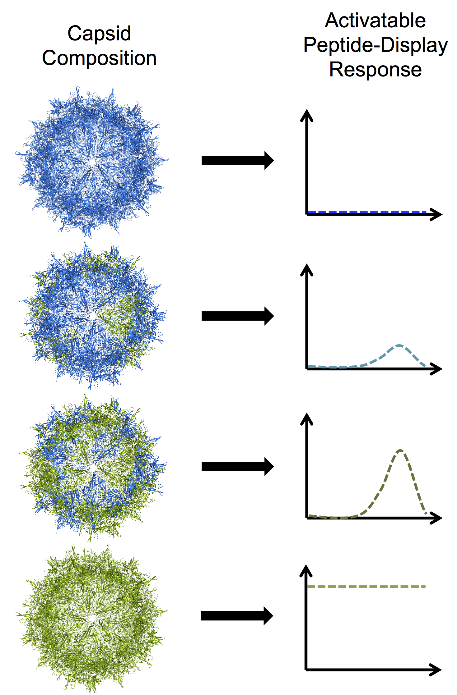
Rice University bioengineers have developed programmable adeno-associated viruses by modifying one of three proteins that assemble into a tough shell called a capsid. In this illustration, blue subunits in the capsid represent the protein VP3 and green subunits represent a truncated mutant of VP2.

Here’s an image illustrating the work,

Rice University bioengineers have developed programmable adeno-associated viruses by modifying one of three proteins that assemble into a tough shell called a capsid. In this illustration, blue subunits in the capsid represent the protein VP3 and green subunits represent a truncated mutant of VP2. From top to bottom: a VP3-only capsid that does not display any peptides; a mosaic capsid with a majority of VP3 and small amount of the VP2 mutant that shows a low level of activable peptide display; a mosaic capsid with equal amounts of VP3 and VP2 mutant that shows a high level of activable peptide display; and a homomeric VP2 mutant capsid with a high level of constant, brush-like peptide display. For a larger version, click on the image. Illustration by Nicole Thadani Courtesy: Rice University

A January 8, 2018 Rice University news release (also on EurekAlert), which originated the news item, expands on the story,

Rice bioengineer Junghae Suh studies the manipulation of nondisease-causing AAVs to deliver helpful cargoes like chemotherapy drugs. Her research has led to the development of viruses that can be triggered by light or by extracellular proteases associated with certain diseases.

AAVs are small — about 25 nanometers — and contain a single strand of DNA inside tough capsids that consist of a mosaic of proteins known as VP1, VP2 and VP3. AAVs have been used to deliver gene-therapy payloads, but nobody has figured out how AAV capsids physically reconfigure themselves when triggered by external stimuli, Suh said. That was the starting point for her lab.

“This virus has intrinsic peptide (small protein) domains hidden inside the capsid,” she said. “When the virus infects a cell, it senses the low pH and other endosomal factors, and these peptide domains pop out onto the surface of the virus capsid.

“This conformational change, which we termed an ‘activatable peptide display,’ is important for the virus because the externalized domains break down the endosomal membrane and allow the virus to escape into the cytoplasm,” Suh said. “In addition, nuclear localization sequences in those domains allow the virus to transit into the nucleus. We believed we could replace that functionality with something else.”

Suh and lead author and Rice graduate student Nicole Thadani think their mutant AAVs can become “biocomputing nanoparticles” that detect and process environmental inputs and produce controllable outputs. Modifying the capsid is the first step.

Of the three natural capsid proteins, only VP1 and VP2 can be triggered to expose their functional peptides, but neither can make a capsid on its own. Shorter VP3s can form capsids by themselves, but do not display peptides. In natural AAVs, VP3 proteins outnumber each of their compadres 10-to-1.

That limits the number of peptides that can be exposed, so Suh, Thadani and their co-authors set out to change the ratio. That led them to truncate VP2 and synthesize mosaic capsids with VP3, resulting in successful alteration of the number of exposed peptides. Based on previous research, they inserted a common hexahistidine tag that made it easy to monitor the surface display of the peptide region.

“We wanted to boost the protein’s activable property beyond what occurs in the native virus capsid,” Thadani said. “Rather than displaying just five copies of the peptide per capsid, now we may be able to display 20 or 30 and get more of the bioactivity that we want.”

They then made a truncated VP2 able to form a capsid on its own. “The results were quite surprising, and not obvious to us,” Suh said. “We chopped down that VP2 component enough to form what we call a homomeric capsid, where the entire capsid is made up of just that mutant subunit. That gave us viruses that appear to have peptide ‘brushes’ that are always on the surface.

“A viral structure like that has never been seen in nature,” she said. “We got a particle with this peptide brush, with loose ends everywhere. Now we want to know if we can use these loose ends to attach other things or carry out other functions.”

Homomeric AAVs display as many as 60 peptides, while mosaic AAVs could be programmed to respond to stimuli specific to particular cells or tissues and display a smaller desired number of peptides, the researchers said.

“Viruses have evolved to invade cells very effectively,” Suh said. “We want to use our virus as a nanoparticle platform to deliver protein- or peptide-based therapeutics more efficiently into cells. We want to harness what nature has already created, tweak it a little bit and use it for our purposes.”

Here’s a link to and a citation for the paper,

Reprogramming the Activatable Peptide Display Function of Adeno-Associated Virus Nanoparticles by Nicole N. Thadani, Christopher Dempsey, Julia Zhao, Sonya M. Vasquez, and Junghae Suh. ACS Nano, Article ASAP DOI: 10.1021/acsnano.7b07804 Publication Date (Web): December 26, 2017

Copyright © 2017 American Chemical Society

This paper is behind a paywall.

Editing the genome with CRISPR ((clustered regularly interspaced short palindromic repeats)-carrying nanoparticles

MIT (Massachusetts Institute of Technology) researchers have developed a new nonviral means of delivering CRISPR ((clustered regularly interspaced short palindromic repeats)-CAS9 gene therapy according to a November 13, 2017 news item on Nanowerk,

In a new study, MIT researchers have developed nanoparticles that can deliver the CRISPR genome-editing system and specifically modify genes in mice. The team used nanoparticles to carry the CRISPR components, eliminating the need to use viruses for delivery.

Using the new delivery technique, the researchers were able to cut out certain genes in about 80 percent of liver cells, the best success rate ever achieved with CRISPR in adult animals.

In a new study, MIT researchers have developed nanoparticles that can deliver the CRISPR genome-editing system and specifically modify genes, eliminating the need to use viruses for delivery. Image: MIT News

A November 13, 2017 MIT news release (also on EurekAlert), which originated the news item, provides more details about the research and a good description of and comparison between using a viral system and using a nanoparticle-based system to deliver CRISPR-CAS9,

“What’s really exciting here is that we’ve shown you can make a nanoparticle that can be used to permanently and specifically edit the DNA in the liver of an adult animal,” says Daniel Anderson, an associate professor in MIT’s Department of Chemical Engineering and a member of MIT’s Koch Institute for Integrative Cancer Research and Institute for Medical Engineering and Science (IMES).

One of the genes targeted in this study, known as Pcsk9, regulates cholesterol levels. Mutations in the human version of the gene are associated with a rare disorder called dominant familial hypercholesterolemia, and the FDA recently approved two antibody drugs that inhibit Pcsk9. However these antibodies need to be taken regularly, and for the rest of the patient’s life, to provide therapy. The new nanoparticles permanently edit the gene following a single treatment, and the technique also offers promise for treating other liver disorders, according to the MIT team.

Anderson is the senior author of the study, which appears in the Nov. 13 [2017] issue of Nature Biotechnology. The paper’s lead author is Koch Institute research scientist Hao Yin. Other authors include David H. Koch Institute Professor Robert Langer of MIT, professors Victor Koteliansky and Timofei Zatsepin of the Skolkovo Institute of Science and Technology [Russia], and Professor Wen Xue of the University of Massachusetts Medical School.

Targeting disease

Many scientists are trying to develop safe and efficient ways to deliver the components needed for CRISPR, which consists of a DNA-cutting enzyme called Cas9 and a short RNA that guides the enzyme to a specific area of the genome, directing Cas9 where to make its cut.

In most cases, researchers rely on viruses to carry the gene for Cas9, as well as the RNA guide strand. In 2014, Anderson, Yin, and their colleagues developed a nonviral delivery system in the first-ever demonstration of curing a disease (the liver disorder tyrosinemia) with CRISPR in an adult animal. However, this type of delivery requires a high-pressure injection, a method that can also cause some damage to the liver.

Later, the researchers showed they could deliver the components without the high-pressure injection by packaging messenger RNA (mRNA) encoding Cas9 into a nanoparticle instead of a virus. Using this approach, in which the guide RNA was still delivered by a virus, the researchers were able to edit the target gene in about 6 percent of hepatocytes, which is enough to treat tyrosinemia.

While that delivery technique holds promise, in some situations it would be better to have a completely nonviral delivery system, Anderson says. One consideration is that once a particular virus is used, the patient will develop antibodies to it, so it couldn’t be used again. Also, some patients have pre-existing antibodies to the viruses being tested as CRISPR delivery vehicles.

In the new Nature Biotechnology paper, the researchers came up with a system that delivers both Cas9 and the RNA guide using nanoparticles, with no need for viruses. To deliver the guide RNAs, they first had to chemically modify the RNA to protect it from enzymes in the body that would normally break it down before it could reach its destination.

The researchers analyzed the structure of the complex formed by Cas9 and the RNA guide, or sgRNA, to figure out which sections of the guide RNA strand could be chemically modified without interfering with the binding of the two molecules. Based on this analysis, they created and tested many possible combinations of modifications.

“We used the structure of the Cas9 and sgRNA complex as a guide and did tests to figure out we can modify as much as 70 percent of the guide RNA,” Yin says. “We could heavily modify it and not affect the binding of sgRNA and Cas9, and this enhanced modification really enhances activity.”

Reprogramming the liver

The researchers packaged these modified RNA guides (which they call enhanced sgRNA) into lipid nanoparticles, which they had previously used to deliver other types of RNA to the liver, and injected them into mice along with nanoparticles containing mRNA that encodes Cas9.

They experimented with knocking out a few different genes expressed by hepatocytes, but focused most of their attention on the cholesterol-regulating Pcsk9 gene. The researchers were able to eliminate this gene in more than 80 percent of liver cells, and the Pcsk9 protein was undetectable in these mice. They also found a 35 percent drop in the total cholesterol levels of the treated mice.

The researchers are now working on identifying other liver diseases that might benefit from this approach, and advancing these approaches toward use in patients.

“I think having a fully synthetic nanoparticle that can specifically turn genes off could be a powerful tool not just for Pcsk9 but for other diseases as well,” Anderson says. “The liver is a really important organ and also is a source of disease for many people. If you can reprogram the DNA of your liver while you’re still using it, we think there are many diseases that could be addressed.”

“We are very excited to see this new application of nanotechnology open new avenues for gene editing,” Langer adds.

The research was funded by the National Institutes of Health (NIH), the Russian Scientific Fund, the Skoltech Center, and the Koch Institute Support (core) Grant from the National Cancer Institute.

Here’s a link to and a citation for the paper,

Structure-guided chemical modification of guide RNA enables potent non-viral in vivo genome editing by Hao Yin, Chun-Qing Song, Sneha Suresh, Qiongqiong Wu, Stephen Walsh, Luke Hyunsik Rhym, Esther Mintzer, Mehmet Fatih Bolukbasi, Lihua Julie Zhu, Kevin Kauffman, Haiwei Mou, Alicia Oberholzer, Junmei Ding, Suet-Yan Kwan, Roman L Bogorad, Timofei Zatsepin, Victor Koteliansky, Scot A Wolfe, Wen Xue, Robert Langer, & Daniel G Anderson. Nature Biotechnology doi:10.1038/nbt.4005 Published online: 13 November 2017

This paper is behind a paywall.

CRISPR-CAS9 and gold

As so often happens in the sciences, now that the initial euphoria has expended itself problems (and solutions) with CRISPR ((clustered regularly interspaced short palindromic repeats))-CAAS9 are being disclosed to those of us who are not experts. From an Oct. 3, 2017 article by Bob Yirka for phys.org,

A team of researchers from the University of California and the University of Tokyo has found a way to use the CRISPR gene editing technique that does not rely on a virus for delivery. In their paper published in the journal Nature Biomedical Engineering, the group describes the new technique, how well it works and improvements that need to be made to make it a viable gene editing tool.

CRISPR-Cas9 has been in the news a lot lately because it allows researchers to directly edit genes—either disabling unwanted parts or replacing them altogether. But despite many success stories, the technique still suffers from a major deficit that prevents it from being used as a true medical tool—it sometimes makes mistakes. Those mistakes can cause small or big problems for a host depending on what goes wrong. Prior research has suggested that the majority of mistakes are due to delivery problems, which means that a replacement for the virus part of the technique is required. In this new effort, the researchers report that they have discovered just a such a replacement, and it worked so well that it was able to repair a gene mutation in a Duchenne muscular dystrophy mouse model. The team has named the new technique CRISPR-Gold, because a gold nanoparticle was used to deliver the gene editing molecules instead of a virus.

An Oct. 2, 2017 article by Abby Olena for The Scientist lays out the CRISPR-CAS9 problems the scientists are trying to solve (Note: Links have been removed),

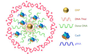
While promising, applications of CRISPR-Cas9 gene editing have so far been limited by the challenges of delivery—namely, how to get all the CRISPR parts to every cell that needs them. In a study published today (October 2) in Nature Biomedical Engineering, researchers have successfully repaired a mutation in the gene for dystrophin in a mouse model of Duchenne muscular dystrophy by injecting a vehicle they call CRISPR-Gold, which contains the Cas9 protein, guide RNA, and donor DNA, all wrapped around a tiny gold ball.

The authors have made “great progress in the gene editing area,” says Tufts University biomedical engineer Qiaobing Xu, who did not participate in the work but penned an accompanying commentary. Because their approach is nonviral, Xu explains, it will minimize the potential off-target effects that result from constant Cas9 activity, which occurs when users deliver the Cas9 template with a viral vector.

Duchenne muscular dystrophy is a degenerative disease of the muscles caused by a lack of the protein dystrophin. In about a third of patients, the gene for dystrophin has small deletions or single base mutations that render it nonfunctional, which makes this gene an excellent candidate for gene editing. Researchers have previously used viral delivery of CRISPR-Cas9 components to delete the mutated exon and achieve clinical improvements in mouse models of the disease.

“In this paper, we were actually able to correct [the gene for] dystrophin back to the wild-type sequence” via homology-directed repair (HDR), coauthor Niren Murthy, a drug delivery researcher at the University of California, Berkeley, tells The Scientist. “The other way of treating this is to do something called exon skipping, which is where you delete some of the exons and you can get dystrophin to be produced, but it’s not [as functional as] the wild-type protein.”

The research team created CRISPR-Gold by covering a central gold nanoparticle with DNA that they modified so it would stick to the particle. This gold-conjugated DNA bound the donor DNA needed for HDR, which the Cas9 protein and guide RNA bound to in turn. They coated the entire complex with a polymer that seems to trigger endocytosis and then facilitate escape of the Cas9 protein, guide RNA, and template DNA from endosomes within cells.

In order to do HDR, “you have to provide the cell [with] the Cas9 enzyme, guide RNA by which you target Cas9 to a particular part of the genome, and a big chunk of DNA, which will be used as a template to edit the mutant sequence to wild-type,” explains coauthor Irina Conboy, who studies tissue repair at the University of California, Berkeley. “They all have to be present at the same time and at the same place, so in our system you have a nanoparticle which simultaneously delivers all of those three key components in their active state.”

Olena’s article carries on to describe how the team created CRISPR-Gold and more.

Additional technical details are available in an Oct. 3, 2017 University of California at Berkeley news release by Brett Israel (also on EurekAlert), which originated the news item (Note: A link has been removed) ,

Scientists at the University of California, Berkeley, have engineered a new way to deliver CRISPR-Cas9 gene-editing technology inside cells and have demonstrated in mice that the technology can repair the mutation that causes Duchenne muscular dystrophy, a severe muscle-wasting disease. A new study shows that a single injection of CRISPR-Gold, as the new delivery system is called, into mice with Duchenne muscular dystrophy led to an 18-times-higher correction rate and a two-fold increase in a strength and agility test compared to control groups.

Diagram of CRISPR-Gold

CRISPR–Gold is composed of 15 nanometer gold nanoparticles that are conjugated to thiol-modified oligonucleotides (DNA-Thiol), which are hybridized with single-stranded donor DNA and subsequently complexed with Cas9 and encapsulated by a polymer that disrupts the endosome of the cell.

Since 2012, when study co-author Jennifer Doudna, a professor of molecular and cell biology and of chemistry at UC Berkeley, and colleague Emmanuelle Charpentier, of the Max Planck Institute for Infection Biology, repurposed the Cas9 protein to create a cheap, precise and easy-to-use gene editor, researchers have hoped that therapies based on CRISPR-Cas9 would one day revolutionize the treatment of genetic diseases. Yet developing treatments for genetic diseases remains a big challenge in medicine. This is because most genetic diseases can be cured only if the disease-causing gene mutation is corrected back to the normal sequence, and this is impossible to do with conventional therapeutics.

CRISPR/Cas9, however, can correct gene mutations by cutting the mutated DNA and triggering homology-directed DNA repair. However, strategies for safely delivering the necessary components (Cas9, guide RNA that directs Cas9 to a specific gene, and donor DNA) into cells need to be developed before the potential of CRISPR-Cas9-based therapeutics can be realized. A common technique to deliver CRISPR-Cas9 into cells employs viruses, but that technique has a number of complications. CRISPR-Gold does not need viruses.

In the new study, research lead by the laboratories of Berkeley bioengineering professors Niren Murthy and Irina Conboy demonstrated that their novel approach, called CRISPR-Gold because gold nanoparticles are a key component, can deliver Cas9 – the protein that binds and cuts DNA – along with guide RNA and donor DNA into the cells of a living organism to fix a gene mutation.

“CRISPR-Gold is the first example of a delivery vehicle that can deliver all of the CRISPR components needed to correct gene mutations, without the use of viruses,” Murthy said.

The study was published October 2 [2017] in the journal Nature Biomedical Engineering.

CRISPR-Gold repairs DNA mutations through a process called homology-directed repair. Scientists have struggled to develop homology-directed repair-based therapeutics because they require activity at the same place and time as Cas9 protein, an RNA guide that recognizes the mutation and donor DNA to correct the mutation.

To overcome these challenges, the Berkeley scientists invented a delivery vessel that binds all of these components together, and then releases them when the vessel is inside a wide variety of cell types, triggering homology directed repair. CRISPR-Gold’s gold nanoparticles coat the donor DNA and also bind Cas9. When injected into mice, their cells recognize a marker in CRISPR-Gold and then import the delivery vessel. Then, through a series of cellular mechanisms, CRISPR-Gold is released into the cells’ cytoplasm and breaks apart, rapidly releasing Cas9 and donor DNA.

Schematic of CRISPR-Gold's method of action

CRISPR-Gold’s method of action (Click to enlarge).

A single injection of CRISPR-Gold into muscle tissue of mice that model Duchenne muscular dystrophy restored 5.4 percent of the dystrophin gene, which causes the disease, to the wild- type, or normal, sequence. This correction rate was approximately 18 times higher than in mice treated with Cas9 and donor DNA by themselves, which experienced only a 0.3 percent correction rate.

Importantly, the study authors note, CRISPR-Gold faithfully restored the normal sequence of dystrophin, which is a significant improvement over previously published approaches that only removed the faulty part of the gene, making it shorter and converting one disease into another, milder disease.

CRISPR-Gold was also able to reduce tissue fibrosis – the hallmark of diseases where muscles do not function properly – and enhanced strength and agility in mice with Duchenne muscular dystrophy. CRISPR-Gold-treated mice showed a two-fold increase in hanging time in a common test for mouse strength and agility, compared to mice injected with a control.

“These experiments suggest that it will be possible to develop non-viral CRISPR therapeutics that can safely correct gene mutations, via the process of homology-directed repair, by simply developing nanoparticles that can simultaneously encapsulate all of the CRISPR components,” Murthy said.

CRISPR-Cas9

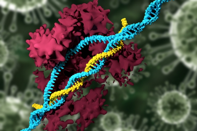
CRISPR in action: A model of the Cas9 protein cutting a double-stranded piece of DNA

The study found that CRISPR-Gold’s approach to Cas9 protein delivery is safer than viral delivery of CRISPR, which, in addition to toxicity, amplifies the side effects of Cas9 through continuous expression of this DNA-cutting enzyme. When the research team tested CRISPR-Gold’s gene-editing capability in mice, they found that CRISPR-Gold efficiently corrected the DNA mutation that causes Duchenne muscular dystrophy, with minimal collateral DNA damage.

The researchers quantified CRISPR-Gold’s off-target DNA damage and found damage levels similar to the that of a typical DNA sequencing error in a typical cell that was not exposed to CRISPR (0.005 – 0.2 percent). To test for possible immunogenicity, the blood stream cytokine profiles of mice were analyzed at 24 hours and two weeks after the CRISPR-Gold injection. CRISPR-Gold did not cause an acute up-regulation of inflammatory cytokines in plasma, after multiple injections, or weight loss, suggesting that CRISPR-Gold can be used multiple times safely, and that it has a high therapeutic window for gene editing in muscle tissue.

“CRISPR-Gold and, more broadly, CRISPR-nanoparticles open a new way for safer, accurately controlled delivery of gene-editing tools,” Conboy said. “Ultimately, these techniques could be developed into a new medicine for Duchenne muscular dystrophy and a number of other genetic diseases.”

A clinical trial will be needed to discern whether CRISPR-Gold is an effective treatment for genetic diseases in humans. Study co-authors Kunwoo Lee and Hyo Min Park have formed a start-up company, GenEdit (Murthy has an ownership stake in GenEdit), which is focused on translating the CRISPR-Gold technology into humans. The labs of Murthy and Conboy are also working on the next generation of particles that can deliver CRISPR into tissues from the blood stream and would preferentially target adult stem cells, which are considered the best targets for gene correction because stem and progenitor cells are capable of gene editing, self-renewal and differentiation.

“Genetic diseases cause devastating levels of mortality and morbidity, and new strategies for treating them are greatly needed,” Murthy said. “CRISPR-Gold was able to correct disease-causing gene mutations in vivo, via the non-viral delivery of Cas9 protein, guide RNA and donor DNA, and therefore has the potential to develop into a therapeutic for treating genetic diseases.”

The study was funded by the National Institutes of Health, the W.M. Keck Foundation, the Moore Foundation, the Li Ka Shing Foundation, Calico, Packer, Roger’s and SENS, and the Center of Innovation (COI) Program of the Japan Science and Technology Agency.

Here’s a link to and a citation for the paper,

Nanoparticle delivery of Cas9 ribonucleoprotein and donor DNA in vivo induces homology-directed DNA repair by Kunwoo Lee, Michael Conboy, Hyo Min Park, Fuguo Jiang, Hyun Jin Kim, Mark A. Dewitt, Vanessa A. Mackley, Kevin Chang, Anirudh Rao, Colin Skinner, Tamanna Shobha, Melod Mehdipour, Hui Liu, Wen-chin Huang, Freeman Lan, Nicolas L. Bray, Song Li, Jacob E. Corn, Kazunori Kataoka, Jennifer A. Doudna, Irina Conboy, & Niren Murthy. Nature Biomedical Engineering (2017) doi:10.1038/s41551-017-0137-2 Published online: 02 October 2017

This paper is behind a paywall.

Hybrid bacterial genes and virus shell combined to create ‘nano reactor’ for hydrogen biofuel

Turning water into fuel may seem like an almost biblical project (e.g., Jesus turning water to wine in the New Testament) but scientists at Indiana University are hopeful they are halfway to their goal. From a Jan. 4, 2016 news item on ScienceDaily,

Scientists at Indiana University have created a highly efficient biomaterial that catalyzes the formation of hydrogen — one half of the “holy grail” of splitting H2O to make hydrogen and oxygen for fueling cheap and efficient cars that run on water.

A Jan. 4, 2016 Indiana University (IU) news release (also on EurekAlert*), which originated the news item, explains further (Note: Links have been removed),

A modified enzyme that gains strength from being protected within the protein shell — or “capsid” — of a bacterial virus, this new material is 150 times more efficient than the unaltered form of the enzyme.

“Essentially, we’ve taken a virus’s ability to self-assemble myriad genetic building blocks and incorporated a very fragile and sensitive enzyme with the remarkable property of taking in protons and spitting out hydrogen gas,” said Trevor Douglas, the Earl Blough Professor of Chemistry in the IU Bloomington College of Arts and Sciences’ Department of Chemistry, who led the study. “The end result is a virus-like particle that behaves the same as a highly sophisticated material that catalyzes the production of hydrogen.”

The genetic material used to create the enzyme, hydrogenase, is produced by two genes from the common bacteria Escherichia coli, inserted inside the protective capsid using methods previously developed by these IU scientists. The genes, hyaA and hyaB, are two genes in E. coli that encode key subunits of the hydrogenase enzyme. The capsid comes from the bacterial virus known as bacteriophage P22.

The resulting biomaterial, called “P22-Hyd,” is not only more efficient than the unaltered enzyme but also is produced through a simple fermentation process at room temperature.

The material is potentially far less expensive and more environmentally friendly to produce than other materials currently used to create fuel cells. The costly and rare metal platinum, for example, is commonly used to catalyze hydrogen as fuel in products such as high-end concept cars.

“This material is comparable to platinum, except it’s truly renewable,” Douglas said. “You don’t need to mine it; you can create it at room temperature on a massive scale using fermentation technology; it’s biodegradable. It’s a very green process to make a very high-end sustainable material.”

In addition, P22-Hyd both breaks the chemical bonds of water to create hydrogen and also works in reverse to recombine hydrogen and oxygen to generate power. “The reaction runs both ways — it can be used either as a hydrogen production catalyst or as a fuel cell catalyst,” Douglas said.

The form of hydrogenase is one of three occurring in nature: di-iron (FeFe)-, iron-only (Fe-only)- and nitrogen-iron (NiFe)-hydrogenase. The third form was selected for the new material due to its ability to easily integrate into biomaterials and tolerate exposure to oxygen.

NiFe-hydrogenase also gains significantly greater resistance upon encapsulation to breakdown from chemicals in the environment, and it retains the ability to catalyze at room temperature. Unaltered NiFe-hydrogenase, by contrast, is highly susceptible to destruction from chemicals in the environment and breaks down at temperatures above room temperature — both of which make the unprotected enzyme a poor choice for use in manufacturing and commercial products such as cars.

These sensitivities are “some of the key reasons enzymes haven’t previously lived up to their promise in technology,” Douglas said. Another is their difficulty to produce.

“No one’s ever had a way to create a large enough amount of this hydrogenase despite its incredible potential for biofuel production. But now we’ve got a method to stabilize and produce high quantities of the material — and enormous increases in efficiency,” he said.

The development is highly significant according to Seung-Wuk Lee, professor of bioengineering at the University of California-Berkeley, who was not a part of the study.

“Douglas’ group has been leading protein- or virus-based nanomaterial development for the last two decades. This is a new pioneering work to produce green and clean fuels to tackle the real-world energy problem that we face today and make an immediate impact in our life in the near future,” said Lee, whose work has been cited in a U.S. Congressional report on the use of viruses in manufacturing.

Beyond the new study, Douglas and his colleagues continue to craft P22-Hyd into an ideal ingredient for hydrogen power by investigating ways to activate a catalytic reaction with sunlight, as opposed to introducing elections using laboratory methods.

“Incorporating this material into a solar-powered system is the next step,” Douglas said.

Here’s a link to and a citation for the paper,

Self-assembling biomolecular catalysts for hydrogen production by Paul C. Jordan, Dustin P. Patterson, Kendall N. Saboda, Ethan J. Edwards, Heini M. Miettinen, Gautam Basu, Megan C. Thielges, & Trevor Douglas. Nature Chemistry (2015) doi:10.1038/nchem.2416 Published online 21 December 2015

This paper is behind a paywall.

*(also on EurekAlert) added on Jan. 5, 2016 at 1550 PST.

Physics, nanopores, viruses, and DNA

A June 17, 2014 news item on Azonano describes a project which could help scientists decode strands of DNA at top speeds,

Nanopores may one day lead a revolution in DNA sequencing. By sliding DNA molecules one at a time through tiny holes in a thin membrane, it may be possible to decode long stretches of DNA at lightning speeds. Scientists, however, haven’t quite figured out the physics of how polymer strands like DNA interact with nanopores. Now, with the help of a particular type of virus, researchers from Brown University have shed new light on this nanoscale physics.

“What got us interested in this was that everybody in the field studied DNA and developed models for how they interact with nanopores,” said Derek Stein, associate professor of physics and engineering at Brown [Brown University, US] who directed the research. “But even the most basic things you would hope models would predict starting from the basic properties of DNA — you couldn’t do it. The only way to break out of that rut was to study something different.”

A June 16, 2014 Brown University news release (also on EurekAlert), which originated the news item, describes the problems with nanopores,

The concept behind nanopore sequencing is fairly simple. A hole just a few billionths of a meter wide is poked in a membrane separating two pools of salty water. An electric current is applied to the system, which occasionally snares a charged DNA strand and whips it through the pore — a phenomenon called translocation. When a molecule translocates, it causes detectable variations in the electric current across the pore. By looking carefully at those variations in current, scientists may be able to distinguish individual nucleotides — the A’s, C’s, G’s and T’s coded in DNA molecules.

The first commercially available nanopore sequencers may only be a few years away, but despite advances in the field, surprisingly little is known about the basic physics involved when polymers interact with nanopores. That’s partly because of the complexities involved in studying DNA. In solution, DNA molecules form balls of random squiggles, which make understanding their physical behavior extremely difficult.

For example, the factors governing the speed of DNA translocation aren’t well understood. Sometimes molecules zip through a pore quickly; other times they slither more slowly, and nobody completely understands why.

One possible explanation is that the squiggly configuration of DNA causes each molecule to experience differences in drag as they’re pulled through the water toward the pore. “If a molecule is crumpled up next to the pore, it has a shorter distance to travel and experiences less drag,” said Angus McMullen, a physics graduate student at Brown and the study’s lead author. “But if it’s stretched out then it would feel drag along the whole length and that would cause it to go slower.”

The news release then goes on to detail a possible solution to the problem of why DNA translocation varies in speed. Answering this question about DNA translocation could lead to faster and more accurate nanopore sequencing,

The drag effect is impossible to isolate experimentally using DNA, but the virus McMullen and his colleagues studied offered a solution.

The researchers looked at fd, a harmless virus that infects e. coli bacteria. Two things make the virus an ideal candidate for study with nanpores. First, fd viruses are all identical clones of each other. Second, unlike squiggly DNA, fd virus is a stiff, rod-like molecule. Because the virus doesn’t curl up like DNA does, the effect of drag on each one should be essentially the same every time.

With drag eliminated as a source of variation in translocation speed, the researchers expected that the only source of variation would be the effect of thermal motion. The tiny virus molecules constantly bump up against the water molecules in which they are immersed. A few random thermal kicks from the rear would speed the virus up as it goes through the pore. A few kicks from the front would slow it down.

The experiments showed that while thermal motion explained much of the variation in translocation speed, it didn’t explain it all. Much to the researchers’ surprise, they found another source of variation that increased when the voltage across the pore was increased.

“We thought that the physics would be crystal clear,” said Jay Tang, associate professor of physics and engineering at Brown and one of the study’s co-authors. “You have this stiff [virus] with well-defined diameter and size and you would expect a very clear-cut signal. As it turns out, we found some puzzling physics we can only partially explain ourselves.”

The researchers can’t say for sure what’s causing the variation they observed, but they have a few ideas.

“It’s been predicted that depending on where [an object] is inside the pore, it might be pulled harder or weaker,” McMullen said. “If it’s in the center of the pore, it pulls a little bit weaker than if it’s right on the edge. That’s been predicted, but never experimentally verified. This could be evidence of that happening, but we’re still doing follow up work.

The new approach using a virus answered questions while leading to new insights and possibilities (from the news release),

A better understanding of translocation speed could improve the accuracy of nanopore sequencing, McMullen says. It would also be helpful in the crucial task of measuring the length of DNA strands. “If you can predict the translocation speed,” McMullen said, “then you can easily get the length of the DNA from how long its translocation was.”

The research also helped to reveal other aspects of the translocation process that could be useful in designing future devices. The study showed that the electrical current tends to align the viruses head first to the pore, but on occasions when they’re not lined up, they tend to bounce around on the edge of the pore until thermal motion aligns them to go through. However, when the voltage was turned too high, the thermal effects were suppressed and the virus became stuck to the membrane. That suggests a sweet spot in voltage where headfirst translocation is most likely.

None of this is observable directly — the system is simply too small to be seen in action. But the researchers could infer what was happening by looking at slight changes in the current across the pore.

“When the viruses miss, they rattle around and we see these little bumps in the current,” Stein said. “So with these little bumps, we’re starting to get an idea of what the molecule is doing before it slides through. Normally these sensors are blind to anything that’s going on until the molecule slides through.”

That would have been impossible to observe using DNA. The floppiness of the DNA molecule allows it to go through a pore in a folded configuration even if it’s not aligned head-on. But because the virus is stiff, it can’t fold to go through. That enabled the researchers to isolate and observe those contact dynamics.

“These viruses are unique,” Stein said. “They’re like perfect little yardsticks.”

In addition to shedding light on basic physics, the work might also have another application. While the fd virus itself is harmless, the bacteria it infects — e. coli — is not. Based on this work, it might be possible to build a nanopore device for detecting the presence of fd, and by proxy, e. coli. Other dangerous viruses — Ebola and Marburg among them — share the same rod-like structure as fd.

“This might be an easy way to detect these viruses,” Tang said. “So that’s another potential application for this.”

Here’s a link to and a citation for the paper,

Stiff filamentous virus translocations through solid-state nanopores by Angus McMullen, Hendrick W. de Haan, Jay X. Tang, & Derek Stein. Nature Communications 5, Article number: 4171 doi:10.1038/ncomms5171 Published 16 June 2014

This paper is behind a paywall.