Tag Archives: tissues

Cellulose biosensor heralds new bioimaging approach to tissue engineering

I keep an eye on how nanocellulose is being used in various applications and I’m not sure that this cellulose biosensor quite fits the bill as nanocellulose, nonetheless, it’s interesting and that’s enough for me. From a December 12, 2018 Sechenov University (Russia) press release on EurekAlert,

I.M. Sechenov First Moscow State Medical University teamed up together with Irish colleagues to develop a new imaging approach for tissue engineering. The team produced so-called ‘hybrid biosensor’ scaffold materials, which are based on cellulose matrices labeled with pH- and calcium-sensitive fluorescent proteins. These materials enable visualization of the metabolism and other important biomarkers in the engineered artificial tissues by microscopy. The results of the work were published in the Acta Biomaterialia journal.
The success of tissue engineering is based on the use of scaffold matrices – materials that support the viability and direct the growth of cells, tissues, and organoids. Scaffolds are important for basic and applied biomedical research, tissue engineering and regenerative medicine, and are promising for development of new therapeutics. However, the ability ‘to see’ what happens within the scaffolds during the tissue growth poses a significant research challenge

“We developed a new approach allowing visualization of scaffold-grown tissue and cells by using labeling with biosensor fluorescent proteins. Due to the high specificity of labeling and the use of fluorescence microscopy FLIM, we can quantify changes in pH and calcium in the vicinity of cells,” says Dr. Ruslan Dmitriev, Group Leader at the University College Cork and the Institute for Regenerative Medicine (I.M. Sechenov First Moscow State Medical University).
To achieve the specific labeling of cellulose matrices, researchers used well-known cellulose-binding proteins. The use of extracellular pH- and calcium-sensitive biosensors allow for analysis of cell metabolism: indeed, the extracellular acidification is directly associated with the balance of cell energy production pathways and the glycolytic flux (release of lactate). It is also a frequent hallmark of cancer and transformed cell types. On the other hand, calcium plays a key role in the extra- and intracellular signaling affecting cell growth and differentiation.

The approach was tested on different types of cellulose matrices (bacterial and produced from decellularised plant tissues) using 3D culture of human colon cancer cells and stem-cell derived mouse small intestinal organoids. The scaffolds informed on changes in the extracellular acidification and were used together with the analysis of real-time oxygenation of intestinal organoids. The resulting data can be presented in the form of colour maps, corresponding to the areas of cell growth within different microenvironments.

“Our results open new prospects in the imaging of tissue-engineered constructs for regenerative medicine. They enable deeper understanding of tissue metabolism in 3D and are also highly promising for commercialisation,” concludes Dr. Dmitriev.

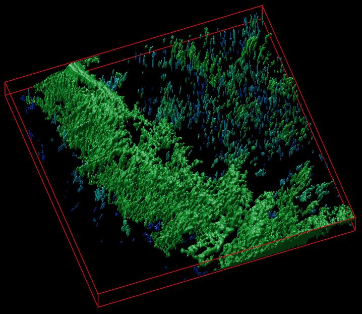
The researchers have provided an image to illustrate their work,

Caption: A 3D reconstruction of a cellulose matrix stained with a pH-sensitive biosensor. Credit: Dr. R. Dmitriev

Here’s a link to and a citation for the paper,

Cellulose-based scaffolds for fluorescence lifetime imaging-assisted tissue engineering by Neil O’Donnell, Irina A. Okkelman, Peter Timashev, Tatyana I.Gromovykh, Dmitri B. Papkovsky, Ruslan I.Dmitriev. Acta Biomaterialia Volume 80, 15 October 2018, Pages 85-96 DOI: https://doi.org/10.1016/j.actbio.2018.09.034


This paper is behind a paywall.

Tractor beams for artificial cells

This particular piece has videos of cells moving around. I won’t be including all of them but they are weirdly fascinating. First, a May 14, 2018 news item on Nanowerk announces the latest in tractor beam news from the Imperial College London (ICL; UK),

Researchers have used lasers to connect, arrange and merge artificial cells, paving the way for networks of artificial cells that act like tissues.

The team say that by altering artificial cell membranes they can now get the cells to stick together like ‘stickle bricks’ – allowing them to be arranged into whole new structures.

Biological cells can perform complex functions, but are difficult to controllably engineer.

Artificial cells, however, can in principle be made to order. Now, researchers from Imperial College London and Loughborough University have demonstrated a new level of complexity with artificial cells by arranging them into basic tissue structures with different types of connectivity.

These structures could be used to perform functions like initiating chemical reactions or moving chemicals around networks of artificial and biological cells. This could be useful in carrying out chemical reactions in ultra-small volumes, in studying the mechanisms through which cells communicate with one another, and in the development of a new generation of smart biomaterials.

A May 14, 2018 ICL press release by Hayley Dunning , which originated the news item, provides more detail,

Cells are the basic units of biology, which are capable of working together as a collective when arranged into tissues. In order to do this, cells must be connected and be capable of exchanging materials with one another.

The team were able to link up artificial cells into a range of new architectures, the results of which are published today in Nature Communications.

The artificial cells have a membrane-like layer as their shell, which the researchers engineered to ‘stick’ to each other. In order to get the cells to come close enough, the team first had to manipulate the cells with ‘optical tweezers’ that act like mini ‘tractor beams’ dragging and dropping cells into any position. Once connected in this way the cells can be moved as one unit.

Lead researcher Dr Yuval Elani, an EPSRC Research Fellow from the Department of Chemistry at Imperial, said: “Artificial cell membranes usually bounce off each other like rubber balls. By altering the biophysics of the membranes in our cells, we got them instead to stick to each other like stickle bricks.

“With this, we were able to form networks of cells connected by ‘biojunctions’. By reinserting biological components such as proteins in the membrane, we could get the cells to communicate and exchange material with one another. This mimics what is seen in nature, so it’s a great step forward in creating biological-like artificial cell tissues.”

Building up complexity

The team were also able to engineer a ‘tether’ between two cells. Here the membranes are not stuck together, but a tendril of membrane material links them so that they can be moved together.

Once they had perfected the cell-sticking process, the team were able to build up more complex arrangements. These include lines of cells, 2D shapes like squares, and 3D shapes like pyramids. Once the cells are stuck together, they can be rearranged, and also pulled by the laser beam as an ensemble

Finally, the team were also able to connect two cells, and then make them merge into one larger cell. This was achieved by coating the membranes with gold nanoparticles.

When the laser beam at the heart of the ‘optical tweezer’ technology was concentrated at the junction between the two cells, the nanoparticles resonated, breaking the membranes at that point. The membrane then reforms as a whole.

Merging cells in this way allowed whatever chemicals they were carrying to mix within the new, larger cell, kicking off chemical reactions. This could be useful, for example, for delivering materials such as drugs into cells, and in changing the composition of cells in real time, getting them to adopt new functions.

Professor Oscar Ces, also from the Department of Chemistry at Imperial, said: “Connecting artificial cells together is a valuable technology in the wider toolkit we are assembling for creating these biological systems using bottom-up approaches.

“We can now start to scale up basic cell technologies into larger tissue-scale networks, with precise control over the kind of architecture we create.”

Here’s one of the videos that has been embedded with ICL press release,

You can see the whole series if you go to the May 14, 2018 ICL press release.

Here’s a link to and a citation for the paper,

Sculpting and fusing biomimetic vesicle networks using optical tweezers by Guido Bolognesi, Mark S. Friddin, Ali Salehi-Reyhani, Nathan E. Barlow, Nicholas J. Brooks, Oscar Ces, & Yuval Elani. Nature Communicationsvolume 9, Article number: 1882 (2018) doi:10.1038/s41467-018-04282-w Published: 14 May 2018

This paper is open access.

Better blood vessel growth for regenerative medicine?

If the organs and tissues grown in labs are to be successfully transplanted into bodies, then growing the blood vessels needed to maintain them becomes very important. A May 24, 2016 news item on ScienceDaily describes a new technique for the growing the vessels,

Growing tissues and organs in the lab for transplantation into patients could become easier after scientists discovered an effective way to produce three-dimensional networks of blood vessels, vital for tissue survival yet a current stumbling block in regenerative medicine.

In addition the technique to grow the blood vessels in a 3D scaffold cuts down on the risk of transplant rejection because it uses cells from the patient. It was developed by researchers from the University of Bath’s Department of Pharmacy and Pharmacology, working with colleagues at Bristol Heart Institute.

A May 24 (?), 2016 University of Bath (UK) press release, which originated the news item, expands on the theme (Note: Links have been removed),

So far the shortage of adequate patient-derived scaffolds that can support blood vessel growth has been a major limitation for regenerative medicine and tissue engineering.

Other methods only allow limited formation of small blood vessels such as capillaries, which makes tissue less likely to successfully transplant into a patient. In addition other methods of tissue growth require the use of animal products, unnecessary in this technique which uses human platelet lysate gel (hPLG) and endothelial progenitor cells (EPCs) – a type of cell which helps maintain blood vessel walls.

Dr Giordano Pula, Lecturer in Pharmacology at the University of Bath and head of the research team making the discovery, said: “A major challenge in tissue engineering and regenerative medicine is providing the new tissue with a network of blood vessels, and linking this to the patient’s existing blood supply; this is vital for the tissue’s survival and integration with adjacent tissues.

Dr Paul De Bank, Senior Lecturer in Pharmaceutics at the University of Bath and co-author of the paper, said: “By embedding EPCs in a gel derived from platelets, both of which can be isolated from the patient’s blood, we have demonstrated the formation of a network of small vessels. What is more, the gel contains a number of different growth factors which can induce existing blood vessels to infiltrate the gel and form connections with the new structures. Combining tissue-specific cells with this EPC-containing gel offers the potential for the formation of fully vascularised, functional tissues or organs, which integrate seamlessly with the patient.

“This discovery has the potential to accelerate the development of regenerative medicine applications.”

Professor Peter Weissberg, Medical Director of the British Heart Foundation, said: “Over a half a million people in the UK are living with heart failure, a disabling condition which can leave people unable to carry out everyday activities such as climbing the stairs or even walking to the shops. This regenerative research brings the British Heart Foundation’s goal to mend a broken heart and beat heart failure one step closer.

“All living tissues, including new heart muscle, need a blood supply. One of the fundamental goals of regenerative medicine is to find ways to grow a new blood supply from scratch. Previous attempts at this using human cells and synthetic scaffolds have met with only limited success.

“The beauty of this new approach is that components of a person’s own blood could be manipulated to create a scaffold on which new blood vessels could grow. This increases the likelihood that the new tissue will be integrated into the patient’s body which, if proven successful with more research, could improve the lives of people affected by heart failure.”

Here’s a link to and a citation for the paper,

Platelet lysate gel and endothelial progenitors stimulate microvascular network formation in vitro: tissue engineering implications by Tiago M. Fortunato, Cristina Beltrami, Costanza Emanueli, Paul A. De Bank & Giordano Pula. Scientific Reports 6, Article number: 25326 (2016)  doi:10.1038/srep25326 Published online: 04 May 2016

This is an open access paper.

One of the criticisms of Paolo Macchiarini’s work with synthetic tracheas centered around blood supply to the cells (from my April 19, 2016 posting; it was part 1 of a 2-part series),

This ground-breaking achievement consisted of bringing to life a dead windpipe from a donor, by putting it in a plastic box, a so-called ‘bioreactor’ together with bone marrow fluid (stem cells). A few weeks later, I [Pierre Delaere*]  wrote a letter to The Lancet, pointing out:

“The main drawback of the proposed reconstruction is the lack of an intrinsic blood supply to the trachea. We know that a good blood supply is the first requirement in all other tissue and organ transplantations. Therefore, the reported success of this technique is questionable” (correspondence by Delaere and Hermans, Lancet 2009).

The excerpt you’ve just seen features part of an open letter Pierre Delaere (a long time Macchiarini critic), published in Leonid Schneider’s blog ‘For Better Science’ in an April 2, 2016 posting.

Getting back to Bath, this is exciting stuff and I hope the research is reproducible.