Tag Archives: Indian Institute of Science (IISc)

Relief from tooth sensitivity with magnetically guided nanobots

An August 11, 2025 Indian Institute of Science (IISc) press release (also on EurekAlert) by Shruti Sharma announces research into improving relief for people with tooth sensitivity, Note: A link has been removed,

Sensitive teeth need tough toothpaste, but technology can also help. Researchers at the Indian Institute of Science (IISc) in collaboration with deep-tech startup Theranautilus have now engineered CalBots – magnetic nanobots that can penetrate deep into dentinal tubules, which are tiny tunnels in teeth that lead to nerve endings. These CalBots can then form durable seals for worn enamel, offering lasting relief from sensitivity in just one application. The study is published in Advanced Science. 

The CalBots use a completely new class of bioceramic cement. While bioceramics are widely used in orthopaedics and dentistry for their mineralising properties, the team wanted a solution tailored for hypersensitivity – a formulation that could travel deeper and last longer. 

“We didn’t want to create a slightly better version of what’s already out there,” says Shanmukh Peddi, first author of the study and postdoctoral researcher at the Centre for Nano Science and Engineering (CeNSE), IISc, and co-founder of Theranautilus. “We wanted a technology that solves a real problem in a way that no one’s attempted before.”

Dental hypersensitivity affects nearly one in four people worldwide. It occurs when microscopic tubules in the dentine – the layer beneath the enamel –become exposed due to erosion or gum recession. These tiny tubules lead directly to nerve endings, which is why even a sip of cold water can cause a sudden, stabbing pain. Most current solutions, such as desensitising toothpastes, offer only surface-level relief and need to be reapplied regularly. 

CalBots, however, are different. These 400 nanometre-sized magnetic particles, loaded with a proprietary calcium silicate-based bioceramic formula, are guided by an external magnetic field deep into the exposed tubules. They can reach depths of up to 300-500 micrometers inside the tubules. Once there, the bots self-assemble into stable, cement-like plugs that block the tubules and recreate a durable seal that mimics the natural environment of the tooth.  

To test their innovation, the team used human teeth extracted for clinical reasons and created conditions where the dentine was exposed. On these samples, they applied CalBots under a magnetic field for 20 minutes, during which the bots sealed the dentinal tubules by forming deep, stable plugs – a result confirmed through high-resolution imaging. Encouraged by this, they progressed to animal trials in collaboration with researchers at IISc’s Center for Neuroscience. It involved giving mice a choice between cold and room temperature water. Healthy mice preferred both equally. But the mice with induced tooth sensitivity avoided the cold water completely. 

“After we treated the sensitive mice with our CalBot solution, they started drinking cold water again – the treatment worked like a charm. We saw 100% behavioural recovery. That was a big moment for us,” Peddi says.

The CalBots are composed entirely of materials classified as ‘Generally Recognised as Safe’ (GRAS), ensuring high biocompatibility. Toxicity tests on mice showed no adverse effects. “This is a compelling demonstration of what nanorobotics can achieve, and how they could significantly impact future healthcare,” says Ambarish Ghosh, Professor at CeNSE and one of the corresponding authors of the study. “We’re excited to see this work progress toward clinical use.” 

While the immediate goal is to relieve sensitivity, the implications of this work extend much further. “We’ve created a regenerative, active nanomaterial – a step towards the kind of ‘tiny mechanical surgeons’ Richard Feynman once envisioned,” says Debayan Dasgupta, former PhD student at CeNSE, co-founder of Theranautilus and one of the corresponding authors.

“This is something we’ve worked towards silently for years,” adds Peddi. “And the fact that we’ve done it here, in India, makes us very happy.” 


I don’t think this will show up at your dentist’s office next week but here’s a sneak peak,

Caption: Microscopic images of CalBots inside teeth. Credit: Shanmukh Peddi, Debayan Dasgupta

Here’s a link to and a citation for the paper,

Directed Self-Assembly of Magnetic Bioceramic Deep Inside Dentinal Tubules May Alleviate Dental Hypersensitivity by Shanmukh Peddi, Prajwal Hegde, Prannay Reddy, Anaxee Barman, Arnab Barik, Debayan Dasgupta, Ambarish Ghosh. Advanced Science Volume 12, Issue 39 October 20, 2025 e07664 DOI: https://doi.org/10.1002/advs.202507664 First published online: 17 July 2025

This paper is open access.

You can find the startup Theranautilus here

Huge leap forward in computing efficiency with Indian Institute of Science’s (IISc) neuromorphic (brainlike) platform

This is pretty thrilling news in a September 11, 2024 Indian Institute of Science (IISc) press release (also on EurekAlert), Note: A link has been removed,

In a landmark advancement, researchers at the Indian Institute of Science (IISc) have developed a brain-inspired analog computing platform capable of storing and processing data in an astonishing 16,500 conductance states within a molecular film. Published today in the journal Nature, this breakthrough represents a huge step forward over traditional digital computers in which data storage and processing are limited to just two states. 

Such a platform could potentially bring complex AI tasks, like training Large Language Models (LLMs), to personal devices like laptops and smartphones, thus taking us closer to democratising the development of AI tools. These developments are currently restricted to resource-heavy data centres, due to a lack of energy-efficient hardware. With silicon electronics nearing saturation, designing brain-inspired accelerators that can work alongside silicon chips to deliver faster, more efficient AI is also becoming crucial.

“Neuromorphic computing has had its fair share of unsolved challenges for over a decade,” explains Sreetosh Goswami, Assistant Professor at the Centre for Nano Science and Engineering (CeNSE), IISc, who led the research team. “With this discovery, we have almost nailed the perfect system – a rare feat.”

The fundamental operation underlying most AI algorithms is quite basic – matrix multiplication, a concept taught in high school maths. But in digital computers, these calculations hog a lot of energy. The platform developed by the IISc team drastically cuts down both the time and energy involved, making these calculations a lot faster and easier.

The molecular system at the heart of the platform was designed by Sreebrata Goswami, Visiting Professor at CeNSE. As molecules and ions wiggle and move within a material film, they create countless unique memory states, many of which have been inaccessible so far. Most digital devices are only able to access two states (high and low conductance), without being able to tap into the infinite number of intermediate states possible.

By using precisely timed voltage pulses, the IISc team found a way to effectively trace a much larger number of molecular movements, and map each of these to a distinct electrical signal, forming an extensive “molecular diary” of different states. “This project brought together the precision of electrical engineering with the creativity of chemistry, letting us control molecular kinetics very precisely inside an electronic circuit powered by nanosecond voltage pulses,” explains Sreebrata Goswami.

Tapping into these tiny molecular changes allowed the team to create a highly precise and efficient neuromorphic accelerator, which can store and process data within the same location, similar to the human brain. Such accelerators can be seamlessly integrated with silicon circuits to boost their performance and energy efficiency. 

A key challenge that the team faced was characterising the various conductance states, which proved impossible using existing equipment. The team designed a custom circuit board that could measure voltages as tiny as a millionth of a volt, to pinpoint these individual states with unprecedented accuracy.

The team also turned this scientific discovery into a technological feat. They were able to recreate NASA’s iconic “Pillars of Creation” image from the James Webb Space Telescope data – originally created by a supercomputer – using just a tabletop computer. They were also able to do this at a fraction of the time and energy that traditional computers would need.

The team includes several students and research fellows at IISc. Deepak Sharma performed the circuit and system design and electrical characterisation, Santi Prasad Rath handled synthesis and fabrication, Bidyabhusan Kundu tackled the mathematical modelling, and Harivignesh S crafted bio-inspired neuronal response behaviour. The team also collaborated with Stanley Williams [also known as R. Stanley Williams], Professor at Texas A&M University and Damien Thompson, Professor at the University of Limerick. 

The researchers believe that this breakthrough could be one of India’s biggest leaps in AI hardware, putting the country on the map of global technology innovation. Navakanta Bhat, Professor at CeNSE and an expert in silicon electronics led the circuit and system design in this project. “What stands out is how we have transformed complex physics and chemistry understanding into groundbreaking technology for AI hardware,” he explains. “In the context of the India Semiconductor Mission, this development could be a game-changer, revolutionising industrial, consumer and strategic applications. The national importance of such research cannot be overstated.” 

With support from the Ministry of Electronics and Information Technology, the IISc team is now focused on developing a fully indigenous integrated neuromorphic chip. “This is a completely home-grown effort, from materials to circuits and systems,” emphasises Sreetosh Goswami. “We are well on our way to translating this technology into a system-on-a-chip.”  

Caption: Using their AI accelerator, the team recreated NASA’s iconic “Pillars of Creation” image from the James Webb Space Telescope data on a simple tabletop computer – achieving this in a fraction of the time and energy required by traditional systems. Credit: CeNSE, IISc

Here’s a link to and a citation for the paper,

Linear symmetric self-selecting 14-bit kinetic molecular memristors by Deepak Sharma, Santi Prasad Rath, Bidyabhusan Kundu, Anil Korkmaz, Harivignesh S, Damien Thompson, Navakanta Bhat, Sreebrata Goswami, R. Stanley Williams & Sreetosh Goswami. Nature volume 633, pages 560–566 (2024) DOI: https://doi.org/10.1038/s41586-024-07902-2 Published online: 11 September 2024 Issue Date: 19 September 2024

This paper is behind a paywall.

Scientists at Indian Institute of Science (IISc) created hybrid nanoparticles made of gold and copper sulfide that can kill cancer cells

It’s been a while since there was a theranostic (diagnosis and therapy combined in one treatment) story here.

Caption: Schematic indicating photo-theranostic potential of TSP-CA Credit: Madhavi Tripathi

A September 11, 2023 news item on phys.org announces the research, Note: A link has been removed,

Scientists at the Indian Institute of Science (IISc) have developed a new approach to potentially detect and kill cancer cells, especially those that form a solid tumor mass. They have created hybrid nanoparticles made of gold and copper sulfide that can kill cancer cells using heat and enable their detection using sound waves, according to a study published in ACS Applied Nano Materials.

A September 11, 2023 Indian Institute of Science (IISC) press release (also on EurekAlert), which originated the news item, provides more detail about the research,

Early detection and treatment are key in the battle against cancer. Copper sulphide nanoparticles have previously received attention for their application in cancer diagnosis, while gold nanoparticles, which can be chemically modified to target cancer cells, have shown anticancer effects. In the current study, the IISc team decided to combine these two into hybrid nanoparticles.  

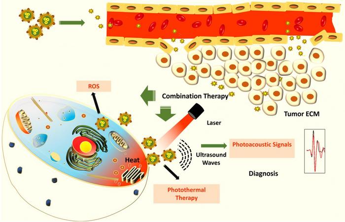
“These particles have photothermal, oxidative stress, and photoacoustic properties,” says Jaya Prakash, Assistant Professor at the Department of Instrumentation and Applied Physics (IAP), IISc, and one of the corresponding authors of the paper. PhD students Madhavi Tripathi and Swathi Padmanabhan are co-first authors.

When light is shined on these hybrid nanoparticles, they absorb the light and generate heat, which can kill cancer cells. These nanoparticles also produce singlet oxygen atoms that are toxic for the cells. “We want both these mechanisms to kill the cancer cell,” Jaya Prakash explains.  

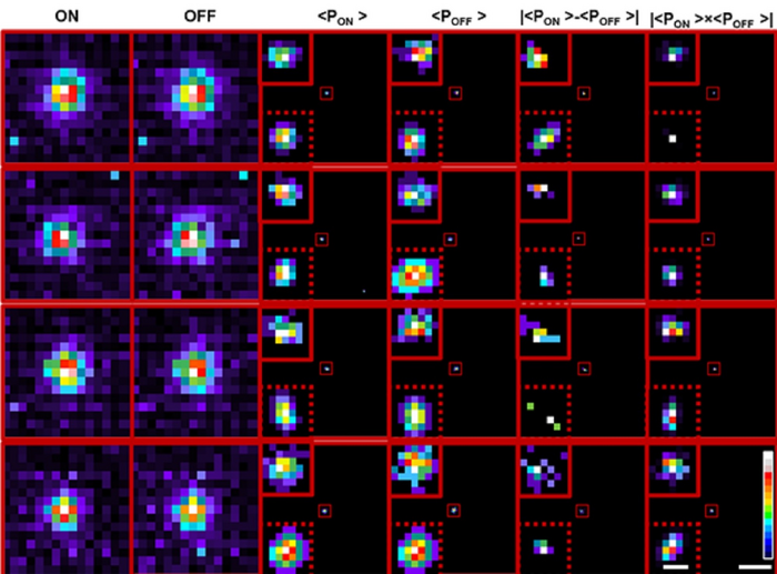
The researchers say that the nanoparticles can also help diagnose certain cancers. Existing methods such as standalone CT and MRI scans require trained radiology professionals to decipher the images. The photoacoustic property of the nanoparticles allows them to absorb light and generate ultrasound waves, which can be used to detect cancer cells with high contrast once the particles reach them. The ultrasound waves generated from the particles allow for a more accurate image resolution as sound waves scatter less when they pass through tissues compared to light. Scans created from the generated ultrasound waves can also provide better clarity and can be used to measure the oxygen saturation in the tumour, boosting their detection.

“You can integrate this with existing systems of detection or treatment,” says Ashok M Raichur, Professor at the Department of Materials Engineering, and another corresponding author. For example, the nanoparticles can be triggered to produce heat by shining a light on them using an endoscope that is typically used for cancer screening. 

Previously developed nanoparticles have limited applications because of their large size. The IISc team used a novel reduction method to deposit tiny seeds of gold onto the copper sulphide surface. The resulting hybrid nanoparticles – less than 8 nm in size – can potentially travel inside tissues easily and reach tumours. The researchers believe that the nanoparticles’ small size would also allow them to leave the human body naturally without accumulating, although extensive studies have to be carried out to determine if they are safe to use inside the human body.  

In the current study, the researchers have tested their nanoparticles on lung cancer and cervical cancer cell lines in the lab. They now plan to take the results forward for clinical development.  

Here’s a link to and a citation for the paper,

Seed-Mediated Galvanic Synthesis of CuS–Au Nanohybrids for Photo-Theranostic Applications by Madhavi Tripathi, Swathi Padmanabhan, Jaya Prakash, and Ashok M. Raichur. ACS Appl. Nano Mater. 2023, 6, 16, 14861–14875 DOI: https://doi.org/10.1021/acsanm.3c02405 Publication Date:August 10, 2023 Copyright © 2023 American Chemical Society

This paper is behind a paywall.

A brainlike (neuromorphic) camera can go beyond diffraction limit of light

Just when I think I’m getting caught up with my backlog along comes something like this. A February 21, 2023 news item on Nanowerk announces research that combines neuromorphic (brainlike) engineering and nanotechnology, Note: A link has been removed,

In a new study, researchers at the Indian Institute of Science (IISc) show how a brain-inspired image sensor can go beyond the diffraction limit of light to detect miniscule objects such as cellular components or nanoparticles invisible to current microscopes. Their novel technique, which combines optical microscopy with a neuromorphic camera and machine learning algorithms, presents a major step forward in pinpointing objects smaller than 50 nanometers in size.

A February 21, 2023 (?) Indian Institute of Science (IISc) press release (also on EurekAlert), which originated the news item, describes the nature of the task and provides some technical details,

Since the invention of optical microscopes, scientists have strived to surpass a barrier called the diffraction limit, which means that the microscope cannot distinguish between two objects if they are smaller than a certain size (typically 200-300 nanometers). Their efforts have largely focused on either modifying the molecules being imaged, or developing better illumination strategies – some of which led to the 2014 Nobel Prize in Chemistry. “But very few have actually tried to use the detector itself to try and surpass this detection limit,” says Deepak Nair, Associate Professor at the Centre for Neuroscience (CNS), IISc, and corresponding author of the study.  

Measuring roughly 40 mm (height) by 60 mm (width) by 25 mm (diameter), and weighing about 100 grams, the neuromorphic camera used in the study mimics the way the human retina converts light into electrical impulses, and has several advantages over conventional cameras. In a typical camera, each pixel captures the intensity of light falling on it for the entire exposure time that the camera focuses on the object, and all these pixels are pooled together to reconstruct an image of the object. In neuromorphic cameras, each pixel operates independently and asynchronously, generating events or spikes only when there is a change in the intensity of light falling on that pixel. This generates sparse and lower amount of data compared to traditional cameras, which capture every pixel value at a fixed rate, regardless of whether there is any change in the scene. This functioning of a neuromorphic camera is similar to how the human retina works, and allows the camera to “sample” the environment with much higher temporal resolution – because it is not limited by a frame rate like normal cameras – and also perform background suppression.  

“Such neuromorphic cameras have a very high dynamic range (>120 dB), which means that you can go from a very low-light environment to very high-light conditions. The combination of the asynchronous nature, high dynamic range, sparse data, and high temporal resolution of neuromorphic cameras make them well-suited for use in neuromorphic microscopy,” explains Chetan Singh Thakur, Assistant Professor at the Department of Electronic Systems Engineering (DESE), IISc, and co-author. 

In the current study, the group used their neuromorphic camera to pinpoint individual fluorescent beads smaller than the limit of diffraction, by shining laser pulses at both high and low intensities, and measuring the variation in the fluorescence levels. As the intensity increases, the camera captures the signal as an “ON” event, while an “OFF” event is reported when the light intensity decreases. The data from these events were pooled together to reconstruct frames. 

To accurately locate the fluorescent particles within the frames, the team used two methods. The first was a deep learning algorithm, trained on about one and a half million image simulations that closely represented the experimental data, to predict where the centroid of the object could be, explains Rohit Mangalwedhekar, former research intern at CNS and first author of the study. A wavelet segmentation algorithm was also used to determine the centroids of the particles separately for the ON and the OFF events. Combining the predictions from both allowed the team to zero in on the object’s precise location with greater accuracy than existing techniques.  

“In biological processes like self-organisation, you have molecules that are alternating between random or directed movement, or that are immobilised,” explains Nair. “Therefore, you need to have the ability to locate the centre of this molecule with the highest precision possible so that we can understand the thumb rules that allow the self-organisation.” The team was able to closely track the movement of a fluorescent bead moving freely in an aqueous solution using this technique. This approach can, therefore, have widespread applications in precisely tracking and understanding stochastic processes in biology, chemistry and physics.  

Caption: Transformation of cumulative probability density of ON and OFF processes allows localisation below the limit of classical single particle detection. Credit: Mangalwedhekar et al

Here’s a link to and a citation for the paper,

Achieving nanoscale precision using neuromorphic localization microscopy by Rohit Mangalwedhekar, Nivedita Singh, Chetan Singh Thakur, Chandra Sekhar Seelamantula, Mini Jose & Deepak Nair. Nature Nanotechnology volume 18, pages 380–389 (2023) DOI: https://doi.org/10.1038/s41565-022-01291-1 Published online: 23 January 2023 Issue Date: April 2023

This paper is behind a paywall.