Tag Archives: pacemakers

Breakthrough for tissue-interfaced bioelectronics

Let’s call this a cold open,

This October 24, 2024 news item on ScienceDaily describes some of what is in the video

The ideal material for interfacing electronics with living tissue is soft, stretchable, and just as water-loving as the tissue itself–in short, a hydrogel. Semiconductors, the key materials for bioelectronics such as pacemakers, biosensors, and drug delivery devices, on the other hand, are rigid, brittle, and water-hating, impossible to dissolve in the way hydrogels have traditionally been built. Scientists have now solved this challenge that has long stymied researchers, reimagining the process of creating hydrogels to build a powerful semiconductor in hydrogel form. The result is a bluish gel that flutters like a sea jelly in water but retains the immense semiconductive ability needed to transmit information between living tissue and machine.

An October 24, 2024 University of Chicago news release (also on EurekAlert) by Paul Dailing, which originated the news item, describes the breakthrough, Note: Links have been removed,

A paper published today in Science from the UChicago Pritzker School of Molecular Engineering (PME) has solved this challenge that has long stymied researchers, reimagining the process of creating hydrogels to build a powerful semiconductor in hydrogel form. Led by Asst. Prof. Sihong Wang’s research group, the result is a bluish gel that flutters like a sea jelly in water but retains the immense semiconductive ability needed to transmit information between living tissue and machine.

The material demonstrated tissue-level moduli as soft as 81 kPa, stretchability of 150% strain, and charge-carrier mobility up to 1.4 cm2 V-1 s-1. This means their material—both semiconductor and hydrogel at the same time—ticks all the boxes for an ideal bioelectronic interface.

“When making implantable bioelectronic devices, one challenge you must address is to make a device with tissue-like mechanical properties,” said Yahao Dai, the first author of the new paper. “That way, when it gets directly interfaced with the tissue, they can deform together and also form a very intimate bio-interface.”

Although the paper mainly focused on the challenges facing implanted medical devices such as biochemical sensors and pacemakers, Dai said the material also has many potential non-surgical applications, like better readings off the skin or improved care for wounds.

“It has very soft mechanical properties and a large degree of hydration similar to living tissue,” said UChicago PME Asst. Prof. Sihong Wang. “Hydrogel is also very porous, so it allows the efficient diffusion transport of different kinds of nutrition and chemicals. All these traits combine to make hydrogel probably the most useful material for tissue engineering and drug delivery.”

‘Let’s change our perspective’

The typical way of making a hydrogel is to take a material, dissolve it in water, and add the gelation chemicals to puff the new liquid into a gel form. Some materials simply dissolve in water, others require researchers to tinker and chemically modify the process, but the core mechanism is the same: No water, no hydrogel.

Semiconductors, however, don’t normally dissolve in water. Rather than find new, time-consuming means of trying to force the process, the UChicago PME team re-examined the question.

“We started to think, ‘Okay, let’s change our perspective,’ and we came up with a solvent exchange process,” Dai said.

Instead of dissolving the semiconductors in water, they dissolved them in an organic solvent that is miscible with water. They then prepared a gel from the dissolved semiconductors and hydrogel precursors. Their gel initially was an organogel, not a hydrogel.

“To eventually turn it into a hydrogel, we then immersed the whole material system into the water to let the organic solvent dissolve out and let the water come in,” Dai said.

An important benefit of such a solvent-exchange-based method is its broad applicability to different types of polymer semiconductors with different functions.

‘One plus one is greater than two’

The hydrogel semiconductor, which the team has patented and is commercializing through UChicago’s Polsky Center for Entrepreneurship and Innovation, is not merging a semiconductor with a hydrogel. It’s one material that is both semiconductor and hydrogel at the same time.

“It’s just one piece that has both semiconducting properties and hydrogel design, meaning that this whole piece is just like any other hydrogel,” Wang said.

Unlike any other hydrogel, however, the new material actually improved biological functions in two areas, creating better results than either hydrogel or semiconductor could accomplish on their own.

First, having a very soft material bond directly with tissue reduces the immune responses and inflammation typically triggered when a medical device is implanted.

Second, because hydrogels are so porous, the new material enables elevated biosensing response and stronger photo-modulation effects. With biomolecules being able to diffuse into the film to have volumetric interactions, the interaction sites for biomarkers-under-detection are significantly increased, which gives rise to higher sensitivity. Besides sensing, the responses to light for therapeutic functions at tissue surfaces also get increased from the more efficient transport of redox-active species. This benefits functions such as light-operated pacemakers or wound dressing that can be more efficiently heated with a flick of light to help speed healing.

“It’s a ‘one plus one is greater than two’ kind of combination,” Wang joked.

Researchers in the lab of UChicago Pritzker School of Engineering Asst. Prof. Sihong Wang (right), including PhD student Yahao Dai (left), have developed a hydrogel that retains the semiconductive ability needed to transmit information between living tissue and machine, which can be used both in implantable medical devices and non-surgical applications. (Photo by John Zich)

Here’s a link to and a citation for the paper,

Soft hydrogel semiconductors with augmented biointeractive functions by Yahao Dai, Shinya Wai, Pengju Li, Naisong Shan, Zhiqiang Cao, Yang Li, Yunfei Wang, Youdi Liu, Wei Liu, Kan Tang, Yuzi Liu, Muchuan Hua, Songsong Li, Nan Li, Shivani Chatterji, H. Christopher Fry, Sean Lee, Cheng Zhang, Max Weires, Sean Sutyak, Jiuyun Shi, Chenhui Zhu, Jie Xu, Xiaodan Gu, Bozhi Tian, and Sihong Wang. Science 24 Oct 2024 Vol 386, Issue 6720 pp. 431-439 DOI: 10.1126/science.adp9314

This paper is behind a paywall.

Plug me in: how to power up ingestible and implantable electroncis

From time to time I’ve featured ‘vampire technology’, a name I vastly prefer to energy harvesting or any of its variants. The focus has usually been on implantable electronic devices such as pacemakers and deep brain stimulators.

In this February 16, 2021 Nanowerk Spotlight article, Michael Berger broadens the focus to include other electronic devices,

Imagine edible medical devices that can be safely ingested by patients, perform a test or release a drug, and then transmit feedback to your smartphone; or an ingestible, Jell-O-like pill that monitors the stomach for up to a month.

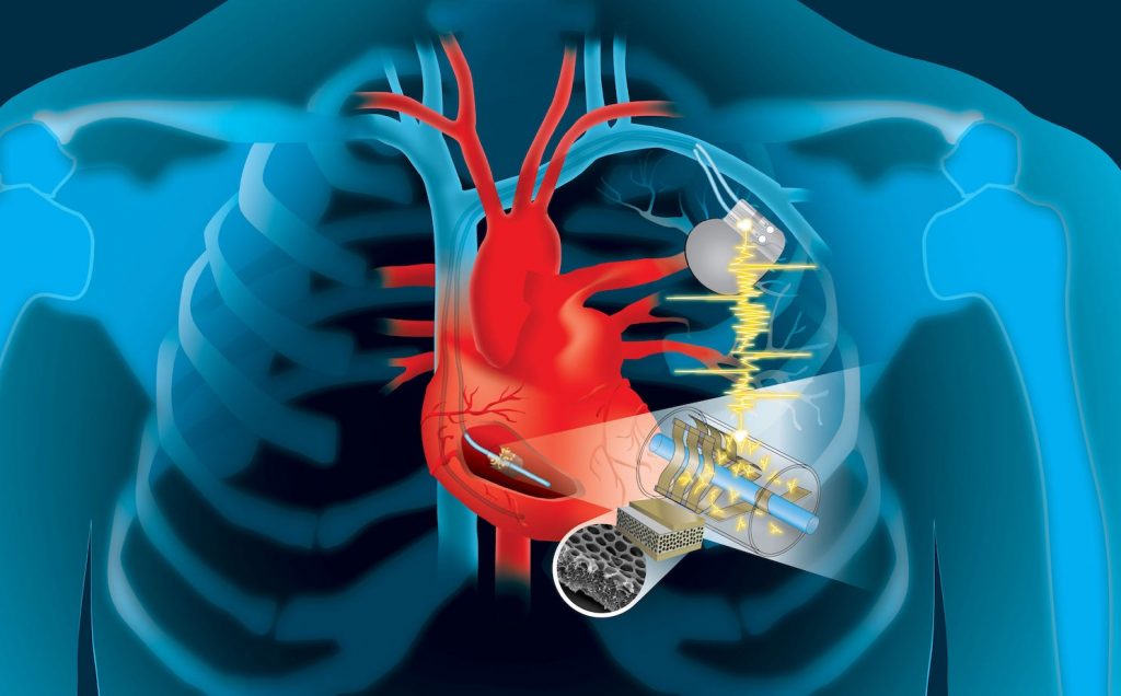
Devices like these, as well as a wide range of implantable biomedical electronic devices such as pacemakers, neurostimulators, subdermal blood sensors, capsule endoscopes, and drug pumps, can be useful tools for detecting physiological and pathophysiological signals, and providing treatments performed inside the body.

Advances in wireless communication enable medical devices to be untethered when in the human body. Advances in minimally invasive or semi-invasive surgical implantation procedures have enabled biomedical devices to be implanted in locations where clinically important biomarkers and physiological signals can be detected; it has also enabled direct administration of medication or treatment to a target location.

However, one major challenge in the development of these devices is the limited lifetime of their power sources. The energy requirements of biomedical electronic devices are highly dependent on their application and the complexity of the required electrical systems.

Berger’s commentary was occasioned by a review article in Advanced Functional Materials (link and citation to follow at the end of this post). Based on this review, the February 16, 2021 Nanowerk Spotlight article provides insight into the current state of affairs and challenges,

Biomedical electronic devices can be divided into three main categories depending on their application: diagnostic, therapeutic, and closed-loop systems. Each category has a different degree of complexity in the electronic system.

… most biomedical electronic devices are composed of a common set of components, including a power unit, sensors, actuators, a signal processing and control unit, and a data storage unit. Implantable and ingestible devices that require a great deal of data manipulation or large quantities of data logging also need to be wirelessly connected to an external device so that data can be transmitted to an external receiver and signal processing, data storage, and display can be performed more efficiently.

The power unit, which is composed of one or more energy sources – batteries, energy-harvesting, and energy transfer – as well as power management circuits, supplies electrical energy to the whole system.

Implantable medical devices such as cardiac pacemakers, neurostimulators and drug delivery devices are major medical tools to support life activity and provide new therapeutic strategies. Most such devices are powered by lithium batteries whose service life is as low as 10 years. Hence, many patients must undergo a major surgery to check the battery performance and replace the batteries as necessary.

In the last few decades, new battery technology has led to increases in the performance, reliability, and lifetime of batteries. However, challenges remain, especially in terms of volumetric energy density and safety.

Electronic miniaturization allows more functionalities to be added to devices, which increases power requirements. Recently, new material-based battery systems have been developed with higher energy densities.

Different locations and organ systems in the human body have access to different types of energy sources, such as mechanical, chemical, and electromagnetic energies.

Energy transfer technologies can deliver energy from outside the body to implanted or ingested devices. If devices are implanted at the locations where there are no accessible endogenous energies, exogenous energies in the form of ultrasonic or electromagnetic waves can penetrate through the biological barriers and wirelessly deliver the energies to the devices.

Both images embedded in the February 16, 2021 Nanowerk Spotlight article are informative. I’m particularly taken with the timeline which follows the development of batteries, energy harvesting/transfer devices, ingestible electronics, and implantable electronics. The first battery was in 1800 followed by ingestible and implantable electronics in the 1950s.

Berger’s commentary ends on this,

Concluding their review, the authors [in Advanced Functional Materials] note that low energy conversion efficiency and power output are the fundamental bottlenecks of energy harvesting and transfer devices. They suggest that additional studies are needed to improve the power output of energy harvesting and transfer devices so that they can be used to power various biomedical electronics.

Furthermore, durability studies of promising energy harvesters should be performed to evaluate their use in long-term applications. For degradable energy harvesting devices, such as friction-based energy harvesters and galvanic cells, improving the device lifetime is essential for use in real-life applications.

Finally, manufacturing cost is another factor to consider when commercializing novel batteries, energy harvesters, or energy transfer devices as power sources for medical devices.

Here’s a link to and a citation for the paper,

Powering Implantable and Ingestible Electronics by So‐Yoon Yang, Vitor Sencadas, Siheng Sean You, Neil Zi‐Xun Jia, Shriya Sruthi Srinivasan, Hen‐Wei Huang, Abdelsalam Elrefaey Ahmed, Jia Ying Liang, Giovanni Traverso. Advanced Functional Materials DOI: https://doi.org/10.1002/adfm.202009289 First published: 04 February 2021

This paper is behind a paywall.

It may be possible to receive a full text PDF of the article from the authors. Try here.

There are others but here are two of my posts about ‘vampire energy’,

Harvesting the heart’s kinetic energy to power implants (July 26, 2019)

Vampire nanogenerators: 2017 (October 19, 2017)

Harvesting the heart’s kinetic energy to power implants

This work comes from Dartmouth College, an educational institution based on the US east coast in the state of New Hampshire. I hardly ever stumble across research from Dartmouth and I assume that’s because they usually focus their interests in areas that are not of direct interest to me,

Rendering of the two designs of the cardiac energy harvesting device. (Cover art by Patricio Sarzosa) Courtesy: Dartmouth College

For a change, we have a point of connection (harvesting biokinetic energy) according to a February 4, 2019 news item on ScienceDaily,

The heart’s motion is so powerful that it can recharge devices that save our lives, according to new research from Dartmouth College.

Using a dime-sized invention developed by engineers at the Thayer School of Engineering at Dartmouth, the kinetic energy of the heart can be converted into electricity to power a wide-range of implantable devices, according to the study funded by the National Institutes of Health.

A February 4, 2019 Dartmouth College news release, which originated the news item, describes the problem and the proposed solution,

Millions of people rely on pacemakers, defibrillators and other live-saving implantable devices powered by batteries that need to be replaced every five to 10 years. Those replacements require surgery which can be costly and create the possibility of complications and infections.

“We’re trying to solve the ultimate problem for any implantable biomedical device,” says Dartmouth engineering professor John X.J. Zhang, a lead researcher on the study his team completed alongside clinicians at the University of Texas in San Antonio. “How do you create an effective energy source so the device will do its job during the entire life span of the patient, without the need for surgery to replace the battery?”

“Of equal importance is that the device not interfere with the body’s function,” adds Dartmouth research associate Lin Dong, first author on the paper. “We knew it had to be biocompatible, lightweight, flexible, and low profile, so it not only fits into the current pacemaker structure but is also scalable for future multi-functionality.”

The team’s work proposes modifying pacemakers to harness the kinetic energy of the lead wire that’s attached to the heart, converting it into electricity to continually charge the batteries. The added material is a type of thin polymer piezoelectric film called “PVDF” and, when designed with porous structures — either an array of small buckle beams or a flexible cantilever — it can convert even small mechanical motion to electricity. An added benefit: the same modules could potentially be used as sensors to enable data collection for real-time monitoring of patients.

The results of the three-year study, completed by Dartmouth’s engineering researchers along with clinicians at UT Health San Antonio, were just published in the cover story for Advanced Materials Technologies.

The two remaining years of NIH funding plus time to finish the pre-clinical process and obtain regulatory approval puts a self-charging pacemaker approximately five years out from commercialization, according to Zhang

“We’ve completed the first round of animal studies with great results which will be published soon,” says Zhang. “There is already a lot of expressed interest from the major medical technology companies, and Andrew Closson, one of the study’s authors working with Lin Dong and an engineering PhD Innovation Program student at Dartmouth, is learning the business and technology transfer skills to be a cohort in moving forward with the entrepreneurial phase of this effort.”

Other key collaborators on the study include Dartmouth engineering professor Zi Chen, an expert on thin structure mechanics, and Dr. Marc Feldman, professor and clinical cardiologist at UT [University of Texas] Health San Antonio.

Here’s a citation and another link for the paper,

Energy Harvesting: Flexible Porous Piezoelectric Cantilever on a Pacemaker Lead for Compact Energy Harvesting by Lin Dong, Xiaomin Han, Zhe Xu, Andrew B. Closson, Yin Liu, Chunsheng Wen, Xi Liu, Gladys Patricia Escobar, Meagan Oglesby, Marc Feldman, Zi Chen, John X. J. Zhang. Adv. Mater. Technol. 1/2019 https://doi.org/10.1002/admt.201970002 First published: 08 January 2019

This paper is open access.

Nanofiber coating for artificial joints and implants

The researchers have a great image to accompany their research, which fit well with Hallowe’en and the Day of the Dead celebrations taking place around the same time as the research was published.

 A titanium implant (blue) without a nanofiber coating in the femur of a mouse. Bacteria are shown in red and responding immune cells in yellow. Credit: Lloyd Miller/Johns Hopkins Medicine

A titanium implant (blue) without a nanofiber coating in the femur of a mouse. Bacteria are shown in red and responding immune cells in yellow.
Credit: Lloyd Miller/Johns Hopkins Medicine

An Oct. 24, 2016 news item on ScienceDaily announces the research on nanofibers,

In a proof-of-concept study with mice, scientists at The Johns Hopkins University show that a novel coating they made with antibiotic-releasing nanofibers has the potential to better prevent at least some serious bacterial infections related to total joint replacement surgery.

An Oct. 24, 2016 Johns Hopkins Medicine news release (also on EurekAlert), provides further details (Note: Links have been removed),

A report on the study, published online the week of Oct. 24 [2016] in Proceedings of the National Academy of Sciences, was conducted on the rodents’ knee joints, but, the researchers say, the technology would have “broad applicability” in the use of orthopaedic prostheses, such as hip and knee total joint replacements, as well pacemakers, stents and other implantable medical devices. In contrast to other coatings in development, the researchers report the new material can release multiple antibiotics in a strategically timed way for an optimal effect.

“We can potentially coat any metallic implant that we put into patients, from prosthetic joints, rods, screws and plates to pacemakers, implantable defibrillators and dental hardware,” says co-senior study author Lloyd S. Miller, M.D., Ph.D., an associate professor of dermatology and orthopaedic surgery at the Johns Hopkins University School of Medicine.

Surgeons and biomedical engineers have for years looked for better ways —including antibiotic coatings — to reduce the risk of infections that are a known complication of implanting artificial hip, knee and shoulder joints.

Every year in the U.S., an estimated 1 to 2 percent of the more than 1 million hip and knee replacement surgeries are followed by infections linked to the formation of biofilms — layers of bacteria that adhere to a surface, forming a dense, impenetrable matrix of proteins, sugars and DNA. Immediately after surgery, an acute infection causes swelling and redness that can often be treated with intravenous antibiotics. But in some people, low-grade chronic infections can last for months, causing bone loss that leads to implant loosening and ultimately failure of the new prosthesis. These infections are very difficult to treat and, in many cases of chronic infection, prostheses must be removed and patients placed on long courses of antibiotics before a new prosthesis can be implanted. The cost per patient often exceeds $100,000 to treat a biofilm-associated prosthesis infection, Miller says.

Major downsides to existing options for local antibiotic delivery, such as antibiotic-loaded cement, beads, spacers or powder, during the implantation of medical devices are that they can typically only deliver one antibiotic at a time and the release rate is not well-controlled. To develop a better approach that addresses those problems, Miller teamed up with Hai-Quan Mao, Ph.D., a professor of materials science and engineering at the Johns Hopkins University Whiting School of Engineering, and a member of the Institute for NanoBioTechnology, Whitaker Biomedical Engineering Institute and Translational Tissue Engineering Center.

Over three years, the team focused on designing a thin, biodegradable plastic coating that could release multiple antibiotics at desired rates. This coating is composed of a nanofiber mesh embedded in a thin film; both components are made of polymers used for degradable sutures.

To test the technology’s ability to prevent infection, the researchers loaded the nanofiber coating with the antibiotic rifampin in combination with one of three other antibiotics: vancomycin, daptomycin or linezolid. “Rifampin has excellent anti-biofilm activity but cannot be used alone because bacteria would rapidly develop resistance,” says Miller. The coatings released vancomycin, daptomycin or linezolid for seven to 14 days and rifampin over three to five days. “We were able to deploy two antibiotics against potential infection while ensuring rifampin was never present as a single agent,” Miller says.

The team then used each combination to coat titanium Kirschner wires — a type of pin used in orthopaedic surgery to fix bone in place after wrist fractures — inserted them into the knee joints of anesthetized mice and introduced a strain of Staphylococcus aureus, a bacterium that commonly causes biofilm-associated infections in orthopaedic surgeries. The bacteria were engineered to give off light, allowing the researchers to noninvasively track infection over time.

Miller says that after 14 days of infection in mice that received an antibiotic-free coating on the pins, all of the mice had abundant bacteria in the infected tissue around the knee joint, and 80 percent had bacteria on the surface of the implant. In contrast, after the same time period in mice that received pins with either linezolid-rifampin or daptomycin-rifampin coating, none of the mice had detectable bacteria either on the implants or in the surrounding tissue.

“We were able to completely eradicate infection with this coating,” says Miller. “Most other approaches only decrease the number of bacteria but don’t generally or reliably prevent infections.”

After the two-week test, each of the rodents’ joints and adjacent bones were removed for further study. Miller and Mao found that not only had infection been prevented, but the bone loss often seen near infected joints — which creates the prosthetic loosening in patients — had also been completely avoided in animals that received pins with the antibiotic-loaded coating.

Miller emphasized that further research is needed to test the efficacy and safety of the coating in humans, and in sorting out which patients would best benefit from the coating — people with a previous prosthesis joint infection receiving a new replacement joint, for example.

The polymers they used to generate the nanofiber coating have already been used in many approved devices by the U.S. Food and Drug Administration, such as degradable sutures, bone plates and drug delivery systems.

Here’s a link to and a citation for the paper,

Polymeric nanofiber coating with tunable combinatorial antibiotic delivery prevents biofilm-associated infection in vivo by Alyssa G. Ashbaugh, Xuesong Jiang, Jesse Zheng, Andrew S. Tsai, Woo-Shin Kim, John M. Thompson, Robert J. Miller, Jonathan H. Shahbazian, Yu Wang, Carly A. Dillen, Alvaro A. Ordonez, Yong S. Chang, Sanjay K. Jain, Lynne C. Jones, Robert S. Sterling, Hai-Quan Mao, and Lloyd S. Miller. PNAS [Proceedings of the National Academy of Sciences] 2016 doi: 10.1073/pnas.1613722113 Published ahead of print October 24, 2016

This paper is behind a paywall.