Tag Archives: silver nanorods

Gold nanotubes for treating mesothelioma?

An October 26, 2020 news item on Nanowerk describes some new research that may lead the way to treatments for people with asbestos-related cancers (e.g., mesothelioma), Note: A link has been removed,

Gold nanotubes – tiny hollow cylinders one thousandth the width of a human hair – could be used to treat mesothelioma, a type of cancer caused by exposure to asbestos, according to a team of researchers at the Universities of Cambridge and Leeds.

In a study published in journal Small (“Exploring High Aspect Ratio Gold Nanotubes as Cytosolic Agents: Structural Engineering and Uptake into Mesothelioma Cells”), the researchers demonstrate that once inside the cancer cells, the nanotubes absorb light, causing them to heat up, thereby killing the cells.

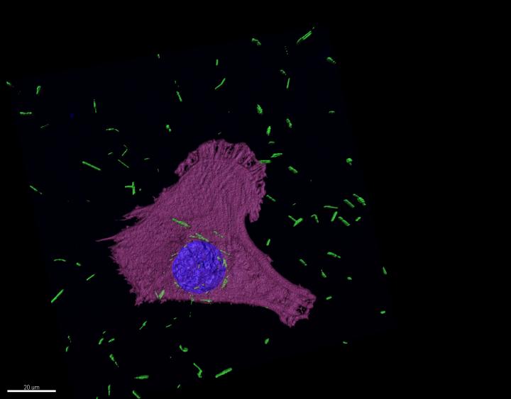
Here`s an image illustrating the research,

Caption: Confocal fluorescence image of gold nanotures (green) in mesothelioma cells. Credit: Arsalan Azad

An October 27, 2020 University of Cambridge press release (also on EurekAlert but published on Oct. 26, 2020), which originated the news item, describes the context for the research and provides a few more technical details,

More than 2,600 people are diagnosed in the UK each year with mesothelioma, a malignant form of cancer caused by exposure to asbestos. Although the use of asbestos is outlawed in the UK now, the country has the world’s highest levels of mesothelioma because it imported vast amounts of asbestos in the post-war years. The global usage of asbestos remains high, particularly in low- and middle-income countries, which means mesothelioma will become a global problem.

“Mesothelioma is one of the ‘hard-to-treat’ cancers, and the best we can offer people with existing treatments is a few months of extra survival,” said Dr Arsalan Azad from the Cambridge Institute for Medical Research at the University of Cambridge. “There’s an important unmet need for new, effective treatments.”

In 2018, the University of Cambridge was awarded £10million from the Engineering and Physical Sciences Research Council to help develop engineering solutions, including nanotech, to find ways to address hard-to-treat cancers.

In a collaboration between the University of Cambridge and University of Leeds, researchers have developed a form of gold nanotubes whose physical properties are ‘tunable’ – in other words, the team can tailor the wall thickness, microstructure, composition, and ability to absorb particular wavelengths of light.

The researchers added the nanotubes to mesothelioma cells cultured in the lab and found that they were absorbed by the cells, residing close to the nucleus, where the cell’s DNA lies. When the team targeted the cells with a laser, the nanotubes absorbed the light and heated up, killing the mesothelioma cell.

Professor Stefan Marciniak, also from the Cambridge Institute for Medical Research, added: “The mesothelioma cells ‘eat’ the nanotubes, leaving them susceptible when we shine light on them. Laser light is able to penetrate deep into tissue without causing damage to surrounding tissue. It then gets absorbed by the nanotubes, which heat up and, we hope in the future, could be used to cause localised cancer-cell killing.”

The team will be developing the work further to ensure the nanotubes are targeted to cancer cells with less effect on normal tissue.

The nanotubes are made in a two-step process. First, solid silver nanorods are created of the desired diameter. Gold is then deposited from solution onto the surface of the silver. As the gold builds-up at the surface, the silver dissolves from the inside to leave a hollow nanotube.

The approach advanced by the Leeds team allows these nanotubes to be developed at room temperature, which should make their manufacture at scale more feasible.

Professor Stephen Evans from the School of Physics and Astronomy at the University of Leeds said: “Having control over the size and shape of the nanotubes allows us to tune them to absorb light where the tissue is transparent and will allow them to be used for both the imaging and treatment of cancers. The next stage will be to load these nanotubes with medicines for enhanced therapies.”

Here`s a link to and a citation for the paper,

Exploring High Aspect Ratio Gold Nanotubes as Cytosolic Agents: Structural Engineering and Uptake into Mesothelioma Cells by Sunjie Ye, Arsalan A. Azad, Joseph E. Chambers, Alison J. Beckett, Lucien Roach, Samuel C. T. Moorcroft, Zabeada Aslam, Ian A. Prior, Alexander F. Markham, P. Louise Coletta, Stefan J. Marciniak, Stephen D. Evans. Small DOI: https://doi.org/10.1002/smll.202003793 First published: 25 October 2020

This paper is open access.

One step diagnosis (nanotechnology-enabled) from University of Georgia (US)

The researchers haven’t tried this out on blood, saliva, or urine yet but this July 21, 2012 news item by Gary Thomas on Azonano hints that will be the next step,

Researchers at the University of Georgia have devised a single-step, quick and accurate technique using nanomaterials to detect pathogens and contaminants. The team demonstrated the capability of the new technique in detecting compounds like protein albumin and lactic acid in extremely diluted mixtures that comprised of dyes and chemicals.

The researchers conclude that the same method can be employed on biological mixtures like blood, saliva, food and urine to detect contaminants and pathogens.

The originating July 19, 2012 news release by Sam Fahmy for the University of Georgia provides more detail,

“The results are unambiguous and quickly give you a high degree of specificity,” said senior author Yiping Zhao, professor of physics in the UGA [University of Georgia] Franklin College of Arts and Sciences and director of the university’s Nanoscale Science and Engineering Center.

Zhao and his co-authors—doctoral students Jing Chen and Justin Abell and professor Yao-wen Huang of the UGA College of Agricultural and Environmental Sciences—used nanotechnology to combine two well-known techniques and create their new diagnostic test. …

The first component of their two-in-one system uses a technique known as surface enhanced Raman spectroscopy, or SERS, which measures the change in frequency of a laser as it scatters off a compound. Every compound displays a series of distinctive changes in frequency, or Raman shifts, that are as unique as a fingerprint. The signal produced by Raman scattering is inherently weak, but Zhao and his colleagues have arrayed silver nanorods 1,000 times finer than the width of a human hair at a precise angle to significantly amplify the signal. In previous studies with Ralph Tripp in the UGA College of Veterinary Medicine and chemist Richard Dluhy in the Franklin College, they demonstrated that the use of SERS with silver nanorods could identify viruses such as HIV and RSV isolated from infected cells.

Here’s why they needed a second technique and how it fits into the picture (from the news release),

“In a clinical setting, the sample that you obtain from patients typically contains bacteria or viruses as well as a lot of fluid—as in blood, urine or saliva—that contains biological agents that interfere with the signal you’re trying to detect,” Zhao said. “To develop a diagnostic that could be used at the point of care, we needed a way to separate those agents.”

Once again, the scientists turned to nanotechnology to create a next-generation diagnostic test. Using traditional thin layer chromatography, or TLC, scientists blot a drop of sample onto a porous surface. They then apply a solvent such as methanol to the sample, and the sample components separate based on how strongly they’re attracted to the solvent and the surface.

Study co-author Justin Abell, a doctoral student in the UGA College of Engineering, explained that TLC typically requires a large sample volume because the compound of interest soaks into the surface in addition to moving along it, like a stain on a rug. The silver nanorod surface that the researchers use, in contrast, allows them to use a miniscule amount of sample in a technique known as ultra-thin layer chromatography.

“In our case, the nanorods are acting as the detection medium but also as the separation medium,” Abell said, “so it’s a two-in-one system.”

To test their method, the researchers used mixtures of dyes, the organic chemical melamine, lactic acid and the protein albumin. In each case, they were able to directly identify the compounds of interest, even in samples diluted to concentrations below 182 nanograms per milliliter-roughly 200 billionths of a gram in a fifth of a teaspoon. And while the detection of viruses using techniques such as polymerase chain reaction can take days or even weeks and requires fluorescent labels, the on-chip method developed by the UGA researchers yields results in less than an hour without the use of molecular labels.

As for future plans to develop this application (from the news release),

The researchers are currently testing their technique with biological samples from Tripp’s lab that contain viruses, and Zhao said preliminary results are promising. He adds that while his team is focused on health and food safety applications, SERS and ultra-thin layer chromatography can be used to detect compounds of all types—everything from forensic materials at a crime scene to environmental pollutants. His team also is working with colleagues across campus to create an online encyclopedia that would allow technicians to identify viruses, bacteria, biomarkers and pharmaceuticals based on their distinctive Raman shifts.

“Every compound has a unique SERS spectrum,” Zhao said, “so this is a very robust technology whose applications are practically endless.”