Tag Archives: Ali K. Yetisen

Edible nano-structured holograms could decorate food one day

Caption Nanostructures (yellowish-green images; scale bar, 5 μm) were patterned onto dried corn syrup films, producing edible, rainbow-colored holograms (scale bar, 2 mm). Credit Adapted from ACS Nano 2021, DOI: 10.1021/acsnano.0c02438

Where food safety is concerned, much of the research I’ve seen is focused on adding senors to the packaging rather than direct application to the foodstuff but this is different, from a February 17, 2021 news item on phys.org,

Holograms are everywhere, from driver’s licenses to credit cards to product packaging. And now, edible holograms could someday enhance foods. Researchers reporting in ACS [American Chemical Society] Nano have developed a laser-based method to print nanostructured holograms on dried corn syrup films. The edible holograms could also be used to ensure food safety, label a product or indicate sugar content, the researchers say.

A February 17, 2021American Chemical Society news release (also on EurekAlert), which originated the news item,

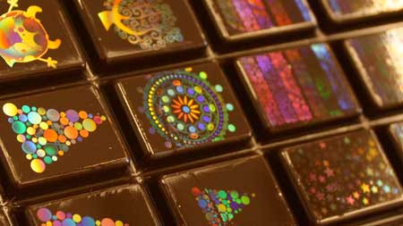
Most holograms are imprinted with lasers onto metal surfaces, such as aluminum, but the materials aren’t edible. For foods, holograms made with nanoparticles have been proposed, but the tiny particles can generate reactive oxygen species, which might be harmful for people to eat. In a different approach, food scientists have molded edible holograms onto chocolate, but the process only works for certain types of the confection, and a different mold is needed for each hologram design. Bader AlQattan, Haider Butt and colleagues wanted to find a safe, fast and versatile way to pattern edible holograms onto a variety of foods.

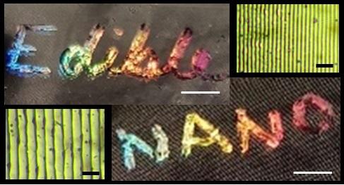
To develop their method, the researchers made a solution of corn syrup, vanilla and water and dried it into a thin film. They coated the film with a fine layer of non-toxic black dye. Then, they used a technique called direct laser interference patterning to etch off most of the dye, leaving behind raised, nanoscale lines that formed a diffraction grating. When struck by light, the nanostructure diffracted the light into a rainbow pattern, with different colors appearing at different angles of viewing. The team could control the intensity and range of colors by varying the spacing between lines in the grating or the sugar content of the corn syrup film. Before edible holograms are ready to hit store shelves, however, the researchers want to adapt the method to a food-grade dye that could replace the synthetic black dye used in these pilot experiments.

Here’s a link to and a citation for the paper,

Direct Printing of Nanostructured Holograms on Consumable Substrates by Bader AlQattan, Joelle Doocey, Murad Ali, Israr Ahmed, Ahmed E. Salih, Fahad Alam, Magdalena Bajgrowicz-Cieslak, Ali K. Yetisen, Mohamed Elsherif, and Haider Butt. ACS Nano 2021, 15, 2, 2340–2349 DOI: https://doi.org/10.1021/acsnano.0c02438 Publication Date:February 1, 2021 Copyright © 2021 American Chemical Society

This paper appears to be open access.

It seems these scientists are also considering the aesthetic possibilities. Ffrom the paper, Note: Links have been removed,

The use of holograms in food could potentially improve sensory appeal [emphasis mine] and, through biosensing, could increase health and safety.(1,2) Holograms can even be used to store information as edible microtags.(3) They are also attractive to the eye as they produce rainbow patterns with light. Using edible holograms on foods, not only as decoration but also to sense harmful bacteria, could improve food quality/lifetime monitoring.(4,5) Food holograms which signify a qualitative information about the sugar contents could be of value in controlling the sugar consumption, that is challenging to be measured at the moment.(6)

As it is, I find food pretty attractive. So, I’m not sure why there’s a need to improve its sensory appeal. On the other hand, I can’t argue with increased food safety.

Should you be interested in more about holograms and their current applications, including chocolate decoration, you can check out Michael Berger’s February 17, 2021 Nanowerk Spotlight article.

Holographic chocolate surfaces. (Image: Morphotonix) [downloaded from https://www.nanowerk.com/spotlight/spotid=57310.php]

What do you think about decorating food with holograms? If you feel inclined, do let me know in the comments.

Stretchy optical materials for implants that could pulse light

An Oct. 17, 2016 Massachusetts Institute of Technology (MIT) news release (also on EurekAlert) by Emily Chu describes research that could lead to long-lasting implants offering preventive health strategies,

Researchers from MIT and Harvard Medical School have developed a biocompatible and highly stretchable optical fiber made from hydrogel — an elastic, rubbery material composed mostly of water. The fiber, which is as bendable as a rope of licorice, may one day be implanted in the body to deliver therapeutic pulses of light or light up at the first sign of disease. [emphasis mine]

The researchers say the fiber may serve as a long-lasting implant that would bend and twist with the body without breaking down. The team has published its results online in the journal Advanced Materials.

Using light to activate cells, and particularly neurons in the brain, is a highly active field known as optogenetics, in which researchers deliver short pulses of light to targeted tissues using needle-like fibers, through which they shine light from an LED source.

“But the brain is like a bowl of Jell-O, whereas these fibers are like glass — very rigid, which can possibly damage brain tissues,” says Xuanhe Zhao, the Robert N. Noyce Career Development Associate Professor in MIT’s Department of Mechanical Engineering. “If these fibers could match the flexibility and softness of the brain, they could provide long-term more effective stimulation and therapy.”

Getting to the core of it

Zhao’s group at MIT, including graduate students Xinyue Liu and Hyunwoo Yuk, specializes in tuning the mechanical properties of hydrogels. The researchers have devised multiple recipes for making tough yet pliable hydrogels out of various biopolymers. The team has also come up with ways to bond hydrogels with various surfaces such as metallic sensors and LEDs, to create stretchable electronics.

The researchers only thought to explore hydrogel’s use in optical fibers after conversations with the bio-optics group at Harvard Medical School, led by Associate Professor Seok-Hyun (Andy) Yun. Yun’s group had previously fabricated an optical fiber from hydrogel material that successfully transmitted light through the fiber. However, the material broke apart when bent or slightly stretched. Zhao’s hydrogels, in contrast, could stretch and bend like taffy. The two groups joined efforts and looked for ways to incorporate Zhao’s hydrogel into Yun’s optical fiber design.

Yun’s design consists of a core material encased in an outer cladding. To transmit the maximum amount of light through the core of the fiber, the core and the cladding should be made of materials with very different refractive indices, or degrees to which they can bend light.

“If these two things are too similar, whatever light source flows through the fiber will just fade away,” Yuk explains. “In optical fibers, people want to have a much higher refractive index in the core, versus cladding, so that when light goes through the core, it bounces off the interface of the cladding and stays within the core.”

Happily, they found that Zhao’s hydrogel material was highly transparent and possessed a refractive index that was ideal as a core material. But when they tried to coat the hydrogel with a cladding polymer solution, the two materials tended to peel apart when the fiber was stretched or bent.

To bond the two materials together, the researchers added conjugation chemicals to the cladding solution, which, when coated over the hydrogel core, generated chemical links between the outer surfaces of both materials.

“It clicks together the carboxyl groups in the cladding, and the amine groups in the core material, like molecular-level glue,” Yuk says.

Sensing strain

The researchers tested the optical fibers’ ability to propagate light by shining a laser through fibers of various lengths. Each fiber transmitted light without significant attenuation, or fading. They also found that fibers could be stretched over seven times their original length without breaking.

Now that they had developed a highly flexible and robust optical fiber, made from a hydrogel material that was also biocompatible, the researchers began to play with the fiber’s optical properties, to see if they could design a fiber that could sense when and where it was being stretched.

They first loaded a fiber with red, green, and blue organic dyes, placed at specific spots along the fiber’s length. Next, they shone a laser through the fiber and stretched, for instance, the red region. They measured the spectrum of light that made it all the way through the fiber, and noted the intensity of the red light. They reasoned that this intensity relates directly to the amount of light absorbed by the red dye, as a result of that region being stretched.

In other words, by measuring the amount of light at the far end of the fiber, the researchers can quantitatively determine where and by how much a fiber was stretched.

“When you stretch a certain portion of the fiber, the dimensions of that part of the fiber changes, along with the amount of light that region absorbs and scatters, so in this way, the fiber can serve as a sensor of strain,” Liu explains.

“This is like a multistrain sensor through a single fiber,” Yuk adds. “So it can be an implantable or wearable strain gauge.”

The researchers imagine that such stretchable, strain-sensing optical fibers could be implanted or fitted along the length of a patient’s arm or leg, to monitor for signs of improving mobility.

Zhao envisions the fibers may also serve as sensors, lighting up in response to signs of disease.

“We may be able to use optical fibers for long-term diagnostics, to optically monitor tumors or inflammation,” he says. “The applications can be impactful.”

Here’s a link to and a citation for the paper,

Highly Stretchable, Strain Sensing Hydrogel Optical Fibers by Jingjing Guo, Xinyue Liu, Nan Jiang, Ali K. Yetisen, Hyunwoo Yuk, Changxi Yang, Ali Khademhosseini, Xuanhe Zhao, and Seok-Hyun Yun. Advanced Materials DOI: 10.1002/adma.201603160 Version of Record online: 7 OCT 2016

© 2016 WILEY-VCH Verlag GmbH & Co. KGaA, Weinheim

This paper is behind a paywall.

Using scientific methods and technology to explore living systems as artistic subjects: bioart

There is a fascinating set of stories about bioart designed to whet your appetite for more (*) in a Nov. 23, 2015 Cell Press news release on EurekAlert (Note: A link has been removed),

Joe Davis is an artist who works not only with paints or pastels, but also with genes and bacteria. In 1986, he collaborated with geneticist Dan Boyd to encode a symbol for life and femininity into an E. coli bacterium. The piece, called Microvenus, was the first artwork to use the tools and techniques of molecular biology. Since then, bioart has become one of several contemporary art forms (including reclamation art and nanoart) that apply scientific methods and technology to explore living systems as artistic subjects. A review of the field, published November 23, can be found in Trends in Biotechnology.

Bioart ranges from bacterial manipulation to glowing rabbits, cellular sculptures, and–in the case of Australian-British artist Nina Sellars–documentation of an ear prosthetic that was implanted onto fellow artist Stelarc’s arm. In the pursuit of creating art, practitioners have generated tools and techniques that have aided researchers, while sometimes crossing into controversy, such as by releasing invasive species into the environment, blurring the lines between art and modern biology, raising philosophical, societal, and environmental issues that challenge scientific thinking.

“Most people don’t know that bioart exists, but it can enable scientists to produce new ideas and give us opportunities to look differently at problems,” says author Ali K. Yetisen, who works at Harvard Medical School and the Wellman Center for Photomedicine, Massachusetts General Hospital. “At the same time there’s been a lot of ethical and safety concerns happening around bioart and artists who wanted to get involved in the past have made mistakes.”

Here’s a sample of Joe Davis’s work,

 Caption This photograph shows polyptich paintings by Joe Davis of his 28-mer Microvenus DNA molecule (2006 Exhibition in Greece at Athens School of Fine Arts). Credit: Courtesy of Joe Davis

This photograph shows polyptich paintings by Joe Davis of his 28-mer Microvenus DNA molecule (2006 Exhibition in Greece at Athens School of Fine Arts). Credit: Courtesy of Joe Davis

The news release goes on to recount a brief history of bioart, which stretches back to 1928 and then further back into the 19th and 18th centuries,

In between experiments, Alexander Fleming would paint stick figures and landscapes on paper and in Petri dishes using bacteria. In 1928, after taking a brief hiatus from the lab, he noticed that portions of his “germ paintings,” had been killed. The culprit was a fungus, penicillin–a discovery that would revolutionize medicine for decades to come.

In 1938, photographer Edward Steichen used a chemical to genetically alter and produce interesting variations in flowering delphiniums. This chemical, colchicine, would later be used by horticulturalists to produce desirable mutations in crops and ornamental plants.

In the late 18th and early 19th centuries, the arts and sciences moved away from traditionally shared interests and formed secular divisions that persisted well into the 20th century. “Appearance of environmental art in the 1970s brought about renewed awareness of special relationships between art and the natural world,” Yetisen says.

To demonstrate how we change landscapes, American sculptor Robert Smithsonian paved a hillside with asphalt, while Bulgarian artist Christo Javacheffa (of Christo and Jeanne-Claude) surrounded resurfaced barrier islands with bright pink plastic.

These pieces could sometimes be destructive, however, such as in Ten Turtles Set Free by German-born Hans Haacke. To draw attention to the excesses of the pet trade, he released what he thought were endangered tortoises back to their natural habitat in France, but he inadvertently released the wrong subspecies, thus compromising the genetic lineages of the endangered tortoises as the two varieties began to mate.

By the late 1900s, technological advances began to draw artists’ attention to biology, and by the 2000s, it began to take shape as an artistic identity. Following Joe Davis’ transgenic Microvenus came a miniaturized leather jacket made of skin cells, part of the Tissue Culture & Art Project (initiated in 1996) by duo Oran Catts and Ionat Zurr. Other examples of bioart include: the use of mutant cacti to simulate appearance of human hair in the place of cactus spines by Laura Cinti of University College London’s C-Lab; modification of butterfly wings for artistic purposes by Marta de Menezes of Portugal; and photographs of amphibian deformation by American Brandon Ballengée.

“Bioart encourages discussions about societal, philosophical, and environmental issues and can help enhance public understanding of advances in biotechnology and genetic engineering,” says co-author Ahmet F. Coskun, who works in the Division of Chemistry and Chemical Engineering at California Institute of Technology.

Life as a Bioartist

Today, Joe Davis is a research affiliate at MIT Biology and “Artist-Scientist” at the George Church Laboratory at Harvard–a place that fosters creativity and technological development around genetic engineering and synthetic biology. “It’s Oz, pure and simple,” Davis says. “The total amount of resources in this environment and the minds that are accessible, it’s like I come to the city of Oz every day.”

But it’s not a one-way street. “My particular lab depends on thinking outside the box and not dismissing things because they sound like science fiction,” says [George M.] Church, who is also part of the Wyss Institute for Biologically Inspired Engineering. “Joe is terrific at keeping us flexible and nimble in that regard.”

For example, Davis is working with several members of the Church lab to perform metagenomics analyses of the dust that accumulates at the bottom of money-counting machines. Another project involves genetically engineering silk worms to spin metallic gold–an homage to the fairy tale of Rumpelstiltskin.

“I collaborate with many colleagues on projects that don’t necessarily have direct scientific results, but they’re excited to pursue these avenues of inquiry that they might not or would not look into ordinarily–they might try to hide it, but a lot of scientists have poetic souls,” Davis says. “Art, like science, has to describe the whole word and you can’t describe something you’re basically clueless about. The most exciting part of these activities is satiating overwhelming curiosity about everything around you.”

The number of bioartists is still small, Davis says, partly because of a lack of federal funding of the arts in general. Accessibility to the types of equipment bioartists want to experiment with can also be an issue. While Davis has partnered with labs over the past few decades, other artists affiliate themselves with community access laboratories that are run by do-it-yourself biologists. One way that universities can help is to create departmental-wide positions for bioartists to collaborate with scientists.

“In the past, there have been artists affiliated with departments in a very utilitarian way to produce figures or illustrations,” Church says. “Having someone like Joe stimulates our lab to come together in new ways and if we had more bioartists, I think thinking out of the box would be a more common thing.”

“In the era of genetic engineering, bioart will gain new meanings and annotations in social and scientific contexts,” says Yetisen. “Bioartists will surely take up new roles in science laboratories, but this will be subject to ethical criticism and controversy as a matter of course.”

Here’s a link to and a citation for the paper,

Bioart by Ali K. Yetisen, Joe Davis, Ahmet F. Coskun, George M. Church, Seok Hyun. Trends in Biotechnology,  DOI: http://dx.doi.org/10.1016/j.tibtech.2015.09.011 Published Online: November 23, 2015

This paper appears to be open access.

*Removed the word ‘featured’ on Dec. 1, 2015 at 1030 hours PDT.