Tag Archives: llamas

Breakthrough with Alpaca nanobodies

Caption: Bryson and Sanchez, two alpacas who produce unusually small antibodies. These ‘nanobodies’ could help highly promising CAR T-cell therapies kill solid tumors, where right now they work only in blood cancers. Credit: Courtesy of Boston Children’s Hospital

Bryson and Sanchez are not the first camelids to grace this blog. ‘Llam’ me lend you some antibodies—antibody particles extracted from camels and llamas, a June 12, 2014 posting, and Llama-derived nanobodies are good for solving crystal structure, a December 14, 2017 posting, both feature news about medical breakthroughs with regard to the antibodies found in Llamas, camels, and other camelids (including alpacas) could enable.

The latest camelid-oriented medical research story is in an April 11, 2019 news item on phys.org (Note: A link has been removed),

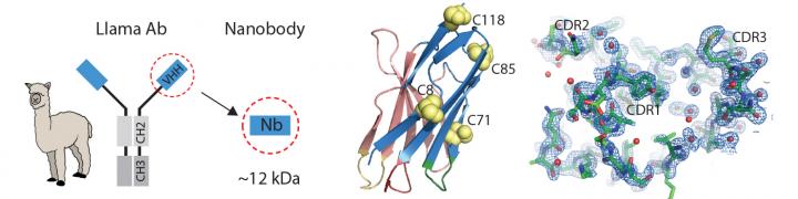
In 1989, two undergraduate students at the Free University of Brussels were asked to test frozen blood serum from camels, and stumbled on a previously unknown kind of antibody. It was a miniaturized version of a human antibody, made up only of two heavy protein chains, rather than two light and two heavy chains. As they eventually reported, the antibodies’ presence was confirmed not only in camels, but also in llamas and alpacas.

Fast forward 30 years. In the journal PNAS [Proceedings of the National Academy of Science] this week [April 8 – 12, 2019], researchers at Boston Children’s Hospital and MIT [Massachusetts Institute of Technology] show that these mini-antibodies, shrunk further to create so-called nanobodies, may help solve a problem in the cancer field: making CAR T-cell therapies work in solid tumors.

An April 11, 2019 Boston Children’s Hospital news release on EurekAlert, which originated the news item, explores the technology,

Highly promising for blood cancers, chimeric antigen receptor (CAR) T-cell therapy genetically engineers a patient’s own T cells to make them better at attacking cancer cells. The Dana-Farber/Boston Children’s Cancer and Blood Disorders Center is currently using CAR T-cell therapy for relapsed acute lymphocytic leukemia (ALL), for example.

But CAR T cells haven’t been good at eliminating solid tumors. It’s been hard to find cancer-specific proteins on solid tumors that could serve as safe targets. Solid tumors are also protected by an extracellular matrix, a supportive web of proteins that acts as a barrier, as well as immunosuppressive molecules that weaken the T-cell attack.

Rethinking CAR T cells

That’s where nanobodies come in. For two decades, they largely remained in the hands of the Belgian team. But that changed after the patent expired in 2013. [emphases mine]

“A lot of people got into the game and began to appreciate nanobodies’ unique properties,” says Hidde Ploegh, PhD, an immunologist in the Program in Cellular and Molecular Medicine at Boston Children’s and senior investigator on the PNAS study.

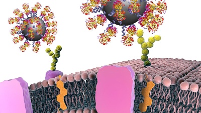
One useful attribute is their enhanced targeting abilities. Ploegh and his team at Boston Children’s, in collaboration with Noo Jalikhani, PhD, and Richard Hynes, PhD at MIT’s Koch Institute for Integrative Cancer Research, have harnessed nanobodies to carry imaging agents, allowing precise visualization of metastatic cancers.

The Hynes team targeted the nanobodies to the tumors’ extracellular matrix, or ECM — aiming imaging agents not at the cancer cells themselves, but at the environment that surrounds them. Such markers are common to many tumors, but don’t typically appear on normal cells.

“Our lab and the Hynes lab are among the few actively pursuing this approach of targeting the tumor micro-environment,” says Ploegh. “Most labs are looking for tumor-specific antigens.”

Targeting tumor protectors

Ploegh’s lab took this idea to CAR T-cell therapy. His team, including members of the Hynes lab, took aim at the very factors that make solid tumors difficult to treat.

The CAR T cells they created were studded with nanobodies that recognize specific proteins in the tumor environment, bearing signals directing them to kill any cell they bound to. One protein, EIIIB, a variant of fibronectin, is found only on newly formed blood vessels that supply tumors with nutrients. Another, PD-L1, is an immunosuppressive protein that most cancers use to silence approaching T cells.

Biochemist Jessica Ingram, PhD of the Dana-Farber Cancer Institute, Ploegh’s partner and a coauthor on the paper, led the manufacturing pipeline. She would drive to Amherst, Mass., to gather T cells from two alpacas, Bryson and Sanchez, inject them with the antigen of interest and harvest their blood for further processing back in Boston to generate mini-antibodies.

Taking down melanoma and colon cancer

Tested in two separate melanoma mouse models, as well as a colon adenocarcinoma model in mice, the nanobody-based CAR T cells killed tumor cells, significantly slowed tumor growth and improved the animals’ survival, with no readily apparent side effects.

Ploegh thinks that the engineered T cells work through a combination of factors. They caused damage to tumor tissue, which tends to stimulate inflammatory immune responses. Targeting EIIIB may damage blood vessels in a way that decreases blood supply to tumors, while making them more permeable to cancer drugs.

“If you destroy the local blood supply and cause vascular leakage, you could perhaps improve the delivery of other things that might have a harder time getting in,” says Ploegh. “I think we should look at this as part of a combination therapy.”

Future directions

Ploegh thinks his team’s approach could be useful in many solid tumors. He’s particularly interested in testing nanobody-based CAR T cells in models of pancreatic cancer and cholangiocarcinoma, a bile duct cancer from which Ingram passed away in 2018.

The technology itself can be pushed even further, says Ploegh.

“Nanobodies could potentially carry a cytokine to boost the immune response to the tumor, toxic molecules that kill tumor and radioisotopes to irradiate the tumor at close range,” he says. “CAR T cells are the battering ram that would come in to open the door; the other elements would finish the job. In theory, you could equip a single T cell with multiple chimeric antigen receptors and achieve even more precision. That’s something we would like to pursue.”

So, the Belgian researchers have a patent for two decades and, after it expires, more researchers could help to take the work further. Hmm …

Moving on, here’s a link to and a citation for the paper,

Nanobody-based CAR T cells that target the tumor microenvironment inhibit the growth of solid tumors in immunocompetent mice by Yushu Joy Xie, Michael Dougan, Noor Jailkhani, Jessica Ingram, Tao Fang, Laura Kummer, Noor Momin, Novalia Pishesha, Steffen Rickelt, Richard O. Hynes, and Hidde Ploegh. PNAS DOI: https://doi.org/10.1073/pnas.1817147116
First published April 1, 2019

This paper is behind a paywall

Llama-derived nanobodies are good for solving crystal structure

This work comes from Denmark, not a locale I associate with llamas (from an Oct. 2, 2017 news item on Nanowerk; Note: A link has been removed),

Aarhus University [Denmark] scientists have developed miniature antibodies (nanobodies) that can be labelled on certain amino acids (Acta Crystallographica Section D, “Introducing site-specific cysteines into nanobodies for mercury labelling allows de novo phasing of their crystal structures”).

This provides a direct route for solving new X-ray crystal structures of protein complexes important for gaining mechanistic understanding of cellular processes, which is important in the development of drugs.

An Oct. 2, 2017 Aarhus University press release on EurekAlert, which originated the news item, provides more detail,

Nanobodies are miniature antibodies derived from naturally circulating heavy-chain only antibodies in llamas. Over the past years, nanobodies and their applications have expanded enormously, both in basic research but also in drug development.

Nanobodies have proven to be well suited as protein stabilizers, which is particularly important during crystallization of a protein where millions of molecules have to arrange in a well-defined lattice. In this way, nanobodies can act as crystallization chaperones.

In an X-ray diffraction experiment, a critical piece of information – called the phases – is lost, which makes it difficult to determine new crystal structures. To overcome this phase problem in crystallography, heavy atoms are needed in the crystal. However, it is challenging to insert heavy atoms into a crystal. The scientists at Aarhus University used a nanobody as the vehicle for introducing mercury atoms. They developed a method to site-specifically label the nanobody with a heavy atom, and in this way, they could overcome the phase problem.

Since the scientists know which specific residues in the nanobodies can be modified and labelled, the technique used at Aarhus University opens for a range of other application. One exciting perspective is the insertion of fluorescent dyes into the nanobody to follow the location and distribution of target proteins in living organisms, which can give essential information on functional and regulatory processes.

Here’s a link to and a citation for the paper,

Introducing site-specific cysteines into nanobodies for mercury labelling allows de novo phasing of their crystal structures by S. B. Hansen, N. S. Laursen, G. R. Andersen and K. R. Andersen. Acta Cryst. (2017). D73 https://doi.org/10.1107/S2059798317013171

This paper is open access.

Here’s an image illustrating the work,

Caption: Nanobodies have proven to be well suited as protein stabilizers, which is particularly important during crystallization of a protein where millions of molecules have to arrange in a well-defined lattice. Credit: Kasper Røjkjær Andersen

‘Llam’ me lend you some antibodies—antibody particles extracted from camels and llamas

Sometimes the urge for wordplay overwhelms me as it did this morning (June 12, 2014) when I saw llamas mentioned in a news item. For anyone unfamiliar with how Canadian English (and I can safely include American English here but am not sure about any other Englishes) is spoken, we leave out consonants in some phrases. For example, ‘let me’ becomes ‘lemme’, which when you’re playing with ‘llama,’ becomes ‘llam’me. As for the verb ‘lend’, I used it for its alliterative quality and used more accurate verb ‘extracted’ later in the headline.

Getting on to the antibodies and the camels and llamas, here’s more from a June 12, 2014 news item on Nanowerk (Note: A link has been removed),

The use of nanoparticles in cancer research is considered as a promising approach in detecting and fighting tumour cells. The method has, however, often failed because the human immune system recognizes the particles as foreign objects and rejects them before they can fulfil their function. Researchers at the Helmholtz-Zentrum Dresden-Rossendorf (HZDR) and at University College Dublin [UCD[ in Ireland have, along with other partners, developed nanoparticles that not only bypass the body’s defence system, but also find their way to the diseased cells (“Diagnostic nanoparticle targeting of the EGF-receptor in complex biological conditions using single-domain antibodies”). This procedure uses fragments from a particular type of antibody that only occurs in camels and llamas. The small particles were even successful under conditions which are very similar to the situation within potential patients’ bodies.

A June 12, 2014 HZDR press release, which originated the news item, supplies a quote from one of the researchers where he explains the problems he and his colleagues were attempting to address,

Describing the current state of research, Dr. Kristof Zarschler of the Helmholtz Virtual Institute NanoTracking at the HZDR explains, “At the moment we must overcome three challenges. First, we need to produce the smallest possible nanoparticles. We then need to modify their surface in a way that the proteins in the human bodies do not envelop them, which would thus render them ineffective. In order to ensure, that the particles do their job, we must also somehow program them to find the diseased cells.” Therefore, the Dresden [HZDR is in Dresden] and Dublin researchers combined expertise to develop nanoparticles made of silicon dioxide with fragments of camel antibodies.

The press release and Zarschler go on to explain the advantages of camel and llama antibodies,

In contrast to conventional antibodies, which consist of two light and two heavy protein chains, those taken from camels and llamas are less complex and are made up of only two heavy chains. “Due to this simplified structure, they are easier to produce than normal antibodies,” explains Zarschler. “We also only need one particular fragment – the portion of the molecule that binds to certain cancer cells – which makes the production of much smaller nanoparticles possible.” By modifying the surface of the nanoparticle, it also gets more difficult for the immune system to recognize the foreign material, which allows the nanoparticles to actually reach their target.

The ultra-small particles should then detect the so-called epidermal growth factor receptor (EGFR) in the human body. In various types of tumours, this molecule is overexpressed and/or exists in a mutated form, which allows the cells to grow and multiply uncontrollably. The Dresden researchers could demonstrate in experiments that nanoparticles that have been combined with the camel antibody fragments can more firmly bind to the cancer cells. “The EGFR is a virtual lock to which our antibody fits like a key,” explains Zarschler.

Most exciting are the experiments the researchers performed with human blood (from the press release),

They even obtained the same results in experiments involving human blood serum – a biologically relevant environment the scientists point out: “This means that we carried out the tests under conditions that are very similar to the reality of the human body,” explains Dr. Holger Stephan, who leads the project. “The problem with many current studies is that artificial conditions are chosen where no disruptive factors exist. While this provides good results, it is ultimately useless because the nanoparticles fail finally in experiments conducted under more complex conditions. In our case, we could at least reduce this error source.”

There are no immediate plans for clinical trials according to the press release,

However, more time is required before the nanoparticles can be utilized in diagnosing human tumours. “The successful tests have brought us one step further,” explains Stephan. “The road, however, to its clinical use is long.” The next aim is to reduce the size of the nanoparticles, which are now approximately fifty nanometres in diameter, to less than ten nanometres. “That would be optimal,” according to Zarschler. “Then they would only remain in the human body for a short period – just long enough to detect the tumour.”

Here’s a link to and a citation for the paper,

Diagnostic nanoparticle targeting of the EGF-receptor in complex biological conditions using single-domain antibodies by K. Zarschler, K. Prapainop, E. Mahon, L. Rocks,  M. Bramini, P. M. Kelly, H. Stephan, and K. A. Dawson. Nanoscale, 2014,6, 6046-6056 DOI: 10.1039/C4NR00595C
First published online 16 Apr 2014

This paper is in an open access journal.

The researchers have provided an illustration of the new antibody particles,

 Title Bild Nanopartikel Copyright 	CBNI, UCD


Title Bild Nanopartikel
With help of proteins, nanoparticles can be produced, which bind specifically to cancer cells, thus making it possible to detect tumours. Copyright CBNI, UCD