Tag Archives: Mike Williams

Harvest water from desert air with carbon nanotube cups (competition for NBD Nano?)

It’s been a while since I’ve seen Pulickel Ajayan’s name in a Rice University (Texas) news release and I wonder if this is the beginning of a series. I’ve noticed that researchers often publish a series of papers within a few months and then become quiet for two or more years as they work in their labs to gather more information.

This time the research from Pulickel’s lab has focused on the use of carbon nanotubes to harvest water from desert air. From a June 12, 2014 news item on Azonano,

If you don’t want to die of thirst in the desert, be like the beetle. Or have a nanotube cup handy.

New research by scientists at Rice University demonstrated that forests of carbon nanotubes can be made to harvest water molecules from arid desert air and store them for future use.

The invention they call a “hygroscopic scaffold” is detailed in a new paper in the American Chemical Society journal Applied Materials and Interfaces.

Researchers in the lab of Rice materials scientist Pulickel Ajayan found a way to mimic the Stenocara beetle, which survives in the desert by stretching its wings to capture and drink water molecules from the early morning fog.

Here’s more about the research from a June 11, 2014 Rice University news release (by Mike Williams?), which originated the news item,

They modified carbon nanotube forests grown through a process created at Rice, giving the nanotubes a superhydrophobic (water-repelling) bottom and a hydrophilic (water loving) top. The forest attracts water molecules from the air and, because the sides are naturally hydrophobic, traps them inside.

“It doesn’t require any external energy, and it keeps water inside the forest,” said graduate student and first author Sehmus Ozden. “You can squeeze the forest to take the water out and use the material again.”

The forests grown via water-assisted chemical vapor deposition consist of nanotubes that measure only a few nanometers (billionths of a meter) across and about a centimeter long.

The Rice team led by Ozden deposited a superhydrophobic layer to the top of the forest and then removed the forest from its silicon base, flipped it and added a layer of hydrophilic polymer to the other side.

In tests, water molecules bonded to the hydrophilic top and penetrated the forest through capillary action and gravity. (Air inside the forest is compressed rather then expelled, the researchers assumed.) Once a little water bonds to the forest canopy, the effect multiplies as the molecules are drawn inside, spreading out over the nanotubes through van der Waals forces, hydrogen bonding and dipole interactions. The molecules then draw more water in.

The researchers tested several variants of their cup. With only the top hydrophilic layer, the forests fell apart when exposed to humid air because the untreated bottom lacked the polymer links that held the top together. With a hydrophilic top and bottom, the forest held together but water ran right through.

But with a hydrophobic bottom and hydrophilic top, the forest remained intact even after collecting 80 percent of its weight in water.

The amount of water vapor captured depends on the air’s humidity. An 8 milligram sample (with a 0.25-square-centimeter surface) pulled in 27.4 percent of its weight over 11 hours in dry air, and 80 percent over 13 hours in humid air. Further tests showed the forests significantly slowed evaporation of the trapped water.

If it becomes possible to grow nanotube forests on a large scale, the invention could become an efficient, effective water-collection device because it does not require an external energy source, the researchers said.

Ozden said the production of carbon nanotube arrays at a scale necessary to put the invention to practical use remains a bottleneck. “If it becomes possible to make large-scale nanotube forests, it will be a very easy material to make,” he said.

This is not the first time researchers have used the Stenocara beetle (also known as the Namib desert beetle) as inspiration for a water-harvesting material. In a Nov. 26, 2012 posting I traced the inspiration  back to 2001 while featuring the announcement of a new startup company,

… US startup company, NBD Nano, which aims to bring a self-filling water bottle based on Namib desert beetle to market,

NBD Nano, which consists of four recent university graduates and was formed in May [2012], looked at the Namib Desert beetle that lives in a region that gets about half an inch of rainfall per year.

Using a similar approach, the firm wants to cover the surface of a bottle with hydrophilic (water-attracting) and hydrophobic (water-repellent) materials.

The work is still in its early stages, but it is the latest example of researchers looking at nature to find inspiration for sustainable technology.

“It was important to apply [biomimicry] to our design and we have developed a proof of concept and [are] currently creating our first fully-functional prototype,” Miguel Galvez, a co-founder, told the BBC.

“We think our initial prototype will collect anywhere from half a litre of water to three litres per hour, depending on local environments.”

You can find out more about NBD Nano here although they don’t give many details about the material they’ve developed. Given that MIT (Massachusetts Institute of Technology) researchers published a  paper about a polymer-based material laced with silicon nanoparticles inspired by the Namib beetle in 2006 and that NBD Nano is based Massachusetts, I believe NBD Nano is attempting to commercialize the material or some variant developed at MIT.

Getting back to Rice University and carbon nanotubes, this is a different material attempting to achieve the same goal, harvesting water from desert air. Here’s a link to and a citation for the latest paper inspired by the Stenocara beetle (Namib beetle),

Anisotropically Functionalized Carbon Nanotube Array Based Hygroscopic Scaffolds by Sehmus Ozden, Liehui Ge , Tharangattu N. Narayanan , Amelia H. C. Hart , Hyunseung Yang , Srividya Sridhar , Robert Vajtai , and Pulickel M Ajayan. ACS Appl. Mater. Interfaces, DOI: 10.1021/am5022717 Publication Date (Web): June 4, 2014

Copyright © 2014 American Chemical Society

This paper is behind a paywall.

One final note, the research at MIT was funded by DARPA (US Defense Advanced Research Projects Agency). According to the news release the Rice University research held interest for similar agencies,

The U.S. Department of Defense and the U.S. Air Force Office of Scientific Research Multidisciplinary University Research Initiative supported the research.

Hitchhikers at the nanoscale show how cells stir themselves

A May 30, 2014 news item on Nanowerk highlights some molecule-tracking research,

Chemical engineers from Rice University and biophysicists from Georg-August Universität Göttingen in Germany and the VU University Amsterdam in the Netherlands have successfully tracked single molecules inside living cells with carbon nanotubes.

Through this new method, the researchers found that cells stir their interiors using the same motor proteins that serve in muscle contraction.

A May 29, 2014 Rice University news release by Mike Williams, which originated the news item, describes the researchers’ work,

The team attached carbon nanotubes to transport molecules known as kinesin motors to visualize and track them as they moved through the cytoplasm of living cells.

Carbon nanotubes are hollow cylinders of pure carbon with one-atom-thick walls. They naturally fluoresce with near-infrared wavelengths when exposed to visible light, a property discovered at Rice by Professor Rick Smalley a decade ago and then leveraged by Rice Professor Bruce Weisman to image carbon nanotubes. When attached to a molecule, the hitchhiking nanotubes serve as tiny beacons that can be precisely tracked over long periods of time to investigate small, random motions inside cells.

“Any probe that can hitch the length and breadth of the cell, rough it, slum it, struggle against terrible odds, win through and still know where its protein is, is clearly a probe to be reckoned with,” said lead author Nikta Fakhri, paraphrasing “The Hitchhiker’s Guide to the Galaxy.” Fakhri, who earned her Rice doctorate in Pasquali’s lab in 2011, is currently a Human Frontier Science Program Fellow at Göttingen.

“In fact, the exceptional stability of these probes made it possible to observe intracellular motions from times as short as milliseconds to as long as hours,” she said.

For long-distance transport, such as along the long axons of nerve cells, cells usually employ motor proteins tied to lipid vesicles, the cell’s “cargo containers.” This process involves considerable logistics: Cargo needs to be packed, attached to the motors and sent off in the right direction.

“This research has helped uncover an additional, much simpler mechanism for transport within the cell interior,” said principal investigator Christoph Schmidt, a professor of physics at Göttingen. “Cells vigorously stir themselves, much in the way a chemist would accelerate a reaction by shaking a test tube. This will help them to move objects around in the highly crowded cellular environment.”

The researchers showed the same type of motor protein used for muscle contraction is responsible for stirring. They reached this conclusion after exposing the cells to drugs that suppressed these specific motor proteins. The tests showed that the stirring was suppressed as well.

The mechanical cytoskeleton of cells consists of networks of protein filaments, like actin. Within the cell, the motor protein myosin forms bundles that actively contract the actin network for short periods. The researchers found random pinching of the elastic actin network by many myosin bundles resulted in the global internal stirring of the cell. Both actin and myosin play a similar role in muscle contraction.

The highly accurate measurements of internal fluctuations in the cells were explained in a theoretical model developed by VU co-author Fred MacKintosh, who used the elastic properties of the cytoskeleton and the force-generation characteristics of the motors.

“The new discovery not only promotes our understanding of cell dynamics, but also points to interesting possibilities in designing ‘active’ technical materials,” said Fakhri, who will soon join the Massachusetts Institute of Technology faculty as an assistant professor of physics. “Imagine a microscopic biomedical device that mixes tiny samples of blood with reagents to detect disease or smart filters that separate squishy from rigid materials.”

There is an accompanying video,

This video is typical of the kind of visual image that nanoscientists look at and provides an interesting contrast to ‘nano art’ where colours and other enhancements are added. as per this example, NanoOrchard, from a May 13, 2014 news item on Nanowerk about the 2014 Materials Research Society spring meeting and their Science as Art competition,

NanoOrchard – Electrochemically overgrown CuNi nanopillars. (Image courtesy of the Materials Research Society Science as Art Competition and Josep Nogues, Institut Catala de Nanociencia i Nanotecnologia (ICN2), Spain, and A. Varea, E. Pellicer, S. Suriñach, M.D. Baro, J. Sort, Univ. Autonoma de Barcelona) [downloaded from http://www.nanowerk.com/nanotechnology-news/newsid=35631.php]

NanoOrchard – Electrochemically overgrown CuNi nanopillars. (Image courtesy of the Materials Research Society Science as Art Competition and Josep Nogues, Institut Catala de Nanociencia i Nanotecnologia (ICN2), Spain, and A. Varea, E. Pellicer, S. Suriñach, M.D. Baro, J. Sort, Univ. Autonoma de Barcelona) [downloaded from http://www.nanowerk.com/nanotechnology-news/newsid=35631.php]

Getting back to the carbon nanotube hitchhikers, here’s a link to and a citation for the paper,

High-resolution mapping of intracellular fluctuations using carbon nanotubes by Nikta Fakhri, Alok D. Wessel, Charlotte Willms, Matteo Pasquali, Dieter R. Klopfenstein, Frederick C. MacKintosh, and Christoph F. Schmidt. Science 30 May 2014: Vol. 344 no. 6187 pp. 1031-1035 DOI: 10.1126/science.1250170

This article is behind a paywall.

One final comment, I am delighted by the researcher’s reference to the Hitchhiker’s Guide to the Galaxy.

Cow blood declumps (stabilizes) gold nanoparticles in a solution

Rice University (Texas, US) researchers have discovered a means of stabilizing gold nanoparticles in a variety of solutions including one of the harshest, salt solutions. From the May 14, 2013 news item on Nanowerk (Note: A link has been removed),

A protein from cow blood has the remarkable ability to keep gold nanoparticles from clumping in a solution. The discovery could lead to improved biomedical applications and contribute to projects that use nanoparticles in harsh environments.

Bovine serum albumin (BSA) forms a protein “corona” around gold nanoparticles that keeps them from aggregating, particularly in high-salt environments like seawater. The new research by the Rice University labs of chemists Stephan Link and Christy Landes was published by the American Chemical Society journal ACS Sustainable Chemistry and Engineering (“Adsorption of a Protein Monolayer via Hydrophobic Interactions Prevents Nanoparticle Aggregation under Harsh Environmental Conditions”).

The May 13, 2012 Rice University news release by Mike Williams, which originated the news item, describes the researchers and the nature of their work,

Link’s primary interest is in the plasmonic properties of nanoparticles. Landes’ work incorporates protein binding and molecular transport. The BSA research combines their unique talents with those of Sergio Dominguez-Medina, a graduate student in Link’s lab who studied to be a physicist at Monterrey Tech and was drawn to this interdisciplinary project during an undergraduate fellowship at Link’s Rice lab.

“Initially, we wanted to look at nanoparticles in solution with something they would encounter frequently in blood: serum albumin,” Landes said. “In our first experiments, Sergio reported the very efficient, reasonably fast and irreversible binding the moment he put nanoparticles into a solution that contained serum albumin.”

“It turned out the salt is actually driving this binding,” Dominguez-Medina said.

Without BSA, gold nanoparticles in a salty solution quickly aggregate and fall to the bottom. “That by itself is undesirable for biomedical or industrial applications, because it could lead to toxicity issues,” he said. “The nanoparticles get more hydrophobic because in the presence of salts, the excess charges on the surface (which discourage clumping) are actually removed.” But if BSA is present, the proteins are drawn to the nanoparticles faster than the particles are drawn to each other.

“Once the protein is bound, it gives a super protection against any type of salt-induced aggregation. We think this could be used for the stabilization of nanoparticles in environments where, right now, it hasn’t been achieved,” Dominguez-Medina said.

He said the discovery also offers the possibility that nanoparticles might be made more compatible for treating humans by using a patient’s own albumin. “Albumin is really easy to purify and the process is well-established,” he said.

Here’s a little more about the plasmonics of the situation and how this discovery about cow blood protein might apply in biomedical and other applications (from the news release),

The ability of gold nanoparticles to absorb and redirect light is at the heart of several breakthrough technologies being developed at Rice and elsewhere. Most notable are a nanoparticle-based cancer treatment now in human testing that was developed by Professor Naomi Halas and former Rice colleague Jennifer West, and Halas’ project to convert solar energy directly into steam for sanitation and water purification.

“The only way nanoparticles exhibit their really nice optical properties in very specific optical frequencies is if they’re separated,” Landes said.

The key words in Landes comment is ‘separated’ (from the news release),

Because pure gold nanoparticles are so hydrophobic, they naturally clump together in a solution unless chemically treated. “A lot of industrial effort goes into keeping stuff off of surfaces, like contact lenses and ship hulls,” she said. “That involves chemically altering the surfaces to prevent unwanted adsorption, or in the case of nanoparticles, unwanted aggregation.”

Protecting the surface is costly, Link said. “But we found we could take nanoparticles prepared in the cheapest way, with a sodium citrate coating that stabilizes the particles by electrostatic repulsion, and add BSA, which coats the particles and makes them really stable.”

Adding the BSA seems logical when one of the scientists explains the reasoning (from the news release),

Albumin is the most common protein in blood, and the bovine version shares 98 percent of its amino acid sequence with human serum albumin. “One of its main purposes, biologically, is to take things that aren’t water-soluble, bind to them and make them soluble,” Landes said. “When you combine it with gold nanoparticles, BSA trades places with the cheap citrate, which isn’t a good protective layer, to form the monolayer corona, which is very strong and protective.”

Aside from obvious biomedical applications (e.g. implants and joint replacements), there are desalination and fuel cell applications (from the news release),

Seawater is the very definition of a harsh environment, Landes said. “One of the problems with desalination applications and, similarly, with fuel cells, is that saline or acidic conditions are very corrosive,” she said. “That’s why you have to use platinum electrodes in fuel cells – not because they’re better than cheaper materials at catalysis, but because they don’t corrode in a harsh environment.” She sees promise for BSA-treated gold nanoparticles in both applications.

The researchers have other plans as well (from the news release),

The researchers are now looking at how well gold nanoparticles retain their albumin corona with repeated use. “Gold is expensive,” Landes said. “But the beauty of it is that if you can reuse it, it only costs you once.”

They also want to use spectroscopy to see how the binding mechanism works in real time, Link said. “We want to study what’s happening at the interface of nanoparticles and biologically relevant media” that may eventually include DNA, RNA and drugs for delivery to cells, he said.

Link plans to see how BSA can be used in combination with gold nanorods. Because nanorods’ plasmonic properties can be tuned, “we can get them into the biological window, which is near-infrared light,” he said. Near-IR from lasers is used to activate, by heating, Halas’ and West’s cancer-killing nanoshells. Nanorods may also offer ways to combine BSA and other useful proteins by coating the tips and sides separately.

For interested parties, here’s a link to and a citation for the published paper,

Adsorption of a Protein Monolayer via Hydrophobic Interactions Prevents Nanoparticle Aggregation under Harsh Environmental Conditions by Sergio Dominguez-Medina, Jan Blankenburg, Jana Olson, Christy F. Landes, and Stephan Link. ACS Sustainable Chem. Eng., Article ASAP DOI: 10.1021/sc400042h
Publication Date (Web): April 3, 2013
Copyright © 2013 American Chemical Society

Unusually for the American Chemical Society (ACS), this paper appears to be open access; I was able to access the full HTML version today, May 14, 2013 at 10:10 am PDT.

Rice U and the US National Institute of Standards and Technology settle into armchairs, the carbon nanotube kind

An armchair carbon nanotube is amongst the most desirable of carbon nanotubes. You’ll have to look carefully to see the resemblance to an armchair,

Armchair carbon nanotubes, so named for the arrangement of atoms that make their ends look like armchairs, are the most desirable among nanotube researchers for their superior electrical properties. Image by Erik Hároz [downloaded from http://news.rice.edu/2013/02/05/essential-armchair-reading-for-nanotube-researchers-2/]

Armchair carbon nanotubes, so named for the arrangement of atoms that make their ends look like armchairs, are the most desirable among nanotube researchers for their superior electrical properties. Image by Erik Hároz [downloaded from http://news.rice.edu/2013/02/05/essential-armchair-reading-for-nanotube-researchers-2/]

The Feb. 6, 2013 news item on phys.org about the armchair carbon nanotubes notes that this latest research is an early outcome from a recently announced (Oct. 2012) partnership between Rice University and the US National Institute of Standards and Technology (NIST). Trom the news item (Note: Links have been removed),

The first fruits of a cooperative venture between scientists at Rice University and the National Institute of Standards and Technology (NIST) have appeared in a paper that brings together a wealth of information for those who wish to use the unique properties of metallic carbon nanotubes.

The feature article published recently in the Royal Society of Chemistry journal Nanoscale gathers research about the separation and fundamental characteristics of armchair carbon nanotubes, which have been of particular interest to researchers trying to tune their electronic and optical properties.

The Rice University Feb. 5, 2013 news release by Mike Williams, which originated the news item, describe s the process the scientists undertook,

This paper, said Rice physicist Junichiro Kono, provides scientists a valuable resource for detailed information about metallic carbon nanotubes, especially armchair nanotubes. “Basically, we summarized all our recent findings as well as all information we could find in the literature about metallic nanotubes, along with detailed accounts of preparation methods for metal-enriched nanotube samples, to show the community just how much we now understand about these one-dimensional metals,” he said.

As part of the lengthy work, the team compiled and published tables of essential statistics, including optical properties, for a variety of metallic nanotubes. “We provide fundamental theoretical backgrounds and then show very detailed experimental results on unique properties of metallic nanotubes,” Kono said. “This paper summarizes what kind of aspects are understood, and what is not, about fundamental optical processes in nanotubes and will make it easier for researchers to identify their spectroscopic features and transition energies.”

For this of us who are less well versed on armchair carbon nanotubes and their electronic and optical properties, the news releases provides some information (Note: Links have been removed),

Nanotubes come in many flavors, depending on their chirality. Chirality is a characteristic akin to the angles at which a flat sheet of paper might align when wrapped into a tube. Cut the tube in half and the atoms at the open edge would line up in the shape of an armchair, a zigzag or some variant. Even though their raw material is identical – chicken-wire-like hexagons of carbon – the chirality makes all the difference in how nanotubes transmit electricity.

Armchairs are the most coveted because they have no band gap; electrons flow through without resistance. Cables made with armchair nanotubes have the potential to move electricity over great distances with virtually no loss. That makes them the gold standard as the basic element of armchair quantum wire. The ongoing development of this very strong, lightweight, high-capacity cable could improve further the record properties of multifunctional carbon nanotube fibers that are being developed by the group of Rice Professor Matteo Pasquali.

For the project-specific work the scientists performed (Note: Links have been removed),

The new work led by Kono and Robert Hauge, a distinguished faculty fellow in chemistry at Rice, along with scientists at NIST and Los Alamos National Laboratory, looks beyond the armchair’s established electrical properties to further detail their potential for electronic, sensing, optical and photonic devices.

“Of course, to get there, we need really good samples,” Kono said. “Many applications will rely on our ability to separate carbon nanotubes and then assemble macroscopically ordered structures consisting of single-chirality nanotubes. Nobody can do that at this point.”

When a batch of nanotubes comes out of a furnace, it’s a jumble of types. That makes detailed analysis of their characteristics — let alone their practical use — a challenge.

But techniques developed in recent years at Rice and by NIST scientist Ming Zheng to purify metallic nanotubes are beginning to change that. Rice graduate student Erik Hároz said recent experiments established “unambiguous evidence” that a process he and Kono are using called density gradient ultracentrifugation can enrich ensemble samples of armchairs. Taking things further, Zheng’s method of DNA-based ion-exchange chromatography provides very small samples of ultrapure armchair nanotubes of a single chirality.

You can read more about the work at phys.org or at Rice University using the links already provided. For those who’d like to read the research,

Fundamental optical processes in armchair carbon nanotubes by Erik H. Hároz ,  Juan G. Duque,  Xiaomin Tu ,  Ming Zheng ,  Angela R. Hight Walker ,  Robert H. Hauge ,  Stephen K. Doorn and Junichiro Kono. Nanoscale, 2013,5, 1411-1439 DOI: 10.1039/C2NR32769D First published on the web 04 Jan 2013

This article is behind a paywall of sorts.  RSC (Royal Society of Chemistry) Publishing (which publishes Nanoscale) has an open access policy but there are various options, from the RSC Publishing’s Open Access Policy webpage,

RSC Open Access statement

Open Access is the term given to making electronic versions of articles accessible to readers, without any subscription or ‘access side’ fees.

RSC supports Open Access models which seek to ensure that scholarly publishing activities operate in a long term sustainable way.

  • Our fundamental goal is to advance the chemical sciences, through the effective dissemination of high quality research content
  • We seek to maximise the dissemination of the research that we publish
  • We support any and all sustainable and fair models of access. We believe that the integrity and archiving of scholarly content must be maintained throughout
  • We support ‘Gold’* Open Access and encourage funding to be made available to support authors during any transition from reader to author side payments
  • We support the author’s ability to choose where they publish their work to the benefit of the advancement of science. We do not wish authors to be discriminated against if they are unable to pay author-side fees
  • We seek to work closely with other parties, including funders and government agencies, to achieve the above goals

RSC Publishing provides authors with the option to make their article Open Access, through payment of a fee on acceptance. Authors following the traditional route still have deposition options – details are on the ‘Deposition and Licence to Publish’ page of the website.

*There are several types of Open Access:

  • Gold Open Access: Publication costs are covered by an ‘Article Processing Fees’ being paid by authors upon acceptance. The final ‘article of record’ is made available to all, immediately, without any barriers to access
  • Green Open Access: A version of the paper (often the author’s manuscript) is made available via a subject or institutional repository. An embargo period is often involved, typically 6-24 months. No payment is made, and publishers should strive to recoup their investment through traditional sales during the embargo period
  • Delayed Open Access: The final version of the paper is made available by the publisher after an embargo period (e.g. publisher deposit the paper in PubMed after 12 months)

It would seem the option for this article is ‘Delayed Open Access’.

James’ bond (Rice University research team creates graphene/nanotube hybrid)

I have to give credit to Mike Williams’ Nov. 27, 2012 Rice University news release for the “James’ bond” phrase used to describe this graphene/nanotube hybrid,

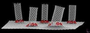
A seamless graphene/nanotube hybrid created at Rice University may be the best electrode interface material possible for many energy storage and electronics applications.

Led by Rice chemist James Tour, researchers have successfully grown forests of carbon nanotubes that rise quickly from sheets of graphene to astounding lengths of up to 120 microns, according to a paper published today by Nature Communications. A house on an average plot with the same aspect ratio would rise into space.

Seven-atom rings (in red) at the transition from graphene to nanotube make this new hybrid material a seamless conductor. The hybrid may be the best electrode interface material possible for many energy storage and electronics applications. Image courtesy of the Tour Group

The Rice hybrid combines two-dimensional graphene, which is a sheet of carbon one atom thick, and nanotubes into a seamless three-dimensional structure. The bonds between them are covalent, which means adjacent carbon atoms share electrons in a highly stable configuration. The nanotubes aren’t merely sitting on the graphene sheet; they become a part of it.

“Many people have tried to attach nanotubes to a metal electrode and it’s never gone very well because they get a little electronic barrier right at the interface,” Tour said. “By growing graphene on metal (in this case copper) and then growing nanotubes from the graphene, the electrical contact between the nanotubes and the metal electrode is ohmic. That means electrons see no difference, because it’s all one seamless material.

In the new work, the team grew a specialized odako that retained the iron catalyst and aluminum oxide buffer but put them on top of a layer of graphene grown separately on a copper substrate. The copper stayed to serve as an excellent current collector for the three-dimensional hybrids that were grown within minutes to controllable lengths of up to 120 microns.

Electron microscope images showed the one-, two- and three-walled nanotubes firmly embedded in the graphene, and electrical testing showed no resistance to the flow of current at the junction.

“The performance we see in this study is as good as the best carbon-based supercapacitors that have ever been made,” Tour said. “We’re not really a supercapacitor lab, and still we were able to match the performance because of the quality of the electrode. It’s really remarkable, and it all harkens back to that unique interface.”

Here’s the citation and a link for the article,

A seamless three-dimensional carbon nanotube graphene hybrid material by Yu Zhu, Lei Li, Chenguang Zhang, Gilberto Casillas,  Zhengzong Sun, Zheng Yan, Gedeng Ruan, Zhiwei Peng, Abdul-Rahman O. Raji, Carter Kittrell, Robert H. Hauge & James M. Tour in Nature Communications 3, Article number:1225 doi:10.1038/ncomms2234 Published 27 November 2012

This article is behind a paywall.

DAVinCI lets Rice University researchers visualize their data big time

The July 13, 2012 news item on physorg.com presents an extraordinary picture (Note: I have removed a link),

The 200-inch wall (measured diagonally) lets users display and analyze images of all types, from atoms to galaxies. This studio is expected to help researchers in Earth science, biomedicine, engineering, art, architecture and other fields gain extraordinarily clear pictures of their data sets, be they bacteria or bridges.

“I can take my 3-D seismic images,  project them here and walk around inside them,” said Alan Levander, Rice’s Carey Croneis Professor of Earth Science and principal investigator of the Data Analysis and Visualization Cyberinfrastructure (DAVinCI) project. “With a tracking device in my hand, I can go through and choose the features that I want to look at.” The DAVinCI project adds to Rice’s extensive supercomputing resources, which also include Blue Gene/P, among the 500 most powerful supercomputers in the United States.

The news item originated in a July 12, 2012 Rice University news release by Mike Williams,

The futuristic wall of 50-inch high-resolution projection monitors supports two- and three-dimensional visualization needs at extremely high resolution and clarity, Odegard [Jan Odegard, executive director of Rice’s Ken Kennedy Institute for Information Technology] said. Backed by custom graphics engines, the wall allows data to be displayed in three dimensions using modern active stereo shutter glasses, often seen in home 3-D TV systems but far more sophisticated than glasses used at a 3-D movie theater.

The shutters are linked wirelessly to the graphic engines so that, in effect, only one eye is open at a time, and it matches the left or right images displayed on the screen. But this all happens very fast, at a frame rate of 120 times a second, so users see no flicker in their images.

Erik Engquist, manager of the lab who joined Rice last year, has been demonstrating the system with geological, molecular and other 3-D data that float in front of the screen and allow viewers to see details that might be invisible on flat images, no matter how big. The system has two other advantages over standard 3-D displays. The 32-megapixel screen can track researchers with an infrared system (also tied into the glasses) and allows them to walk around inside an image. Researchers can also interact with the data by turning them this way and that in midair to get a different perspective and interpret the data quantitatively.

“If you have a 10-dimensional data space — which is not uncommon — you can’t visualize it in 10 dimensions, but you can visualize any three at a time,” Levander said. “You can walk through complicated multidimensional space looking at what are called ‘hypercubes.’ You can interact with them and look for correlations in complex systems.”

Engquist, an applied mathematician, said the 16 projection monitors were chosen for their display brilliance and their narrow borders that leave only a thin strip of black between individual screens. “It’s far less intrusive than if we had used regular TV monitors, which have a large bezel,” he said. “If the images have a black background, you barely see the lines; in fact, after a while you don’t really notice them, since your focus will be on the data.”

Here’s a video about DAVinCI produced by Rice University,

The official opening for this project is Sept. 5, 2012 but researchers are already working with this new equipment (or playing with a fabulous new toy which brings to mind the Star Trek holodeck). Rice University has made an online technical manual, Getting Started on DAVinCI available. You need to be familiar with the Linux operating system and comfortable with writing short scripts (i.e., have rudimentary programming skills).


The relationship of silver ions & nanoparticles, Nietzsche, and Rice University

My hat’s off to Mike Williams for introducing Nietzsche into a news item about silver nanoparticles and toxicity. Here’s the line from his July 11, 2012 Rice University news release (Note: I have removed some links),

Their work comes with a Nietzsche-esque warning: Use enough. If you don’t kill them, you make them stronger.

Scientists have long known that silver ions, which flow from nanoparticles when oxidized, are deadly to bacteria. Silver nanoparticles are used just about everywhere, including in cosmetics, socks, food containers, detergents, sprays and a wide range of other products to stop the spread of germs.

But scientists have also suspected silver nanoparticles themselves may be toxic to bacteria, particularly the smallest of them at about 3 nanometers. Not so, according to the Rice team that reported its results this month in the American Chemical Society journal Nano Letters.

This next bit describing the research is an example of what I find so compelling (curiosity and persistence) about science,

To figure that out, the researchers had to strip the particles of their powers. “Our original expectation was that the smaller a particle is, the greater the toxicity,” said Zongming Xiu, a Rice postdoctoral researcher and lead author of the paper. Xiu set out to test nanoparticles, both commercially available and custom-synthesized from 3 to 11 nanometers, to see whether there was a correlation between size and toxicity.

“We could not get consistent results,” he said. “It was very frustrating and really weird.”

Here’s what they did next, what they found, and the implications of their findings,

Xiu decided to test nanoparticle toxicity in an anaerobic environment – that is, sealed inside a chamber with no exposure to oxygen — to control the silver ions’ release. He found that the filtered particles were a lot less toxic to microbes than silver ions.

Working with the lab of Rice chemist Vicki Colvin, the team then synthesized silver nanoparticles inside the anaerobic chamber to eliminate any chance of oxidation. “We found the particles, even up to a concentration of 195 parts per million, were still not toxic to bacteria,” Xiu said. “But for the ionic silver, a concentration of about 15 parts per billion would kill all the bacteria present. That told us the particle is 7,665 times less toxic than the silver ions, indicating a negligible toxicity.”

“The point of that experiment,” Alvarez [Pedro Alvarez, George R. Brown Professor and chair of Rice’s Civil and Environmental Engineering Department] said, “was to show that a lot of people were obtaining data that was confounded by a release of ions, which was occurring during exposure they perhaps weren’t aware of.”

Alvarez suggested the team’s anaerobic method may be used to test many other kinds of metallic nanoparticles for toxicity and could help fine-tune the antibacterial qualities of silver particles. In their tests, the Rice researchers also found evidence of homesis; [e.g..,] E. coli became stimulated by silver ions when they encountered doses too small to kill them.

“Ultimately, we want to control the rate of (ion) release to obtain the desired concentrations that just do the job,” Alvarez said. “You don’t want to overshoot and overload the environment with toxic ions while depleting silver, which is a noble metal, a valuable resource – and a somewhat expensive disinfectant. But you don’t want to undershoot, either.”

He said the finding should shift the debate over the size, shape and coating of silver nanoparticles. [emphasis mine] “Of course they matter,” Alvarez said, “but only indirectly, as far as these variables affect the dissolution rate of the ions. The key determinant of toxicity is the silver ions. So the focus should be on mass-transfer processes and controlled-release mechanisms.”

Interestingly, this is a joint US-UK effort (US Environmental Protection Agency and the U.K. Natural Environment Research Council). H/T to Will Soutter’s July 12, 2012 news item on Azonano for the information about this latest silver nanoparticle research from Rice University. The July 11, 2012 news item on Nanowerk also features information about the silver nanoparticles, ions, and Rice University.

I have mentioned Vicki Colvin’s work previously including this Jan. 28, 2011 posting about a UK/US joint environmental research effort. I have also mentioned Pedro Alvarez a few times including this Aug. 2, 2010 posting about nanomaterials and the construction industry.