Tag Archives: Jiuyun Shi

Breakthrough for tissue-interfaced bioelectronics

Let’s call this a cold open,

This October 24, 2024 news item on ScienceDaily describes some of what is in the video

The ideal material for interfacing electronics with living tissue is soft, stretchable, and just as water-loving as the tissue itself–in short, a hydrogel. Semiconductors, the key materials for bioelectronics such as pacemakers, biosensors, and drug delivery devices, on the other hand, are rigid, brittle, and water-hating, impossible to dissolve in the way hydrogels have traditionally been built. Scientists have now solved this challenge that has long stymied researchers, reimagining the process of creating hydrogels to build a powerful semiconductor in hydrogel form. The result is a bluish gel that flutters like a sea jelly in water but retains the immense semiconductive ability needed to transmit information between living tissue and machine.

An October 24, 2024 University of Chicago news release (also on EurekAlert) by Paul Dailing, which originated the news item, describes the breakthrough, Note: Links have been removed,

A paper published today in Science from the UChicago Pritzker School of Molecular Engineering (PME) has solved this challenge that has long stymied researchers, reimagining the process of creating hydrogels to build a powerful semiconductor in hydrogel form. Led by Asst. Prof. Sihong Wang’s research group, the result is a bluish gel that flutters like a sea jelly in water but retains the immense semiconductive ability needed to transmit information between living tissue and machine.

The material demonstrated tissue-level moduli as soft as 81 kPa, stretchability of 150% strain, and charge-carrier mobility up to 1.4 cm2 V-1 s-1. This means their material—both semiconductor and hydrogel at the same time—ticks all the boxes for an ideal bioelectronic interface.

“When making implantable bioelectronic devices, one challenge you must address is to make a device with tissue-like mechanical properties,” said Yahao Dai, the first author of the new paper. “That way, when it gets directly interfaced with the tissue, they can deform together and also form a very intimate bio-interface.”

Although the paper mainly focused on the challenges facing implanted medical devices such as biochemical sensors and pacemakers, Dai said the material also has many potential non-surgical applications, like better readings off the skin or improved care for wounds.

“It has very soft mechanical properties and a large degree of hydration similar to living tissue,” said UChicago PME Asst. Prof. Sihong Wang. “Hydrogel is also very porous, so it allows the efficient diffusion transport of different kinds of nutrition and chemicals. All these traits combine to make hydrogel probably the most useful material for tissue engineering and drug delivery.”

‘Let’s change our perspective’

The typical way of making a hydrogel is to take a material, dissolve it in water, and add the gelation chemicals to puff the new liquid into a gel form. Some materials simply dissolve in water, others require researchers to tinker and chemically modify the process, but the core mechanism is the same: No water, no hydrogel.

Semiconductors, however, don’t normally dissolve in water. Rather than find new, time-consuming means of trying to force the process, the UChicago PME team re-examined the question.

“We started to think, ‘Okay, let’s change our perspective,’ and we came up with a solvent exchange process,” Dai said.

Instead of dissolving the semiconductors in water, they dissolved them in an organic solvent that is miscible with water. They then prepared a gel from the dissolved semiconductors and hydrogel precursors. Their gel initially was an organogel, not a hydrogel.

“To eventually turn it into a hydrogel, we then immersed the whole material system into the water to let the organic solvent dissolve out and let the water come in,” Dai said.

An important benefit of such a solvent-exchange-based method is its broad applicability to different types of polymer semiconductors with different functions.

‘One plus one is greater than two’

The hydrogel semiconductor, which the team has patented and is commercializing through UChicago’s Polsky Center for Entrepreneurship and Innovation, is not merging a semiconductor with a hydrogel. It’s one material that is both semiconductor and hydrogel at the same time.

“It’s just one piece that has both semiconducting properties and hydrogel design, meaning that this whole piece is just like any other hydrogel,” Wang said.

Unlike any other hydrogel, however, the new material actually improved biological functions in two areas, creating better results than either hydrogel or semiconductor could accomplish on their own.

First, having a very soft material bond directly with tissue reduces the immune responses and inflammation typically triggered when a medical device is implanted.

Second, because hydrogels are so porous, the new material enables elevated biosensing response and stronger photo-modulation effects. With biomolecules being able to diffuse into the film to have volumetric interactions, the interaction sites for biomarkers-under-detection are significantly increased, which gives rise to higher sensitivity. Besides sensing, the responses to light for therapeutic functions at tissue surfaces also get increased from the more efficient transport of redox-active species. This benefits functions such as light-operated pacemakers or wound dressing that can be more efficiently heated with a flick of light to help speed healing.

“It’s a ‘one plus one is greater than two’ kind of combination,” Wang joked.

Researchers in the lab of UChicago Pritzker School of Engineering Asst. Prof. Sihong Wang (right), including PhD student Yahao Dai (left), have developed a hydrogel that retains the semiconductive ability needed to transmit information between living tissue and machine, which can be used both in implantable medical devices and non-surgical applications. (Photo by John Zich)

Here’s a link to and a citation for the paper,

Soft hydrogel semiconductors with augmented biointeractive functions by Yahao Dai, Shinya Wai, Pengju Li, Naisong Shan, Zhiqiang Cao, Yang Li, Yunfei Wang, Youdi Liu, Wei Liu, Kan Tang, Yuzi Liu, Muchuan Hua, Songsong Li, Nan Li, Shivani Chatterji, H. Christopher Fry, Sean Lee, Cheng Zhang, Max Weires, Sean Sutyak, Jiuyun Shi, Chenhui Zhu, Jie Xu, Xiaodan Gu, Bozhi Tian, and Sihong Wang. Science 24 Oct 2024 Vol 386, Issue 6720 pp. 431-439 DOI: 10.1126/science.adp9314

This paper is behind a paywall.

‘Nanotraps’ for catching and destroying coronavirus

‘Nanotraps’ are not vaccines although they do call the immune system into play. They represent a different way for dealing with COVID-19. (This work reminds of my June 24, 2020 posting Tiny sponges lure coronavirus away from lung cells where the researchers have a similar approach with what they call ‘nanosponges’.)

An April 27, 2021 news item on Nanowerk makes the announcement,

Researchers at the Pritzker School of Molecular Engineering (PME) at the University of Chicago have designed a completely novel potential treatment for COVID-19: nanoparticles that capture SARS-CoV-2 viruses within the body and then use the body’s own immune system to destroy it.

These “Nanotraps” attract the virus by mimicking the target cells the virus infects. When the virus binds to the Nanotraps, the traps then sequester the virus from other cells and target it for destruction by the immune system.

In theory, these Nanotraps could also be used on variants of the virus, leading to a potential new way to inhibit the virus going forward. Though the therapy remains in early stages of testing, the researchers envision it could be administered via a nasal spray as a treatment for COVID-19.

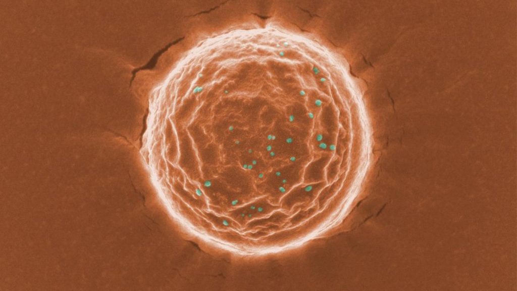
A scanning electron microscope image of a nanotrap (orange) binding a simulated SARS-CoV-2 virus (dots in green). Scientists at the University of Chicago created these nanoparticles as a potential treatment for COVID-19. Image courtesy Chen and Rosenberg et al.

An April 27, 2021 University of Chicago news release (also on EurekAlert) by Emily Ayshford, which originated the news item, describes the work in more detail,

“Since the pandemic began, our research team has been developing this new way to treat COVID-19,” said Asst. Prof. Jun Huang, whose lab led the research. “We have done rigorous testing to prove that these Nanotraps work, and we are excited about their potential.”

Designing the perfect trap

To design the Nanotrap, the research team – led by postdoctoral scholar Min Chen and graduate student Jill Rosenberg – looked into the mechanism SARS-CoV-2 uses to bind to cells: a spike-like protein on its surface that binds to a human cell’s ACE2 receptor protein.

To create a trap that would bind to the virus in the same way, they designed nanoparticles with a high density of ACE2 proteins on their surface. Similarly, they designed other nanoparticles with neutralizing antibodies on their surfaces. (These antibodies are created inside the body when someone is infected and are designed to latch onto the coronavirus in various ways).

Both ACE2 proteins and neutralizing antibodies have been used in treatments for COVID-19, but by attaching them to nanoparticles, the researchers created an even more robust system for trapping and eliminating the virus.

Made of FDA [US Food and Drug Administration]-approved polymers and phospholipids, the nanoparticles are about 500 nanometers in diameter – much smaller than a cell. That means the Nanotraps can reach more areas inside the body and more effectively trap the virus.

The researchers tested the safety of the system in a mouse model and found no toxicity. They then tested the Nanotraps against a pseudovirus – a less potent model of a virus that doesn’t replicate – in human lung cells in tissue culture plates and found that they completely blocked entry into the cells.

Once the pseudovirus bound itself to the nanoparticle – which in tests took about 10 minutes after injection – the nanoparticles used a molecule that calls the body’s macrophages to engulf and degrade the Nanotrap. Macrophages will generally eat nanoparticles within the body, but the Nanotrap molecule speeds up the process. The nanoparticles were cleared and degraded within 48 hours.

The researchers also tested the nanoparticles with a pseudovirus in an ex vivo lung perfusion system – a pair of donated lungs that is kept alive with a ventilator – and found that they completely blocked infection in the lungs.

They also collaborated with researchers at Argonne National Laboratory to test the Nanotraps with a live virus (rather than a pseudovirus) in an in vitro system. They found that their system inhibited the virus 10 times better than neutralizing antibodies or soluble ACE2 alone.

A potential future treatment for COVID-19 and beyond

Next the researchers hope to further test the system, including more tests with a live virus and on the many virus variants.

“That’s what is so powerful about this Nanotrap,” Rosenberg said. “It’s easily modulated. We can switch out different antibodies or proteins or target different immune cells, based on what we need with new variants.”

The Nanotraps can be stored in a standard freezer and could ultimately be given via an intranasal spray, which would place them directly in the respiratory system and make them most effective.

The researchers say it is also possible to serve as a vaccine by optimizing the Nanotrap formulation, creating an ultimate therapeutic system for the virus.

“This is the starting point,” Huang said. “We want to do something to help the world.”

The research involved collaborators across departments, including chemistry, biology, and medicine.

Here’s a link to and a citation for the paper,

Nanotraps for the containment and clearance of SARS-CoV-2 by Min Chen, Jillian Rosenberg, Xiaolei Cai, Andy Chao Hsuan Lee, Jiuyun Shi, Mindy Nguyen, Thirushan Wignakumar, Vikranth Mirle, Arianna Joy Edobor, John Fung, Jessica Scott Donington, Kumaran Shanmugarajah, Yiliang Lin, Eugene Chang, Glenn Randall, Pablo Penaloza-MacMaster, Bozhi Tian, Maria Lucia Madariaga, Jun Huang. Matter, April 19, 2021, DOI: https://doi.org/10.1016/j.matt.2021.04.005

This paper appears to be open access.