Tag Archives: synchrotron

Lighting the way to improvements for the bond between dental implants and bone

A July 3, 2018 Canadian Light Source news release by Colleen MacPherson describes an investigation into how dental implants and bones interact with the hope of making dental implantation safer and more certain,

Research carried out recently at the Canadian Light Source (CLS) [also known as a synchrotron] in Saskatoon [Saskatchewan, Canada] has revealed promising information about how to build a better dental implant, one that integrates more readily with bone to reduce the risk of failure.

“There are millions of dental and orthopedic implants placed every year in North America and a certain number of them always fail, even in healthy people with healthy bone,” said Kathryn Grandfield, assistant professor in the Department of Materials Science and Engineering at McMaster University in Hamilton [Ontario, Canada].

A dental implant restores function after a tooth is lost or removed. It is usually a screw shaped implant that is placed in the jaw bone and acts as the tooth roots, while an artificial tooth is placed on top. The implant portion is the artificial root that holds an artificial tooth in place.

Grandfield led a study that showed altering the surface of a titanium implant improved its connection to the surrounding bone. It is a finding that may well be applicable to other kinds of metal implants, including engineered knees and hips, and even plates used to secure bone fractures.

About three million people in North America receive dental implants annually. While the failure rate is only one to two percent, “one or two percent of three million is a lot,” she said. Orthopedic implants fail up to five per cent of the time within the first 10 years; the expected life of these devices is about 20 to 25 years, she added.

“What we’re trying to discover is why they fail, and why the implants that are successful work. Our goal is to understand the bone-implant interface in order to improve the design of implants.”

Grandfield’s research team, which included post-doctoral fellow Xiaoyue Wang and McMaster colleague Adam Hitchcock from the Department of Chemistry and Chemical Biology. The team members used the soft X-ray spectromicroscopy beamline at the CLS as well as facilities at the Canadian Centre for Electron Microscopy in Hamilton to examine a failed dental implant that had to be removed, along with a small amount of surrounding bone, from a patient. Prior to implantation, a laser beam was used to alter the implant, to roughen the surface, creating what looked like “little volcanoes” on the surface. After removal from the patient, the point of connection between bone and metal was then carefully studied to understand how the implant behaved.

“What we found was that the surface modification changed the chemistry of the implant. The modification created an oxide layer, but not a bad oxide layer like rust but a better, more beneficial layer that helps integrate with bone material.”

The research results were published in Advanced Materials Interfaces in May [2018], ensuring the findings are available “to implant companies interested in using nanotechnology to change the structure of the implants they produce,” said Grandfield.

The next steps in the research will be to apply the surface modification technique to other types of implants “to be able to understand fully how they function.” Grandfield added the research done at the CLS involved healthy bone “so I’d be really interested in seeing the response when bone is a bit more compromised by age or disease, like osteoporosis. We need to find the best surface modifications … because the technology we have today to treat patients with healthier bone may not be sufficient with compromised bone.”

Here’s a link to (even though it’s in the news release text) and a citation for the paper,

Biomineralization at Titanium Revealed by Correlative 4D Tomographic and Spectroscopic Methods by Xiaoyue Wang, Brian Langelier, Furqan A. Shah, Andreas Korinek, Matthieu Bugnet, Adam P. Hitchcock, Anders Palmquist, Kathryn Grandfield. Advnaced Materials Interfaces https://doi-org.proxy.lib.sfu.ca/10.1002/admi.201800262 First published: 16 May 2018

This paper is behind a paywall.

Ghostbusters* (all female version) and science

It was delightful to learn that there is science underlying Paul Feig’s upcoming all female version (remake) of the movie Ghostbusters in a March 4, 2016 article by Darian Alexander for Slate.com (Note: Links have been removed),

With Thursday’s [March 4, 2016] release of the first trailer for Paul Feig’s Ghostbusters, fans finally got a good look at the highly anticipated reboot. The clip offered a peak into the movie’s setup, its setpieces, and its overall tone. But there’s one topic it left mysterious: the science.

Well, in a new and pretty fascinating marketing tie-in, the studio made a video going deep on the science of proton packs. Tucked inconspicuously into the trailer footage (at around the 1:05 mark) was a short shot of an equation-filled whiteboard. Appearing somewhat mysteriously atop it was a url: ParanormalStudiesLab.com.

The Paranormal Studies Lab site (part of Sony’s publicity campaign for the film) doesn’t have a great deal of information at this time but there is this video featuring scientist James Maxwell (not to be confused with James Clerk Maxwell whose 150-year-old theory mashing up magnetism, electricity and optics is being celebrated as noted in my Nov. 27, 2015 posting),

By the way, there is a real paranormal studies laboratory at the University of Virginia according to a Feb. 10, 2014 article by Jake Flanagin for the The Atlantic,

The market for stories of paranormal academe is a rich one. There’s Heidi Julavits’s widely acclaimed 2012 novel The Vanishers, which takes place at a New England college for aspiring Sylvia Brownes. And, of course, there’s Professor X’s School for Gifted Youngsters—Marvel’s take on Andover or Choate—where teachers read minds and students pass like ghosts through ivy-covered walls.

The Division of Perceptual Studies (DOPS) at the University of Virginia’s School of Medicine is decidedly less fantastic than either Julavits’s or Marvel’s creations, but it’s nevertheless a fascinating place. Founded in 1967 by Dr. Ian Stevenson—originally as the Division of Personality Studies—its mission is “the scientific empirical investigation of phenomena that suggest that currently accepted scientific assumptions and theories about the nature of mind or consciousness, and its relation to matter, may be incomplete.”

What sorts of “phenomena” qualify? Largely your typical catalog of Forteana: ESP, poltergeists, near-death experiences, out-of-body experiences, “claimed memories of past lives.” So yes: In 2014, there is a center for paranormal research at a totally legitimate (and respected) American institution of higher learning. But unlike the X-Mansion, or other fictional psy-schools, DOPS doesn’t employ any practicing psychics. The teachers can’t read minds, and the students don’t walk through walls. DOPS is home to a small group of hardworking, impressively credentialed scientists with minds for stats and figures.

Finally, for anyone unfamiliar with the original Ghostbusters movie, it was made in 1984 and featured four comedians in the lead roles, Bill Murray, Dan Ackroyd, Harold Ramis, and Rick Moranis, according to IMDB.com. Feig’s 2016 version features four female comedians: Melissa McCarthy, Kristen Wiig, Kate McKinnon, Leslie Jones.

*’Ghostbuster’ corrected to ‘Ghostbusters’ on March 14, 2016.

*ETA Oct. 17, 2016: L. E. Carmichael has written up a Ghostbusters review in an Oct. 17, 2016 posting on her eponymous blog.*

Shades of 1939! Advance in x-ray imaging of nanomaterials

The technique was first suggested in 1939 but wasn’t feasible until the advent of computers and their algorithms. Researchers at the University College of London have found a way to improve the quality of 3-D images of nanomaterials. From the Aug. 7, 2012 news release on EurekAlert,

A new advance in X-ray imaging has revealed the dramatic three-dimensional shape of gold nanocrystals, and is likely to shine a light on the structure of other nano-scale materials.

Described today in Nature Communications, the new technique improves the quality of nanomaterial images, made using X-ray diffraction, by accurately correcting distortions in the X-ray light.

Dr Jesse Clark, lead author of the study from the London Centre for Nanotechnology [at the University College of London] said: “With nanomaterials playing an increasingly important role in many applications, there is a real need to be able to obtain very high quality three dimensional images of these samples.

“Up until now we have been limited by the quality of our X-rays. Here we have demonstrated that with imperfect X-ray sources we can still obtain very high quality images of nanomaterials.”

You can see the differences for yourself in this image provided by the researchers,
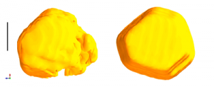
Figure: Shown on the left is the three dimensional image of a gold nanocrystal obtained previously while on the right is the image using the newly developed method. The features of the nanocrystal are vastly improved in the image on the left. The black scale bar is 100 nanometres (1 nanometre = 1 billionth of a meter). Downloaded from http://www.london-nano.com/research-and-facilities/highlight/advance-in-x-ray-imaging-shines-light-on-nanomaterials

The researchers have also provided two videos, the first features the current standard 3-D image of a gold nanocrystal and the second features the improved image,

Standard 3-D

Improved 3-D

The Aug. 7, 2012 news release originated from an article (Aug. 2012?) by Ian Robinson and Jesse Clark for the London Centre for Nanotechnology (part of the University College of London) giving context for the research and describing the technique (Note: I have removed a link),

Up until now, most nanomaterial imaging has been done using electron microscopy. X-ray imaging is an attractive alternative as X-rays penetrate further into the material than electrons and can be used in ambient or controlled environments.

However, making lenses that focus X-rays is very difficult. As an alternative, scientists use the indirect method of coherent diffraction imaging (CDI), where the diffraction pattern of the sample is measured (without lenses) and inverted to an image by computer.

Nobel Prize winner Lawrence Bragg suggested this method in 1939 but had no way to determine the missing phases of the diffraction, which are today provided by computer algorithms.

CDI can be performed very well at the latest synchrotron X-ray sources such as the UK’s Diamond Light Source which have much higher coherent flux than earlier machines.  CDI is gaining momentum in the study of nanomaterials, but, until now, has suffered from poor synchimage quality, with broken or non-uniform density.  This had been attributed to imperfect coherence of the X-ray light used.

The dramatic three-dimensional images of gold nanocrystals presented in this study demonstrate that this distortion can be corrected by appropriate modelling of the coherence function.

Professor Ian Robinson, London Centre for Nanotechnology and author of the paper said: “The corrected images are far more interpretable that ever obtained previously and will likely lead to new understanding of structure of nanoscale materials.”

The method should also work for free-electron-laser, electron- and atom-based diffractive imaging.

That mention of the UK’s Diamond Light Source reminded me of the Canadian Light Source located in Saskatoon, Saskatchewan. I imagine this work will open up some possibilities for the researchers there.

For those who would like to read more about the work, here’s a citation for the article,

High resolution three dimensional partially coherent diffraction imaging, Nature Communications.  J.N. Clark, X. Huang, R. Harder, & I.K. Robinson Nature Communications 3, Article number: 993 doi:10.1038/ncomms1994

This article is behind a paywall.

Canadian scientists get more light in deal with the US Argonne National Laboratory

Canada’s synchrotron, Canadian Light Source (based in Saskatchewan), has signed a new three-year deal with the US Dept. of Energy’s Argonne National Laboratory’s Advanced Photon Source (APS)  that will give Canadian scientists more access to the APS facilities, according to the June 18, 2012 news item at the  Nanowerk website,

Seeking to solve some of today’s greatest global problems, scientists using x-ray light source facilities at national research laboratories in the United States and Canada are sharing more expertise.

The Canadian Light Source (CLS) and the Advanced Photon Source (APS) at the U.S. Department of Energy’s (DOE’s) Argonne National Laboratory agreed in January 2012 to a Partner User Proposal that cements a stronger working relationship between the two facilities for the next three years. These two premier light sources use different but complementary x-ray techniques to probe materials in order to understand chemical and structural behavior.

Tone Kunz’s June 18, 2012 news release for the APS provides details about the deal,

This new agreement will provide Canadian scientists with more research time to use the x-ray light source facilities and more time on a larger number of APS beamlines. Using varied x-ray and imaging capabilities will broaden the range of experiments Canadians may undertake at the APS to augment their research done at the Canadian Light Source. X-ray science offers potential solutions to a broad range of problems in surface, material, environmental and earth sciences, condensed matter physics, chemistry, and geosciences.

Since the Sector 20 beamlines became fully operational, scientists from Canada and other areas who have used these beamlines at the APS have produced an average of 51 scientific publications a year. This research includes the study of more effective mineral exploration strategies, ways to mitigate mine waste and mercury contamination, and novel ways to fabricate nanomaterials for use in fuel cells, batteries, and LEDs.

I had not realized how longstanding the  CLS/APS relationship has been,

Before the Canadian Light Source began operation in 2004, a Canadian group led by Daryl Crozier of Simon Fraser University, working in partnership with colleagues at the University of Washington and the Pacific Northwest National Laboratory, helped found the Sector 20 beamlines at the APS as part of the Pacific Northwest Consortium Collaborative Access Team, or PNC-CAT. Parts of this team were included in the X-ray Science Division of the APS when it was formed.

This long-standing partnership has led to scientifically significant upgrades to the beamline. The new agreement will provide the valuable manpower and expertise to allow the APS to continue to push the innovation envelope. [emphasis mine]

As I was reading Kunz’s news release I kept asking, what’s in it for the APS? Apparently they need more “manpower and expertise.” Unfortunately, their future plans are a little shy of detail,

Scientists from the APS and the Canadian Light Source will work together on R&D projects to improve light-source technology. In particular, scientists will upgrade even further the two beamlines at Sector 20 in four key areas. This will provide a unique capability to prepare and measure in situ films and interfaces, a new technique to create quantitative three-dimensional chemical maps of samples, and improved forms of spectroscopy to expand the range of elements and types of environments that can be examined.

What are the four key areas? For that matter, what is Sector 20? I suspect some of my readers have similar questions about my postings. It’s easy (especially if you write frequently) to forget that your readers may not be as familiar as you are with the subject matter.

(I wrote about the CLS and another deal with a synchrotron in the UK in my May 31, 2011 posting.)

Canadian Light Source and Diamond Light (UK) Synchrotrons

Two synchrotrons, Canadian Light Source (CLS [Saskatoon, Saskatchewan]) and Diamond Light Source (near Oxford, England) have signed a memorandum of understanding (MOU). From the May 31, 2011 news item on physorg.com,

Making the power of synchrotron light available to more businesses, building new experimental equipment and developing new capabilities are three of the areas of collaboration in a trans-Atlantic memorandum of understanding (MOU) signed between Diamond Light Source Ltd. near Oxford and Canadian Light Source Inc. (CLS) in Saskatoon.

The agreement paves the way for the two synchrotron light sources to work together on joint projects related to their industrial science programmes, such as exchanges of staff, marketing materials, and coordinating access for clients to capabilities that are available at one synchrotron but not the other.

“Diamond and the CLS have been working closely together for some time,” said Josef Hormes, Executive Director of the CLS. “Now that we have this formal agreement, I am looking forward to a very bright future where the expertise of both our facilities can be combined to accomplish momentous things for fundamental and industrial science.

They don’t mention nanotechnology but synchrotrons can be used for subnanometre measurement and nanofabrication (National Light Source Synchrotron 2009 seminar with Dr. Lin Wang).  You can find out more about synchrotrons at the CLS Education webpage,

A synchrotron is a source of brilliant light that scientists can use to gather information about the structural and chemical properties of materials at the molecular level.

A synchrotron produces the light by using powerful electro-magnets and radio frequency waves to accelerate electrons to nearly the speed of light. Energy is added to the electrons as they accelerate so that, when the magnets alter their course, they naturally emit a very brilliant, highly focused light. Different spectra of light, such as Infrared, Ultraviolet, and X-rays, are directed down beamlines where researchers choose the desired wavelength to study their samples. The researchers observe the interaction between the light and the matter in their sample at the endstations (small laboratories).

This tool can be used to probe the matter and analyze a host of physical, chemical, geological, and biological processes. Information obtained by scientists can be used to help design new drugs, examine the structure of surfaces to develop more effective motor oils, build smaller, more powerful computer chips, develop new materials for safer medical implants, and help with clean-up of mining wastes, to name just a few applications.

Quick Facts:

  • More than 40 synchrotron light sources have been built around the world. The Canadian synchrotron is competitive with the brightest facilities in Japan, the U.S. and Europe.
  • As of 2009, more than 2000 scientists have used the CLS.
  • More than 3,000 academic, industrial, and government researchers a year from across Canada and from other countries are expected to use the facility once the full complement of beamlines is developed. Beamlines carry the synchrotron light to scientific work stations capable of operating 24 hours per day, 7 days per week, approximately 42 weeks of the year.
  • Initially, the CLS will focus on research in three key areas:
    • mining, natural resources and the environment
    • advanced materials, information technologies and micro systems
    • biotechnology, pharmaceuticals and medicine
  • The first synchrotrons were additions to facilities built to study subatomic physics. Synchrotron light was an annoyance to the researchers because it meant their electron beams lost energy every time they went through a bending magnet. However, the remarkable qualities of this light were soon recognized, and researchers began to come up with ways to use it.

Currently, CLSI has more than 130 employees. The work force of scientists, engineers, technicians, and administrators is growing to match additional CLSI users. Located in the midst of a research cluster on the north end of the University of Saskatchewan, next to Innovation Place, one of Canada’s leading high-tech industrial parks, CLSI strengthens Saskatoon’s reputation as “Science City” as a much-needed national R&D facility.

Intriguingly, they don’t mention the word radiation until the 2nd to last section, Salute to safety. In fact, it wasn’t easy finding the Education webpage; it’s not accessible from the Home page as it’s rendered on my computer screen. (I found it by using a search engine.)

New international nanotechnology safety study and a Canadian synchrotron conference

There’s a new report on nanotechnology safety studies, the ‘EMERGNANO report‘. The researchers surveyed environment, health, and safety studies internationally, determined which ones fit their criteria, and have  now provided an assessment of the findings. Short story: there are no conclusive findings which is troublesome given the number of nanomaterial-based products that are making their way into the international marketplace. Michael Berger on Nanowerk News offers an excellent assessment of the situation vis a vis technophobic and technophilic approaches to emerging technologies and their attendant safety issues,New technologies are always polarizing society – some only see the inherent dangers, others only see the opportunities. Since these two groups usually are the loudest, everybody else inbetween has a hard time to get their message across and with objective information and facts. Nanotechnologies are no different. The nay-sayers call for a total moratorium everytime scientific research with concerning conclusions is published while opportunistic hypsters are only interested in selling more products or reports and ridicule even the faintest objections and concerns as uninformed panicmongering.

For more, please go here. I notice that Andrew Maynard (mentioned frequently here due to his 2020 Science blog and his position as Chief Science Advisor for the Project on Emerging Nanotechnologies) is one of the authors.

There’s a nanotechnology-type conference being held in Saskatoon, Saskatchewan, Canada this week (June 17 – 18, 2009). They have a big synchroton facility there and, I believe, it is the only such facility in Canada, which according to their video, is one of the most advanced such facilities in the world. The 12th annual meeting features a public lecture, ‘Science Fiction as a Mirror for Reality‘, by  Robert J. Sawyer, an internationally renowned Canadian science fiction author. For details about the conference,go here. For information about the synchroton in Saskatoon, go here. For information about Robert J. Sawyer, go here. (Media release noting the event can be found on Nanowerk News.)