Monthly Archives: November 2022

Growing electronics on trees

An April 26, 2022 news item on phys.org caught my eye with its mention of nanocellulose, trees, and electronics,

Electronics can grow on trees thanks to nanocellulose paper semiconductors

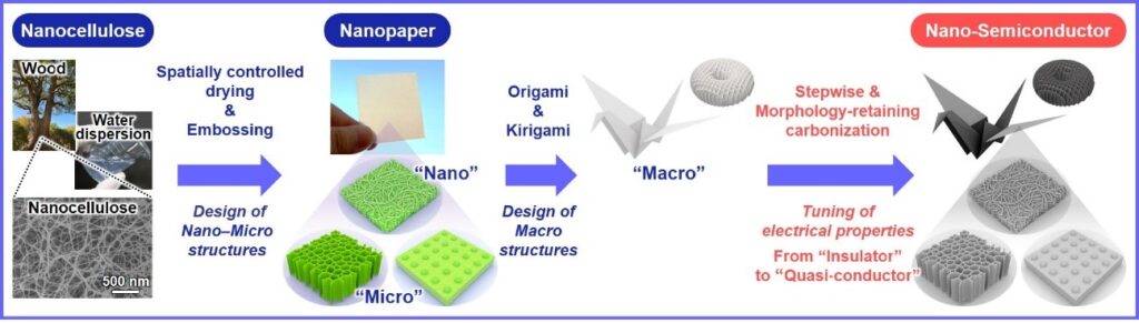
Semiconducting nanomaterials with 3D network structures have high surface areas and a lot of pores that make them excellent for applications involving adsorbing, separating, and sensing. However, simultaneously controlling the electrical properties and creating useful micro- and macro-scale structures, while achieving excellent functionality and end-use versatility, remains challenging. Now, Osaka University researchers, in collaboration with The University of Tokyo, Kyushu University, and Okayama University, have developed a nanocellulose paper semiconductor that provides both nano−micro−macro trans-scale designability of the 3D structures and wide tunability of the electrical properties. Their findings are published in ACS Nano.

Cellulose is a natural and easy to source material derived from wood. Cellulose nanofibers (nanocellulose) can be made into sheets of flexible nanocellulose paper (nanopaper) with dimensions like those of standard A4. Nanopaper does not conduct an electric current; however, heating can introduce conducting properties. Unfortunately, this exposure to heat can also disrupt the nanostructure.

The researchers have therefore devised a treatment process that allows them to heat the nanopaper without damaging the structures of the paper from the nanoscale up to the macroscale.

Caption: Schematic diagram of the preparation of the wood nanocellulose-derived nano-semiconductor with customizable electrical properties and 3D structures Credit: 2022 Koga et al. Nanocellulose paper semiconductor with a 3D network structure and its nano−micro−macro trans-scale design. ACS Nano

An April 28, 2022 Osaka University news release (also on EurekAlert), which originated the news item, provides more detail about the work

“An important property for the nanopaper semiconductor is tunability because this allows devices to be designed for specific applications,” explains study author Hirotaka Koga. “We applied an iodine treatment that was very effective for protecting the nanostructure of the nanopaper. Combining this with spatially controlled drying meant that the pyrolysis treatment did not substantially alter the designed structures and the selected temperature could be used to control the electrical properties.”

The researchers used origami (paper folding) and kirigami (paper cutting) techniques to provide playful examples of the flexibility of the nanopaper at the macrolevel. A bird and box were folded, shapes including an apple and snowflake were punched out, and more intricate structures were produced by laser cutting. This demonstrated the level of detail possible, as well as the lack of damage caused by the heat treatment.

Examples of successful applications showed nanopaper semiconductor sensors incorporated into wearable devices to detect exhaled moisture breaking through facemasks and moisture on the skin. The nanopaper semiconductor was also used as an electrode in a glucose biofuel cell and the energy generated lit a small bulb.

“The structure maintenance and tunability that we have been able to show is very encouraging for the translation of nanomaterials into practical devices,” says Associate Professor Koga. “We believe that our approach will underpin the next steps in sustainable electronics made entirely from plant materials.”

About Osaka University

Osaka University was founded in 1931 as one of the seven imperial universities of Japan and is now one of Japan’s leading comprehensive universities with a broad disciplinary spectrum. This strength is coupled with a singular drive for innovation that extends throughout the scientific process, from fundamental research to the creation of applied technology with positive economic impacts. Its commitment to innovation has been recognized in Japan and around the world, being named Japan’s most innovative university in 2015 (Reuters 2015 Top 100) and one of the most innovative institutions in the world in 2017 (Innovative Universities and the Nature Index Innovation 2017). Now, Osaka University is leveraging its role as a Designated National University Corporation selected by the Ministry of Education, Culture, Sports, Science and Technology to contribute to innovation for human welfare, sustainable development of society, and social transformation.

Here’s a link to and a citation for the paper,

Nanocellulose Paper Semiconductor with a 3D Network Structure and Its Nano–Micro–Macro Trans-Scale Design by Hirotaka Koga, Kazuki Nagashima, Koichi Suematsu, Tsunaki Takahashi, Luting Zhu, Daiki Fukushima, Yintong Huang, Ryo Nakagawa, Jiangyang Liu, Kojiro Uetani, Masaya Nogi, Takeshi Yanagida, and Yuta Nishina. ACS Nano 2022, XXXX, XXX, XXX-XXX DOI: https://doi.org/10.1021/acsnano.1c10728 Publication Date:April 26, 2022 © 2022 The Authors. Published by American Chemical Society

The paper appears to be open access.

In person conference highlights for Navigating Uncertainty; Targeting Sustainability (CSPC 2022) in Ottawa, Canada (Nov. 16 – 18, 2022)

Unless something very exciting happens, I think this will be my last post about the 2022 edition of the Canadian Science Policy Conference (CSPC 2022). From an October 27, 2022 CSPC announcement (received via email), here are some of the highlights for people attending the November 16 – 18, 2022 conference in person,

Conversation with Hon. François-Philippe Champagne, Minister of Innovation, Science and Industry

Remarks by Hon. Kirsty Duncan, Chair of the Standing Committee on Science and Research of the House of Commons

CRCC [Canada Research Coordinating Committee] Panel:
CRCC Progress Report – Moving Forward 

Plenary Sessions: 
Canadian Universities, News Frontier and Societal Challenges
-Steven Liss, Simon Kennedy, Stephen Toope, Sophie D’Amours, Elicia Maine

A Path to Process Innovation and Enhanced Productivity in Canada 
-Iain Stewart, Dan Breznitz, Éric Baril, Andrea Johnston

Breakfast Session: Conversation with New Advisory Panel on the Federal Research Support System
-Frédéric Bouchard, Gilles Patry and Vianne Timmons

Luncheon Session: Conversation with Dr. Mona Nemer, Canada’s Chief Science Advisor 

INGA [International Network for Government Science Advice (INGSA)] North America Chapter Workshop (RSVP Required)

Special Performance: The Anniversary, A Play (RSVP Required)

The Canada Research Coordinating Committee (CRCC) is new to me. So, I went looking for more information,

The Canada Research Coordinating Committee (CRCC) advances federal research priorities and the coordination of policies and programs of Canada’s research funding agencies and the Canada Foundation for Innovation. It provides a senior strategic forum for sharing information, building consensus and making decisions on forward-looking initiatives that strengthen Canada’s research enterprise, foster world-leading research, and advance the social and economic well-being of Canadians.

Details about the play can be found in my August 31, 2022 post titled: Navigating Uncertainty; Targeting Sustainability—the Canadian Science Policy Conference (Nov. 16 – 18, 2022). Scroll down about 40% of the way to find The Anniversary: A play.

I covered the new Advisory Panel on the Federal Research Support System in an October 13, 2022 post titled: Are we spending money on the right research? Government of Canada launches Advisory Panel.

The International Network for Government Science Advice (INGSA) has been mentioned here a few times, notably in an August 31, 2021 post titled: 4th International Conference on Science Advice to Governments (INGSA2021) August 30 – September 2, 2021; it was held here in Canada. I had a follow up the next day in a September 1, 2021 post.

You can find the CSPC 2022 website here.

Visualization of RNA structures at near-atomic resolution enabled by nanotechnology

The illustration that accompanies the research is both fascinating and baffling as its caption,

Caption: This illustration is inspired by the Paleolithic rock painting in the Lascaux cave, signifying the acronym of our method, ROCK. Figuratively, the patterns of the rock art in the background (brown) are the 2D projections of the engineered dimeric construct of the Tetrahymena group I intron, while the main object in the front (blue) is the reconstructed 3D cryo-EM map of the dimer, with one monomer in focus and refined to the high resolution that allowed the collaborators to build an atomic model of the RNA. Credit: Wyss Institute at Harvard University

This May 2, 2022 news item on ScienceDaily announces the research into RNA molecules made possible by ROCK (the technology being illustrated in the above),

We live in a world made and run by RNA [ribonucleic acid], the equally important sibling of the genetic molecule DNA. In fact, evolutionary biologists hypothesize that RNA existed and self-replicated even before the appearance of DNA and the proteins encoded by it. Fast forward to modern day humans: science has revealed that less than 3% of the human genome is transcribed into messenger RNA (mRNA) molecules that in turn are translated into proteins. In contrast, 82% of it is transcribed into RNA molecules with other functions many of which still remain enigmatic.

To understand what an individual RNA molecule does, its 3D structure needs to be deciphered at the level of its constituent atoms and molecular bonds. Researchers have routinely studied DNA and protein molecules by turning them into regularly packed crystals that can be examined with an X-ray beam (X-ray crystallography) or radio waves (nuclear magnetic resonance). However, these techniques cannot be applied to RNA molecules with nearly the same effectiveness because their molecular composition and structural flexibility prevent them from easily forming crystals.

Now, a research collaboration led by Wyss Core Faculty member Peng Yin, Ph.D. at the Wyss Institute for Biologically Inspired Engineering at Harvard University, and Maofu Liao, Ph.D. at Harvard Medical School (HMS), has reported a fundamentally new approach to the structural investigation of RNA molecules. ROCK, as it is called, uses an RNA nanotechnological technique that allows it to assemble multiple identical RNA molecules into a highly organized structure, which significantly reduces the flexibility of individual RNA molecules and multiplies their molecular weight. Applied to well-known model RNAs with different sizes and functions as benchmarks, the team showed that their method enables the structural analysis of the contained RNA subunits with a technique known as cryo-electron microscopy (cryo-EM). Their advance is reported in Nature Methods.

A May 2, 2022 Wyss Institute for Biologically Inspired Engineering at Harvard University news release (also on EurekAlert) by Benjamin Boettner, which originated the news item, delves further into the imaging technology, Note: Links have been removed,

“ROCK is breaking the current limits of RNA structural investigations and enables 3D structures of RNA molecules to be unlocked that are difficult or impossible to access with existing methods, and at near-atomic resolution,” said Yin, who together with Liao led the study. “We expect this advance to invigorate many areas of fundamental research and drug development, including the burgeoning field of RNA therapeutics.” Yin also is a leader of the Wyss Institute’s Molecular Robotics Initiative and Professor in the Department of Systems Biology at HMS.

Gaining control over RNA

Yin’s team at the Wyss Institute has pioneered various approaches that enable DNA and RNA molecules to self-assemble into large structures based on different principles and requirements, including DNA bricks and DNA origami. They hypothesized that such strategies could also be used to assemble naturally occurring RNA molecules into highly ordered circular complexes in which their freedom to flex and move is highly restricted by specifically linking them together. Many RNAs fold in complex yet predictable ways, with small segments base-pairing with each other. The result often is a stabilized “core” and “stem-loops” bulging out into the periphery. 

“In our approach we install ‘kissing loops’ that link different peripheral stem-loops belonging to two copies of an identical RNA in a way that allows a overall stabilized ring to be formed, containing multiple copies of the RNA of interest,” said Di Liu, Ph.D., one of two first-authors and a Postdoctoral Fellow in Yin’s group. “We speculated that these higher-order rings could be analyzed with high resolution by cryo-EM, which had been applied to RNA molecules with first success.”

Picturing stabilized RNA

In cryo-EM, many single particles are flash-frozen at cryogenic temperatures to prevent any further movements, and then visualized with an electron microscope and the help of computational algorithms that compare the various aspects of a particle’s 2D surface projections and reconstruct its 3D architecture. Peng and Liu teamed up with Liao and his former graduate student François Thélot, Ph.D., the other co-first author of the study. Liao with his group has made important contributions to the rapidly advancing cryo-EM field and the experimental and computational analysis of single particles formed by specific proteins.

“Cryo-EM has great advantages over traditional methods in seeing high-resolution details of biological molecules including proteins, DNAs and RNAs, but the small size and moving tendency of most RNAs prevent successful determination of RNA structures. Our novel method of assembling RNA multimers solves these two problems at the same time, by increasing the size of RNA and reducing its movement,” said Liao, who also is Associate Professor of Cell Biology at HMS. “Our approach has opened the door to rapid structure determination of many RNAs by cryo-EM.” The integration of RNA nanotechnology and cryo-EM approaches led the team to name their method “RNA oligomerization-enabled cryo-EM via installing kissing loops” (ROCK).

To provide proof-of-principle for ROCK, the team focused on a large intron RNA from Tetrahymena, a single-celled organism, and a small intron RNA from Azoarcus, a nitrogen-fixing bacterium, as well as the so-called FMN riboswitch. Intron RNAs are non-coding RNA sequences scattered throughout the sequences of freshly-transcribed RNAs and have to be “spliced” out in order for the mature RNA to be generated. The FMN riboswitch is found in bacterial RNAs involved in the biosynthesis of flavin metabolites derived from vitamin B2. Upon binding one of them, flavin mononucleotide (FMN), it switches its 3D conformation and suppresses the synthesis of its mother RNA.  

“The assembly of the Tetrahymena group I intron into a ring-like structure made the samples more homogenous, and enabled the use of computational tools leveraging the symmetry of the assembled structure. While our dataset is relatively modest in size, ROCK’s innate advantages allowed us to resolve the structure at an unprecedented resolution,” said Thélot. “The RNA’s core is resolved at 2.85 Å [one Ångström is one ten-billions (US) of a meter and the preferred metric used by structural biologists], revealing detailed features of the nucleotide bases and sugar backbone. I don’t think we could have gotten there without ROCK – or at least not without considerably more resources.” 

Cryo-EM also is able to capture molecules in different states if they, for example, change their 3D conformation as part of their function. Applying ROCK to the Azoarcus intron RNA and the FMN riboswitch, the team managed to identify the different conformations that the Azoarcus intron transitions through during its self-splicing process, and to reveal the relative conformational rigidity of the ligand-binding site of the FMN riboswitch.

“This study by Peng Yin and his collaborators elegantly shows how RNA nanotechnology can work as an accelerator to advance other disciplines. Being able to visualize and understand the structures of many naturally occurring RNA molecules could have tremendous impact on our understanding of many biological and pathological processes across different cell types, tissues, and organisms, and even enable new drug development approaches,” said Wyss Founding Director Donald Ingber, M.D., Ph.D., who is also the Judah Folkman Professor of Vascular Biology at Harvard Medical School and Boston Children’s Hospital, and Professor of Bioengineering at the Harvard John A. Paulson School of Engineering and Applied Sciences.

The study was also authored by Joseph Piccirilli, Ph.D., an expert in RNA chemistry and biochemistry and Professor at The University of Chicago. It was supported by the National Science Foundation (NSF; grant# CMMI-1333215, CCMI-1344915, and CBET-1729397), Air Force Office of Scientific Research (AFOSR; grant MURI FATE, #FA9550-15-1-0514), National Institutes of Health (NIH; grant# 5DP1GM133052, R01GM122797, and R01GM102489), and the Wyss Institute’s Molecular Robotics Initiative.

Here’s a link to and a citation for the paper,

Sub-3-Å cryo-EM structure of RNA enabled by engineered homomeric self-assembly by Di Liu, François A. Thélot, Joseph A. Piccirilli, Maofu Liao & Peng Yin. Nature Methods (2022) DOI: https://doi.org/10.1038/s41592-022-01455-w Published: 02 May 2022

This paper is behind a paywall.