I have two items concerning butterflies. The first is a bioengineering project at Yale University where they changed the colour of a butterfly’s wings from brown to violet (from an Aug. 5, 2014 news item on ScienceDaily),
Yale University scientists have chosen the most fleeting of mediums for their groundbreaking work on biomimicry: They’ve changed the color of butterfly wings.
In so doing, they produced the first structural color change in an animal by influencing evolution. The discovery may have implications for physicists and engineers trying to use evolutionary principles in the design of new materials and devices.
An Aug.5, 2014 Yale University news release (also on EurekAlert), which originated the news item,
“What we did was to imagine a new target color for the wings of a butterfly, without any knowledge of whether this color was achievable, and selected for it gradually using populations of live butterflies,” said Antónia Monteiro, a former professor of ecology and evolutionary biology at Yale, now at the National University of Singapore.
In this case, Monteiro and her team changed the wing color of the butterfly Bicyclus anynana from brown to violet. They needed only six generations of selection.
The news release goes on to explain the interest in structural colour,
Little is known about how structural colors in nature evolved, although researchers have studied such mechanisms extensively in recent years. Most attempts at biomimicry involve finding a desirable outcome in nature and simply trying to copy it in the laboratory.
“Today, materials engineers are making complex materials to perform multiple functions. The parameter space for the design of such materials is huge, so it is not easy to search for the optimal design,” said Hui Cao, chair of Yale’s Department of Applied Physics, who also worked on the study. “This is why we can learn from nature, which has obtained the optimal solutions in many cases via natural evolution over millions of years.”
Indeed, the scientists explained, natural selection algorithms can select for multiple characteristics simultaneously — which is standard operating procedure in the natural world.
A bit of technical information is also included in the news release,
The desired color for the butterfly wings was achieved by changing the relative thickness of the wing scales — specifically, those of the lower lamina. It took less than a year of selective breeding to produce the color change from brown to violet.
One reason Bicyclus anynana was chosen for the experiment, Monteiro said, was because it has cousin species that have evolved violet colors on their wings twice independently. By reproducing such a change in the lab, the Yale team showed that butterfly populations harbor high levels of genetic variation regulating scale thickness that lets them react quickly to new selective conditions.
“We just thought if natural selection has been able to modify wing colors in members of this genus of butterfly, perhaps so can we,” Monteiro said.
Here’s a link to and a citation for the paper,
Artificial selection for structural color on butterfly wings and comparison with natural evolution by Bethany R. Wasik, Seng Fatt Liew, David A. Lilien, April J. Dinwiddie, Heeso Noh, Hui Cao, and Antónia Monteiro. PNAS doi: 10.1073/pnas.1402770111 Published online August 4, 2014
This seems to be an open access paper (I was able to access the six page paper, albeit in a small font, by clicking on an Adobe reader icon).
I have not been able to find an image of the newly violet-coloured Bicyclus anynana butterfly but Yale University has provided an image of the pre-bioengineered version,
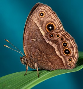
This image shows a male Bicyclus anynana, prior to the wing color change. (Below) This image shows the color change from brown to violet, over six generations of breeding. (Photographs courtesy of Antónia Monteiro)
One of my favourite pieces on structural colour was written for The Scientist and was featured here in a Feb. 7, 2013 posting. Interestingly, Yale University is mentioned in that posting too.
This second butterfly piece focuses on its feeding habits and possible medical applications. From an Aug. 5, 2014 news item on ScienceDaily,
New discoveries about how butterflies feed could help engineers develop tiny probes that siphon liquid out of single cells for a wide range of medical tests and treatments, according to Clemson University researchers.
The National Science Foundation recently awarded the project $696,514. It was the foundation’s third grant to the project, bringing the total since 2009 to more than $3 million.
The research has brought together Clemson’s materials scientists and biologists who have been focusing on the proboscis, the mouthpart that many insects used for feeding.
For materials scientists, the goal is to develop what they call “fiber-based fluidic devices,” among them probes that could eventually allow doctors to pluck a single defective gene out of a cell and replace it with a good one, said Konstantin Kornev, a Clemson materials physics professor. “If someone were programmed to have an illness, it would be eliminated,” he said.
An Aug. 5, 2014 Clemson University media release by Paul Alongi (also on EurekAlert), which originated the news item, explains that this latest research is one of the first steps in a long journey,
… Much remains unknown about how insects use tiny pores and channels in the proboscis to sample and handle fluid.
“It’s like the proverbial magic well,” said Clemson entomology professor Peter Adler. “The more we learn about the butterfly proboscis, the more it has for us to learn about it.”
Kornev said he was attracted to butterflies for their ability to draw various kinds of liquids.
“It can be very thick like nectar and honey or very thin like water,” he said. “They do that easily. That’s a challenge for engineers.”
Researchers want the probe to be able to take fluid out of a single cell, which is 10 times smaller than the diameter of a human hair, Kornev said. The probe also will need to differentiate between different types of fluids, he said.
The technology could be used for medical devices, nanobioreactors that make complex materials and flying “micro-air vehicles” the size of an insect.
“It opens up a huge number of applications,” Kornev said. “We are actively seeking collaboration with cell biologists, medical doctors and other professionals who might find this research exciting and helpful in their applications.”
The study also is breaking new ground in biology. While scientists had a fundamental idea of how butterflies feed, it was less complete than it is now, Adler said.
Scientists have long known that butterflies use the proboscis to suck up fluid, similar to how humans use a drinking straw, Adler said. But the study found that the butterfly proboscis also acts as a sponge, he said.
“It’s a dual mechanism,” Adler said. “As they move the proboscis around, it can help sponge up the liquid and then facilitate the delivery of the liquid so that it can then be sucked up.”
As part of the study, researchers observed butterflies on flowers at the Cherry Farm Insectary just south of the main campus on the shore of Hartwell Lake. Butterflies were raised in the lab and recorded on video as they fed.
Researchers are turning their attention to smaller insects, such as flies, moths and mosquitoes, but the focus will remain on the proboscis.
In the next phase of the study, researchers would like to understand how the proboscis forms.
Larvae enter the pupa without a proboscis and emerge as a butterfly with one. Understanding what happens in the pupa could help develop the probes, Adler said.
Another challenge is figuring out how to keep the probe from getting covered with organic material when it’s inserted into the body, he said.
That’s why researchers are beginning to turn their focus to an insect almost everyone else shoos away.
“It seems the flies are able to pierce an animal’s tissue, take up the blood and not get the proboscis gummed up and covered with bacteria,” Adler said.
Tanju Karanfil, associate dean of research and graduate studies in the College of Engineering and Science, said the study has underscored the importance of breaking down silos that separate researchers from different departments so they can work for the common good.
“The most interesting work happens at the intersection of disciplines,” he said. “In this case, biologists and engineers have come together with different perspectives to answer common questions.
I have a link (which takes you to a correction for the text) and a citation for the paper,
Paradox of the drinking-straw model of the butterfly proboscis by Chen-Chih Tsai, Daria Monaenkova, Charles Beard, Peter Adler, and Konstantin Kornev. J. Exp. Biol. 217, 2130-2138. Original article: doi: 10.1242/jeb.097998 June 15, 2014 J Exp Biol 217, 2130-2138 Correction: doi: 10.1242/jeb.109447 July 1, 2014
The article is behind a paywall but you can view the correction in its entirety.

![AGELESS BRILLIANCE: Although the pigment-derived leaf color of this decades-old specimen of the African perennial Pollia condensata has faded, the fruit still maintains its intense metallic-blue iridescence.COURTESY OF P.J. RUDALL [downloaded from http://www.the-scientist.com/?articles.view/articleNo/34200/title/Color-from-Structure/]](http://www.frogheart.ca/wp-content/uploads/2014/06/AgelessColour_-Pollia_condensata_Herbarium2.jpg)