Tag Archives: colloidal gold

There’s more than one black gold

‘Black gold’ is a phrase I associate with oil, signifying its importance and desirability. These days, this analogic phrase can describe a material according to a July 24, 2015 news item on Nanowerk,

If colloidal gold [gold in solution] self-assembles into the form of larger vesicles, a three-dimensional state can be achieved that is called “black gold” because it absorbs almost the entire spectrum of visible light. How this novel intense plasmonic state can be established and what its characteristics and potential medical applications are is explored by Chinese scientists and reported in the journal Angewandte Chemie …

A July 24, 2015 Wiley (Angewandte Chemie) press release, which originated the news item, provides more details,

Metal nanostructures can self-assemble into superstructures that offer intriguing new spectroscopic and mechanical properties. Plasmonic coupling plays a particular role in this context. For example, it has been found that plasmonic metal nanoparticles help to scatter the incoming light across the surface of the Si substrate at resonance wavelengths, therefore enhancing the light absorbing potential and thus the effectivity of solar cells.

On the other hand, plasmonic vesicles are the promising theranostic platform for biomedical applications, a notion which inspired Yue Li and Cuncheng Li of the Chinese Academy of Science, Hefei, China, and the University of Jinan, China, as well as collaborators to prepare plasmonic colloidosomes composed of gold nanospheres.

As the method of choice, the scientists have designed an emulsion-templating approach based on monodispersed gold nanospheres as building blocks, which arranged themselves into large spherical vesicles in a reverse emulsion system.

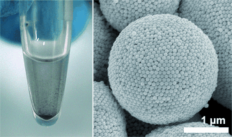
The resulting plasmonic vesicles were of micrometer-size and had a shell composed of hexagonally close-packed colloidal nanosphere particles in bilayer or, for the very large superspheres, multilayer arrangement, which provided the enhanced stability.

“A key advantage of this system is that such self-assembly can avoid the introduction of complex stabilization processes to lock the nanoparticles together”, the authors explain.

The hollow spheres exhibited an intense plasmonic resonance in their three-dimensionally packed structure and had a dark black appearance compared to the brick red color of the original gold nanoparticles. The “black gold” was thus characterized by a strong broadband absorption in the visible light and a very regular vesicle superstructure. In medicine, gold vesicles are intensively discussed as vehicles for the drug delivery to tumor cells, and, therefore, it could be envisaged to exploit the specific light-matter interaction of such plasmonic vesicle structures for medical use, but many other applications are also feasible, as the authors propose: “The presented strategy will pave a way to achieve noble-metal superstructures for biosensors, drug delivery, photothermal therapy, optical microcavity, and microreaction platforms.” This will prove the flexibility and versatility of the noble-metal nanostructures.

Here’s a link to and a citation for the paper,

Black Gold: Plasmonic Colloidosomes with Broadband Absorption Self-Assembled from Monodispersed Gold Nanospheres by Using a Reverse Emulsion System by Dilong Liu, Dr. Fei Zhou, Cuncheng Li, Tao Zhang, Honghua Zhang, Prof. Weiping Cai, and Prof. Yue Li. Angewandte Chemie International Edition Article first published online: 25 JUN 2015 DOI: 10.1002/anie.201503384

© 2015 WILEY-VCH Verlag GmbH & Co. KGaA, Weinheim

This article is behind a paywall.

There is an image illustrating the work but, sadly, the gold doesn’t look black,

BlackGold

© Wiley-VCH

That’s it!

Nanotechnology and the ancient Romans: the Lycurgus Cup

The Lycurgus Cup has long fascinated me. It’s an ancient piece of art that is nanotechnology-enhanced so that depending on how the light hits it, the cup glows either green or red.

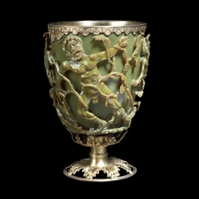
The Lycurgus Cup 1958,1202.1 in reflected light. Scene showing Lycurgus being enmeshed by Ambrosia, now transformed into a vine-shoot. Department of Prehistory and Europe, The British Museum. Height: 16.5 cm (with modern metal mounts), diameter: 13.2 cm. © The Trustees of the British Museum

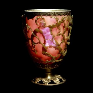
I must admit to a preference for the red simply because I can better see the designs.

The Lycurgus Cup 1958,1202.1 in transmitted light. Scene showing Lycurgus being enmeshed by Ambrosia, now transformed into a vine-shoot. Department of Prehistory and Europe, The British Museum. Height: 16.5 cm (with modern metal mounts), diameter: 13.2 cm. © The Trustees of the British Museum

The *History of the Ancient World website (as Nov. 21, 2013 the link has been changed to the Université de Strasbourg,, Matière Condensée et Nanophysique website) recently featured a 2007 article about the Lycurgus Cup by Ian Freestone, Nigel Meeks, Margaret Sax and Catherine Higgitt for the Gold Bulletin, Vol. 40:4 (2007),

The Lycurgus Cup represents one of the outstanding achievements of the ancient glass industry. This late Roman cut glass vessel is extraordinary in several respects, firstly in the method of fabrication and the exceptional workmanship involved and secondly in terms of the unusual optical effects displayed by the glass.

The Lycurgus Cup is one of a class of Roman vessels known as cage cups or diatreta, where the decoration is in openwork which stands proud from the body of the vessel, to which it is linked by shanks or bridges Typically these openwork “cages” comprise a lattice of linked circles, but a small number have figurative designs, although none of these is as elaborate or as well preserved as the Lycurgus Cup. Cage cups are generally dated to the fourth century A.D. and have been found across the Roman Empire, but the number recovered is small, and probably only in the region of 50-100 examples are known. They are among the most technically sophisticated glass objects produced before the modern era.

The article itself can be viewed or downloaded from here. The cup as noted can be two different colours,

The glass of the cup is dichroic; in direct light it resembles jade with an opaque greenish-yellow tone, but when light shines through the glass (transmitted light) it turns to a translucent ruby colour

The presence of colloidal metals (gold-silver) give the glass at least some of its unusual optical properties according to the authors. Although reading between the lines, it seems that even today we can’t duplicate what those 4th century Roman glassmakers achieved.

The Lycurgus Cup demonstrates a short-lived technology developed in the fourth century A.D. by Roman glass-workers. They discovered that glass could be coloured red and unusual colour change effects generated by the addition of a precious metal bearing material when the glass was molten. We now understand that these effects are due to the development of nanoparticles in the glass. However, the inability to control the colourant process meant that relatively few glasses of this type were produced, and even fewer survive. The Cup is the outstanding example of this technology in every respect – its outstanding cut work and red-green dichroism render it a unique record.

There you have it, ancient Roman nanotechnology.

*Nov. 21, 2013 I changed the link to the article as the History of the Ancient World website is no longer hosting this article.

ETA July 21, 2014: April Holloway has written a piece about the Lycurgus Cup which has been published in two places,

Epoch Times: http://www.theepochtimes.com/n3/807475-1600-year-old-goblet-shows-that-the-romans-used-nanotechnology/ (this features an image of the green and red ‘cups’ side-by-side with a ‘nano’ background)

Ancient Origins: http://www.ancient-origins.net/news-history-archaeology/1600-year-old-goblet-shows-romans-used-nanotechnology-00793#!bjfuCU (image of the green and red ‘cups’ side-by-side featured)

HIV testing, nano gold, and Uganda; not so obsolete?; new nanotube manufacturing technique from McGill University

There’s a portable blood-testing machine, designed by US-based PointCare, which can give a print-out detailing a patient’s immune status in 10 minutes. The machine was designed for use in third-world or developing world clinics such as the one in Uganda which is described in this BBC story.

One of the problems doctors and medical staff had with equipment for testing HIV patients’ immune system was that the chemicals used as reagents in the testing process were too easily perishable in the high heat common in a lot of countries. PointCare soved the problem this way (from the BBC article):

Dr Hansen [from PointCare] invented a test that uses chemical reagent that can be freeze-dried and stored in temperatures of over 40C.

CD4 screening tests use antibodies – molecular tags that recognise and latch onto a chemical marker on the surface of the cell. By attaching to the cells, they act as flags distinguishing CD4 cells from other white blood cells.

But these antibodies need to be “labelled”, so they can be detected by a machine.

Traditionally, antibodies are labelled using fluorescent markers, but these fluorescent chemicals perish if they are not kept refrigerated. So they’re useless for a medical team operating from a temporary clinic in the heat of an African summer.

Dr Hansen developed a new label. “We use colloidal gold,” explains Dr Krauledat [community physician]. “It’s true nanotechnology – extremely tiny gold particles attached to the anti-CD4 antibody.”

Do go and read the full story because there’s more to it than I’ve included. Meanwhile I had another look at those lithography stories (SFU’s new maskwriting facilities and RAPID) that I was posting about last week. While the new RAPID technique may make the use of ultra-violet light obsolete, they still haven’t approached the nanoscale. The measurement mentioned is “… 2500 times smaller than a human hair” [more here]. The measurement usually mentioned when discussing the nanoscale  is between 1/100,000 ro 1/60,000 (nobody seems to agree on the exact measurement, you can check here) of the width of a human hair equals 1 nanometre.  Weirdly, the Simon Fraser University (SFU) release notes that the new facilities will be able to create structures “… under 20 nanometres about 10,000 times smaller than the diameter of a human hair” [more here]. If I’m doing the math correctly, wouldn’t that be between 1/50,0000 and 1/30,000 of the human hair? I know it’s a little fussy but once a technical writer, always a technical writer and that kind of detail can make a big difference.

Researchers led by Dr. Hanadi Sleiman and Dr. Gonzalo Cosa at McGill University (Montreal, Canada) have developed a new way to manufacture nanotubes using DNA, in short they are DNA nanotubes. The longer story is here and the shorter story is here.