Tag Archives: DESY

It’s a golden world

I have a number of stories concerning gold where researchers seemed to have had an extraordinarily rich set of findings within the last month. One of these is especially interesting in light of what I published yesterday (August 11, 2025 “Turning lead into gold (for approximately a microsecond“) about an event in May 2025.

I will be providing my usual citations and links but will not be tagging all the researchers (there are far too many) other than those mentioned in the news releases.

Two from SLAC (SLAC National Accelerator Laboratory, originally named the Stanford Linear Accelerator Center in California)

While both projects took place at SLAC, there’s virtually no crossover between the team members and the findings are of an entirely different nature.

Defying the limits and surviving the entropy catastrophe

An August 11, 2025 news item on ScienceDaily announces that physics limits have been defied,

Scientists have simultaneously broken a temperature record, overturned a long-held theory and utilized a new laser spectroscopy method for dense plasmas in a groundbreaking article published on July 23 in the journal Nature.

In their research article, “Superheating gold beyond the predicted entropy catastrophe threshold,” physicists revealed they were able to heat gold to over 19,000 Kelvin (33,740 degrees Fahrenheit), over 14 times its melting point, without it losing its solid, crystalline structure.

A July 23, 2025 University of Nevada news release, which originated the news item, delves further into the topic,

“This is possibly the hottest crystalline material ever recorded,” Thomas White, lead author and Clemons-Magee Endowed Professor in Physics at the University of Nevada, Reno said.

This result overturns the long-held theoretical limit known as the entropy catastrophe. The entropy catastrophe theory states that solids cannot remain stable above approximately three times their melting temperature without spontaneously melting. The melting point of gold, 1,337 Kelvin (1,947 degrees Fahrenheit), was far more than tripled in this experiment utilizing an extremely powerful laser at Stanford University’s SLAC National Accelerator Laboratory.

“I was expecting the gold to heat quite significantly before melting, but I wasn’t expecting a fourteen-fold temperature increase,” White said.

To heat the gold, researchers at the University of Nevada, Reno, SLAC National Accelerator Laboratory, the University of Oxford, Queen’s University Belfast, the European XFEL and the University of Warwick designed an experiment to heat a thin gold foil using a laser fired for 50 quadrillionths of a second (one millionth of a billionth). The speed with which the gold was heated seems to be the reason the gold remained solid. The findings suggest that the limit of superheating solids may be far higher – or nonexistent – if heating occurs quickly enough. The new methods used in this study open the field of high energy density physics to more exploration, including in areas of planetary physics and fusion energy research.

White and his team expected that the gold would melt at its melting point, but to measure the temperature inside the gold foil, they would need a very special thermometer.

“We used the Linac Coherent Light Source, a 3-kilometer-long X-ray laser at SLAC, as essentially the world’s largest thermometer,” White said. “This allowed us to measure the temperature inside the dense plasma for the first time, something that hasn’t been possible before.”

“This development paves the way for temperature diagnostics across a broad range of high-energy-density environments,” Bob Nagler, staff scientist at SLAC and coauthor on the paper, said. “In particular, it offers the only direct method currently available for probing the temperature of warm dense states encountered during the implosion phase of inertial fusion energy experiments. As such, it is poised to make a transformative contribution to our understanding and control of fusion-relevant plasma conditions.”

Along with the experimental designers, the research article is the result of a decade of work and collaboration between Columbia University, Princeton University, the University of Padova and the University of California, Merced.

“It’s extremely exciting to have these results out in the world, and I’m really looking forward to seeing what strides we can make in the field with these new methods,” White said.

The research, funded by the National Nuclear Security Administration, will open new doors in studies of superheated materials.

“The National Nuclear Security Administrations’ Academics Program is a proud supporter of the groundbreaking innovation and continued learning that Dr. White and his team are leading for furthering future critical research areas beneficial to the Nuclear Security Enterprise,” Jahleel Hudson, director at the Techology and Partnerships Office of the NNSA said.

White and his colleagues returned to the Linac Coherent Light Source in July to measure the temperature inside hot compressed iron and are using those results to gain insights into the interiors of planets.

Several of White’s graduate students and one undergraduate student were coauthors on the study, including doctoral student Travis Griffin, undergraduate student Hunter Stramel, Daniel Haden, a former postdoctoral scholar in White’s lab, Jacob Molina, a former undergraduate student currently pursuing his doctoral degree at Princeton University and Landon Morrison, a former undergraduate student pursuing his master’s degree at the University of Oxford. Jeremy Iratcabal, research assistant professor in the Department of Physics, was also a coauthor on the paper.

“I’m incredibly grateful for the opportunity to contribute to such cutting-edge science using billion-dollar experimental platforms alongside world-class collaborators,” Griffin said. “This discovery highlights the power of this technique, and I’m excited by the possibilities it opens for the future of high-energy-density physics and fusion research. After graduation, I’ll be continuing this work as a staff scientist at the European XFEL.”

SLAC issued a July 23, 2025 news release (by Erin Woodward) of its own and UK’s University of Warwick also issued a July 23, 2025.

Here’s a link to and a citation for the paper,

Superheating gold beyond the predicted entropy catastrophe threshold by Thomas G. White, Travis D. Griffin, Daniel Haden, Hae Ja Lee, Eric Galtier, Eric Cunningham, Dimitri Khaghani, Adrien Descamps, Lennart Wollenweber, Ben Armentrout, Carson Convery, Karen Appel, Luke B. Fletcher, Sebastian Goede, J. B. Hastings, Jeremy Iratcabal, Emma E. McBride, Jacob Molina, Giulio Monaco, Landon Morrison, Hunter Stramel, Sameen Yunus, Ulf Zastrau, Siegfried H. Glenzer, Gianluca Gregori, Dirk O. Gericke & Bob Nagler. Nature volume 643, pages 950–954 (2025) DOI: https://doi.org/10.1038/s41586-025-09253-y Published: 23 July 2025 Issue Date: 24 July 2025

This paper is open access.

Gold’s secret chemistry

An August 11, 2025 news item on ScienceDaily announces how researchers at SLAC unexpectedly created gold hydride,

Scientists at SLAC unexpectedly created gold hydride, a compound of gold and hydrogen, while studying diamond formation under extreme pressure and heat. This discovery challenges gold’s reputation as a chemically unreactive metal and opens doors to studying dense hydrogen, which could help us understand planetary interiors and fusion processes. The results also suggest that extreme conditions can produce exotic, previously unknown compounds, offering exciting opportunities for future high-pressure chemistry research.

Serendipitously and for the first time, an international research team led by scientists at the U.S. Department of Energy’s SLAC National Accelerator Laboratory formed solid binary gold hydride, a compound made exclusively of gold and hydrogen atoms.

An August 4, 2025 SLAC news release by Chris Patrick, which originated the news release, provides more details, Note: Links have been removed,

The researchers were studying how long it takes hydrocarbons, compounds made of carbon and hydrogen, to form diamonds under extremely high pressure and heat. In their experiments at the European XFEL (X-ray Free-Electron Laser) in Germany, the team studied the effect of those extreme conditions in hydrocarbon samples with an embedded gold foil, which was meant to absorb the X-rays and heat the weakly absorbing hydrocarbons. To their surprise, they not only saw the formation of diamonds, but also discovered the formation of gold hydride. 

“It was unexpected because gold is typically chemically very boring and unreactive – that’s why we use it as an X-ray absorber in these experiments,” said Mungo Frost, staff scientist at SLAC who led the study. “These results suggest there’s potentially a lot of new chemistry to be discovered at extreme conditions where the effects of temperature and pressure start competing with conventional chemistry, and you can form these exotic compounds.”

The results, published in Angewandte Chemie International Edition, provide a glimpse of how the rules of chemistry change under extreme conditions like those found inside certain planets or hydrogen-fusing stars.

Studying dense hydrogen

In their experiment, the researchers first squeezed their hydrocarbon samples to pressures greater than those within Earth’s mantle using a diamond anvil cell. Then, they heated the samples to over 3,500 degrees Fahrenheit by hitting them repeatedly with X-ray pulses from the European XFEL. The team recorded and analyzed how the X-rays scattered off the samples, which allowed them to resolve the structural transformations within.

As expected, the recorded scattering patterns showed that the carbon atoms had formed a diamond structure. But the team also saw unexpected signals that were due to hydrogen atoms reacting with the gold foil to form gold hydride. 

Under the extreme conditions created in the study, the researchers found hydrogen to be in a dense, “superionic” state, where the hydrogen atoms flowed freely through the gold’s rigid atomic lattice, increasing the conductivity of the gold hydride. 

Hydrogen, which is the lightest element of the periodic table, is tricky to study with X-rays because it scatters X-rays only weakly. Here, however, the superionic hydrogen interacted with the much heavier gold atoms, and the team was able to observe hydrogen’s impact on how the gold lattice scattered X-rays. “We can use the gold lattice as a witness for what the hydrogen is doing,” Mungo said. 

The gold hydride offers a way to study dense atomic hydrogen under conditions that might also apply to other situations that are experimentally not directly accessible. For example, dense hydrogen makes up the interiors of certain planets, so studying it in the lab could teach us more about those foreign worlds. It could also provide new insights into nuclear fusion processes inside stars like our sun and help develop technology to harness fusion energy here on Earth.

Exploring new chemistry

In addition to paving the way for studies of dense hydrogen, the research also offers an avenue for exploring new chemistry. Gold, which is commonly regarded as an unreactive metal, was found to form a stable hydride at extremely high pressure and temperature. In fact, it appears to be only stable at those extreme conditions as when it cools down, the gold and hydrogen separate. The simulations also showed that more hydrogen could fit in the gold lattice at higher pressure.

The simulation framework could also be extended beyond gold hydride. “It’s important that we can experimentally produce and model these states under these extreme conditions,” said Siegfried Glenzer, High Energy Density Division director and professor for photon science at SLAC and the study’s principal investigator. “These simulation tools could be applied to model other exotic material properties in extreme conditions.” 

The team also included researchers from Rostock University, DESY, European XFEL, Helmholtz-Zentrum Dresden-Rossendorf, Frankfurt University and Bayreuth University, all in Germany; the University of Edinburgh, UK; the Carnegie Institution for Science, Stanford University and the Stanford Institute for Materials and Energy Sciences (SIMES). Parts of this work were supported by the DOE Office of Science.

Here’s a link to and a citation for the paper,

Synthesis of Gold Hydride at High Pressure and High Temperature by Mungo Frost, Kilian Abraham, Alexander F. Goncharov, R. Stewart McWilliams, Rachel J. Husband, Michal Andrzejewski, Karen Appel, Carsten Baehtz, Armin Bergermann, Danielle Brown, Elena Bykova, Anna Celeste, Eric Edmund, Nicholas J. Hartley, Konstantin Glazyrin, Heinz Graafsma, Nicolas Jaisle, Zuzana Konôpková, Torsten Laurus, Yu Lin, Bernhard Massani, Maximilian Schörner, Maximilian Schulze, Cornelius Strohm, Minxue Tang, Zena Younes, Gerd Steinle-Neumann, Ronald Redmer, Siegfried H. Glenzer. Angewandte Chemie International Edition DOI: https://doi.org/10.1002/anie.202505811 First published: 04 August 2025

This paper is behind a paywall.

Gold and a quantum revolution?

An August 11, 2025 news item on ScienceDaily announces joint research from Pennsylvania State University (Penn State) and Colorado State University,

The efficiency of quantum computers, sensors and other applications often relies on the properties of electrons, including how they are spinning. One of the most accurate systems for high performance quantum applications relies on tapping into the spin properties of electrons of atoms trapped in a gas, but these systems are difficult to scale up for use in larger quantum devices like quantum computers. Now, a team of researchers from Penn State and Colorado State has demonstrated how a gold cluster can mimic these gaseous, trapped atoms, allowing scientists to take advantage of these spin properties in a system that can be easily scaled up.

A July 22, 2025 Penn State news release (also on EurekAlert) by Gail McCormick, which originated the news item, reveals more about the work which resulted in two published papers, Note: Links have been removed,

“For the first time, we show that gold nanoclusters have the same key spin properties as the current state-of-the-art methods for quantum information systems,” said Ken Knappenberger, department head and professor of chemistry in the Penn State Eberly College of Science and leader of the research team. “Excitingly, we can also manipulate an important property called spin polarization in these clusters, which is usually fixed in a material. These clusters can be easily synthesized in relatively large quantities, making this work a promising proof-of-concept that gold clusters could be used to support a variety of quantum applications.”

Two papers describing the gold clusters and confirming their spin properties appeared in ACS Central Science, ACS Central Science and The Journal of Physical Chemistry Letters.

“An electron’s spin not only influences important chemical reactions, but also quantum applications like computation and sensing,” said Nate Smith, graduate student in chemistry in the Penn State Eberly College of Science and first author of one of the papers. “The direction an electron spins and its alignment with respect to other electrons in the system can directly impact the accuracy and longevity of quantum information systems.”

Much like the Earth spins around its axis, which is tilted with respect to the sun, an electron can spin around its axis, which can be tilted with respect to its nucleus. But unlike Earth, an electron can spin clockwise or counterclockwise. When many electrons in a material are spinning in the same direction and their tilts are aligned, the electrons are considered correlated, and the material is said to have a high degree of spin polarization. 

“Materials with electrons that are highly correlated, with a high degree of spin polarization, can maintain this correlation for a much longer time, and thus remain accurate for much longer,” Smith said.

The current state-of-the-art system for high accuracy and low error in quantum information systems involve trapped atomic ions — atoms with an electric charge — in a gaseous state. This system allows electrons to be excited to different energy levels, called Rydberg states, which have very specific spin polarizations that can last for a long period of time. It also allows for the superposition of electrons, with electrons existing in multiple states simultaneously until they are measured, which is a key property for quantum systems. 

“These trapped gaseous ions are by nature dilute, which makes them very difficult to scale up,” Knappenberger said. “The condensed phase required for a solid material, by definition, packs atoms together, losing that dilute nature. So, scaling up provides all the right electronic ingredients, but these systems become very sensitive to interference from the environment. The environment basically scrambles all the information that you encoded into the system, so the rate of error becomes very high. In this study, we found that gold clusters can mimic all the best properties of the trapped gaseous ions with the benefit of scalability.”

Scientists have heavily studied gold nanostructures for their potential use in optical technology, sensing, therapeutics and to speed up chemical reactions, but less is known about their magnetic and spin-dependent properties. In the current studies, the researchers specifically explored monolayer-protected clusters, which have a core of gold and are surrounded by other molecules called ligands. The researchers can precisely control the construction of these clusters and can synthesize relatively large amounts at one time. 

“These clusters are referred to as super atoms, because their electronic character is like that of an atom, and now we know their spin properties are also similar,” Smith said. “We identified 19 distinguishable and unique Rydberg-like spin-polarized states that mimic the super-positions that we could do in the trapped, gas-phase dilute ions. This means the clusters have the key properties needed to carry out spin-based operations.”

The researchers determined the spin polarization of the gold clusters using a similar method used with traditional atoms. While one type of gold cluster had 7% spin polarization, a cluster with different a ligand approached 40% spin polarization, which Knappenberger said is competitive with some of the leading two-dimensional quantum materials.

“This tells us that the spin properties of the electron are intimately related to the vibrations of the ligands,” Knappenberger said. “Traditionally, quantum materials have a fixed value of spin polarization that cannot be significantly changed, but our results suggest we can modify the ligand of these gold clusters to tune this property widely.”

The research team plans to explore how different structures within the ligands impact spin polarization and how they could be manipulated to fine tune spin properties.

“The quantum field is generally dominated by researchers in physics and materials science, and here we see the opportunity for chemists to use our synthesis skills to design materials with tunable results,” Knappenberger said. “This is a new frontier in quantum information science.”

In addition to Smith and Knappenberger, the research team includes Juniper Foxley, graduate student in chemistry at Penn State; Patrick Herbert, who earned a doctoral degree in chemistry at Penn State in 2019; Jane Knappenberger, researcher in the Penn State Eberly College of Science; as well as Marcus Tofanelli and Christopher Ackerson at Colorado State

Funding from the Air Force Office of Scientific Research and the U.S. National Science Foundation supported this research.

At Penn State, researchers are solving real problems that impact the health, safety and quality of life of people across the commonwealth, the nation and around the world.

For decades, federal support for research has fueled innovation that makes our country safer, our industries more competitive and our economy stronger. Recent federal funding cuts threaten this progress.

Learn more about the implications of federal funding cuts to our future at Research or Regress. [Research or Regress can found here]

Here are links to and citation for the paper,

The Influence of Passivating Ligand Identity on Au25(SR)18 Spin-Polarized Emission by Nathanael L. Smith, Patrick J. Herbert, Marcus A. Tofanelli, Jane A. Knappenberger, Christopher J. Ackerson, Kenneth L. Knappenberger Jr. The Journal of Physical Chemistry Letters 2025, 16, 20, 5168–5172 DOI: https://doi.org/10.1021/acs.jpclett.5c00723 Published May 15, 2025 Copyright © 2025 American Chemical Society

This paper is behind a paywall.

Diverse Superatomic Magnetic and Spin Properties of Au144(SC8H9)60 Clusters by Juniper Foxley, Marcus Tofanelli, Jane A. Knappenberger, Christopher J. Ackerson, Kenneth L. Knappenberger Jr ACS Central Science 2025, XXXX, XXX, XXX-XXX DOI: https://doi.org/10.1021/acscentsci.5c00139
Published May 29, 2025 © 2025 The Authors. Published by American Chemical Society. This publication is licensed under CC-BY 4.0 .

This paper is open access.

Lead into gold, the second time around

There are reasons why news releases are issued twice and/or months after a research paper was published. Whoever is scanning for news may have missed it or it was a big news day and science was not top of mind or e.g., a number of teams are publishing research in your field and are generating a lot of interest and you hope your institution will benefit from it.

This August 11, 2025 news item on ScienceDaily resuscitates a story from May 2025,

Nuclear physicists working at the Large Hadron Collider recently made headlines by achieving the centuries-old dream of alchemists (and nightmare of precious-metals investors): They transformed lead into gold.

At least for a fraction of a second. The scientists reported their results in Physical Reviews.

The accomplishment at the Large Hadron Collider, the 17-mile particle accelerator buried under the French-Swiss border, happened within a sophisticated and sensitive detector called ALICE, a scientific instrument roughly the size of a McMansion.

A July 30, 2025 University of Kansas news release (also on EurekAlert), which originated the August 11, 2025 news item, adds new details about the work, Note: A link has been removed,

It was scientists from the University of Kansas, working on the ALICE experiment, who developed the technique that tracked “ultra-peripheral” collisions between protons and ions that made gold in the LHC.

“Usually in collider experiments, we make the particles crash into each other to produce lots of debris,” said Daniel Tapia Takaki, professor of physics and leader of KU’s group at ALICE. “But in ultra-peripheral collisions, we’re interested in what happens when the particles don’t hit each other. These are near misses. The ions pass close enough to interact — but without touching. There’s no physical overlap.”

The ions racing around the LHC tunnel are heavy nuclei with many protons, each generating powerful electric fields. When accelerated, these charged ions emit photons — they shine light.

“When you accelerate an electric charge to near light speeds, it starts shining,” Tapia Takaki said. “One ion can shine light that essentially takes a picture of the other. When that light is energetic enough, it can probe deep inside the other nucleus, like a high-energy flashbulb.”

The KU researcher said during these UPC “flashes” surprising interactions can occur, including the rate event that sparked worldwide attention.

“Sometimes, the photons from both ions interact with each other — what we call photon-photon collisions,” he said. “These events are incredibly clean, with almost nothing else produced. They contrast with typical collisions where we see sprays of particles flying everywhere.”

However, the ALICE detector and the LHC were designed to collect data on head-on collisions that result in messy sprays of particles.

“These clean interactions were hard to detect with earlier setups,” Tapia Takaki said. “Our group at KU pioneered new techniques to study them. We built up this expertise years ago when it was not a popular subject.”

These methods allowed for the news-making discovery that the LHC team transmuted lead into gold momentarily via ultra-peripheral collisions where lead ions lose three protons (turning the speck of lead into a gold speck) for a fraction of a second.

Tapia Takaki’s KU co-authors on the paper are graduate student Anna Binoy; graduate student Amrit Gautam; postdoctoral researcher Tommaso Isidori; postdoctoral research assistant Anisa Khatun; and research scientist Nicola Minafra.

The KU team at the LHC ALICE experiment plans to continue studying the ultra-peripheral collisions. Tapia Takaki said that while the creation of gold fascinated the public, the potential of understanding the interactions goes deeper.

“This light is so energetic, it can knock protons out of the nucleus,” he said. “Sometimes one, sometimes two, three or even four protons. We can see these ejected protons directly with our detectors.”

Each proton removed changes the elements: One gives thallium, two gives mercury, three gives gold.

“These new nuclei are very short-lived,” he said. “They decay quickly, but not always immediately. Sometimes they travel along the beamline and hit parts of the collider — triggering safety systems.”

That’s why this research matters beyond the headlines.

“With proposals for future colliders even larger than the LHC — some up to 100 kilometers in Europe and China — you need to understand these nuclear byproducts,” Tapia Takaki said. “This ‘alchemy’ may be crucial for designing the next generation of machines.”

This work was supported by the U.S. Department of Energy Office of Science, Office of Nuclear Physics.

Here’s a new link and citation, which includes each team member’s name, for the paper,

Proton emission in ultraperipheral Pb-Pb collisions at sNN=5.02 TeV by S. Acharya, A. Agarwal, G. Aglieri Rinella, L. Aglietta, M. Agnello, N. Agrawal, Z. Ahammed, S. Ahmad, S. U. Ahn, I. Ahuja, A. Akindinov, V. Akishina, M. Al-Turany, D. Aleksandrov, B. Alessandro, H. M. Alfanda, R. Alfaro Molina, B. Ali, A. Alici, N. Alizadehvandchali, A. Alkin, J. Alme, G. Alocco, T. Alt, A. R. Altamura, I. Altsybeev, J. R. Alvarado, C. O. R. Alvarez, M. N. Anaam, C. Andrei, N. Andreou, A. Andronic, E. Andronov, V. Anguelov, F. Antinori, P. Antonioli, N. Apadula, L. Aphecetche, H. Appelshäuser, C. Arata, S. Arcelli, R. Arnaldi, J. G. M. C. A. Arneiro, I. C. Arsene, M. Arslandok, A. Augustinus, R. Averbeck, D. Averyanov, M. D. Azmi, H. Baba, A. Badalà, J. Bae, Y. Bae, Y. W. Baek, X. Bai, R. Bailhache, Y. Bailung, R. Bala, A. Baldisseri, B. Balis, Z. Banoo, V. Barbasova, F. Barile, L. Barioglio, M. Barlou, B. Barman, G. G. Barnaföldi, L. S. Barnby, E. Barreau, V. Barret, L. Barreto, C. Bartels, K. Barth, E. Bartsch, N. Bastid, S. Basu, G. Batigne, D. Battistini, B. Batyunya, D. Bauri, J. L. Bazo Alba, I. G. Bearden, C. Beattie, P. Becht, D. Behera, I. Belikov, A. D. C. Bell Hechavarria, F. Bellini, R. Bellwied, S. Belokurova, L. G. E. Beltran, Y. A. V. Beltran, G. Bencedi, A. Bensaoula, S. Beole, Y. Berdnikov, A. Berdnikova, L. Bergmann, M. G. Besoiu, L. Betev, P. P. Bhaduri, A. Bhasin, B. Bhattacharjee, L. Bianchi, J. Bielčík, J. Bielčíková, A. P. Bigot, A. Bilandzic, A. Binoy, G. Biro, S. Biswas, N. Bize, J. T. Blair, D. Blau, M. B. Blidaru, N. Bluhme, C. Blume, F. Bock, T. Bodova, J. Bok, L. Boldizsár, M. Bombara, P. M. Bond, G. Bonomi, H. Borel, A. Borissov, A. G. Borquez Carcamo, E. Botta, Y. E. M. Bouziani, D. C. Brandibur, L. Bratrud, P. Braun-Munzinger, M. Bregant, M. Broz, G. E. Bruno, V. D. Buchakchiev, M. D. Buckland, D. Budnikov, H. Buesching, S. Bufalino, P. Buhler, N. Burmasov, Z. Buthelezi, A. Bylinkin, S. A. Bysiak, J. C. Cabanillas Noris, M. F. T. Cabrera, H. Caines, A. Caliva, E. Calvo Villar, J. M. M. Camacho, P. Camerini, F. D. M. Canedo, S. L. Cantway, M. Carabas, A. A. Carballo, F. Carnesecchi, R. Caron, L. A. D. Carvalho, J. Castillo Castellanos, M. Castoldi, F. Catalano, S. Cattaruzzi, R. Cerri, I. Chakaberia, P. Chakraborty, S. Chandra, S. Chapeland, M. Chartier, S. Chattopadhay, M. Chen, T. Cheng, C. Cheshkov, D. Chiappara, V. Chibante Barroso, D. D. Chinellato, E. S. Chizzali, J. Cho, S. Cho, P. Chochula, Z. A. Chochulska, D. Choudhury, S. Choudhury, P. Christakoglou, C. H. Christensen, P. Christiansen, T. Chujo, M. Ciacco, C. Cicalo, F. Cindolo, M. R. Ciupek, G. Clai, F. Colamaria, J. S. Colburn, D. Colella, A. Colelli, M. Colocci, M. Concas, G. Conesa Balbastre, Z. Conesa del Valle, G. Contin, J. G. Contreras, M. L. Coquet, P. Cortese, M. R. Cosentino, F. Costa, S. Costanza, P. Crochet, M. M. Czarnynoga, A. Dainese, G. Dange, M. C. Danisch, A. Danu, P. Das, S. Das, A. R. Dash, S. Dash, A. De Caro, G. de Cataldo, J. de Cuveland, A. De Falco, D. De Gruttola, N. De Marco, C. De Martin, S. De Pasquale, R. Deb, R. Del Grande, L. Dello Stritto, W. Deng, K. C. Devereaux, G. G. A. de Souza, P. Dhankher, D. Di Bari, A. Di Mauro, B. Di Ruzza, B. Diab, R. A. Diaz, Y. Ding, J. Ditzel, R. Divià, Ø. Djuvsland, U. Dmitrieva, A. Dobrin, B. Dönigus, J. M. Dubinski, A. Dubla, P. Dupieux, N. Dzalaiova, T. M. Eder, R. J. Ehlers, F. Eisenhut, R. Ejima, D. Elia, B. Erazmus, F. Ercolessi, B. Espagnon, G. Eulisse, D. Evans, S. Evdokimov, L. Fabbietti, M. Faggin, J. Faivre, F. Fan, W. Fan, A. Fantoni, M. Fasel, G. Feofilov, A. Fernández Téllez, L. Ferrandi, M. B. Ferrer, A. Ferrero, C. Ferrero, A. Ferretti, V. J. G. Feuillard, V. Filova, D. Finogeev, F. M. Fionda, E. Flatland, F. Flor, A. N. Flores, S. Foertsch, I. Fokin, S. Fokin, U. Follo, E. Fragiacomo, E. Frajna, U. Fuchs, N. Funicello, C. Furget, A. Furs, T. Fusayasu, J. J. Gaardhøje, M. Gagliardi, A. M. Gago, T. Gahlaut, C. D. Galvan, S. Gami, D. R. Gangadharan, P. Ganoti, C. Garabatos, J. M. Garcia, T. García Chávez, E. Garcia-Solis, S. Garetti, C. Gargiulo, P. Gasik, H. M. Gaur, A. Gautam, M. B. Gay Ducati, M. Germain, R. A. Gernhaeuser, C. Ghosh, M. Giacalone, G. Gioachin, S. K. Giri, P. Giubellino, P. Giubilato, A. M. C. Glaenzer, P. Glässel, E. Glimos, D. J. Q. Goh, V. Gonzalez, P. Gordeev, M. Gorgon, K. Goswami, S. Gotovac, V. Grabski, L. K. Graczykowski, E. Grecka, A. Grelli, C. Grigoras, V. Grigoriev, S. Grigoryan, F. Grosa, J. F. Grosse-Oetringhaus, R. Grosso, D. Grund, N. A. Grunwald, G. G. Guardiano, R. Guernane, M. Guilbaud, K. Gulbrandsen, J. J. W. K. Gumprecht, T. Gündem, T. Gunji, W. Guo, A. Gupta, R. Gupta, R. Gupta, K. Gwizdziel, L. Gyulai, C. Hadjidakis, F. U. Haider, S. Haidlova, M. Haldar, H. Hamagaki, Y. Han, B. G. Hanley, R. Hannigan, J. Hansen, M. R. Haque, J. W. Harris, A. Harton, M. V. Hartung, H. Hassan, D. Hatzifotiadou, P. Hauer, L. B. Havener, E. Hellbär, H. Helstrup, M. Hemmer, T. Herman, S. G. Hernandez, G. Herrera Corral, S. Herrmann, K. F. Hetland, B. Heybeck, H. Hillemanns, B. Hippolyte, I. P. M. Hobus, F. W. Hoffmann, B. Hofman, M. Horst, A. Horzyk, Y. Hou, P. Hristov, P. Huhn, L. M. Huhta, T. J. Humanic, A. Hutson, D. Hutter, M. C. Hwang, R. Ilkaev, M. Inaba, G. M. Innocenti, M. Ippolitov, A. Isakov, T. Isidori, M. S. Islam, S. Iurchenko, M. Ivanov, M. Ivanov, V. Ivanov, K. E. Iversen, M. Jablonski, B. Jacak, N. Jacazio, P. M. Jacobs, S. Jadlovska, J. Jadlovsky, S. Jaelani, C. Jahnke, M. J. Jakubowska, M. A. Janik, T. Janson, S. Ji, S. Jia, T. Jiang, A. A. P. Jimenez, F. Jonas, D. M. Jones, J. M. Jowett, J. Jung, M. Jung, A. Junique, A. Jusko, J. Kaewjai, P. Kalinak, A. Kalweit, A. Karasu Uysal, D. Karatovic, N. Karatzenis, O. Karavichev, T. Karavicheva, E. Karpechev, M. J. Karwowska, U. Kebschull, M. Keil, B. Ketzer, J. Keul, S. S. Khade, A. M. Khan, S. Khan, A. Khanzadeev, Y. Kharlov, A. Khatun, A. Khuntia, Z. Khuranova, B. Kileng, B. Kim, C. Kim, D. J. Kim, D. Kim, E. J. Kim, J. Kim, J. Kim, J. Kim, M. Kim, S. Kim, T. Kim, K. Kimura, S. Kirsch, I. Kisel, S. Kiselev, A. Kisiel, J. L. Klay, J. Klein, S. Klein, C. Klein-Bösing, M. Kleiner, T. Klemenz, A. Kluge, C. Kobdaj, R. Kohara, T. Kollegger, A. Kondratyev, N. Kondratyeva, J. Konig, S. A. Konigstorfer, P. J. Konopka, G. Kornakov, M. Korwieser, S. D. Koryciak, C. Koster, A. Kotliarov, N. Kovacic, V. Kovalenko, M. Kowalski, V. Kozhuharov, G. Kozlov, I. Králik, A. Kravčáková, L. Krcal, M. Krivda, F. Krizek, K. Krizkova Gajdosova, C. Krug, M. Krüger, D. M. Krupova, E. Kryshen, V. Kučera, C. Kuhn, P. G. Kuijer, T. Kumaoka, D. Kumar, L. Kumar, N. Kumar, S. Kumar, S. Kundu, P. Kurashvili, A. B. Kurepin, A. Kuryakin, S. Kushpil, V. Kuskov, M. Kutyla, A. Kuznetsov, M. J. Kweon, Y. Kwon, S. L. La Pointe, P. La Rocca, A. Lakrathok, M. Lamanna, S. Lambert, A. R. Landou, R. Langoy, P. Larionov, E. Laudi, L. Lautner, R. A. N. Laveaga, R. Lavicka, R. Lea, H. Lee, I. Legrand, G. Legras, J. Lehrbach, A. M. Lejeune, T. M. Lelek, R. C. Lemmon, I. León Monzón, M. M. Lesch, P. Lévai, M. Li, P. Li, X. Li, B. E. Liang-Gilman, J. Lien, R. Lietava, I. Likmeta, B. Lim, H. Lim, S. H. Lim, V. Lindenstruth, C. Lippmann, D. Liskova, D. H. Liu, J. Liu, G. S. S. Liveraro, I. M. Lofnes, C. Loizides, S. Lokos, J. Lömker, X. Lopez, E. López Torres, C. Lotteau, P. Lu, Z. Lu, F. V. Lugo, J. R. Luhder, G. Luparello, Y. G. Ma, M. Mager, A. Maire, E. M. Majerz, M. V. Makariev, M. Malaev, G. Malfattore, N. M. Malik, S. K. Malik, D. Mallick, N. Mallick, G. Mandaglio, S. K. Mandal, A. Manea, V. Manko, F. Manso, G. Mantzaridis, V. Manzari, Y. Mao, R. W. Marcjan, G. V. Margagliotti, A. Margotti, A. Marín, C. Markert, C. F. B. Marquez, P. Martinengo, M. I. Martínez, G. Martínez García, M. P. P. Martins, S. Masciocchi, M. Masera, A. Masoni, L. Massacrier, O. Massen, A. Mastroserio, S. Mattiazzo, A. Matyja, F. Mazzaschi, M. Mazzilli, Y. Melikyan, M. Melo, A. Menchaca-Rocha, J. E. M. Mendez, E. Meninno, A. S. Menon, M. W. Menzel, M. Meres, L. Micheletti, D. Mihai, D. L. Mihaylov, K. Mikhaylov, N. Minafra, D. Miśkowiec, A. Modak, B. Mohanty, M. Mohisin Khan, M. A. Molander, M. M. Mondal, S. Monira, C. Mordasini, D. A. Moreira De Godoy, I. Morozov, A. Morsch, T. Mrnjavac, V. Muccifora, S. Muhuri, J. D. Mulligan, A. Mulliri, M. G. Munhoz, R. H. Munzer, H. Murakami, S. Murray, L. Musa, J. Musinsky, J. W. Myrcha, B. Naik, A. I. Nambrath, B. K. Nandi, R. Nania, E. Nappi, A. F. Nassirpour, V. Nastase, A. Nath, S. Nath, C. Nattrass, M. N. Naydenov, A. Neagu, A. Negru, E. Nekrasova, L. Nellen, R. Nepeivoda, S. Nese, N. Nicassio, B. S. Nielsen, E. G. Nielsen, S. Nikolaev, V. Nikulin, F. Noferini, S. Noh, P. Nomokonov, J. Norman, N. Novitzky, A. Nyanin, J. Nystrand, M. R. Ockleton, S. Oh, A. Ohlson, V. A. Okorokov, J. Oleniacz, A. Onnerstad, C. Oppedisano, A. Ortiz Velasquez, J. Otwinowski, M. Oya, K. Oyama, S. Padhan, D. Pagano, G. Paić, S. Paisano-Guzmán, A. Palasciano, I. Panasenko, S. Panebianco, C. Pantouvakis, H. Park, J. Park, S. Park, J. E. Parkkila, Y. Patley, R. N. Patra, B. Paul, H. Pei, T. Peitzmann, X. Peng, M. Pennisi, S. Perciballi, D. Peresunko, G. M. Perez, Y. Pestov, M. T. Petersen, V. Petrov, M. Petrovici, S. Piano, M. Pikna, P. Pillot, O. Pinazza, L. Pinsky, C. Pinto, S. Pisano, M. Płoskoń, M. Planinic, D. K. Plociennik, M. G. Poghosyan, B. Polichtchouk, S. Politano, N. Poljak, A. Pop, S. Porteboeuf-Houssais, V. Pozdniakov, I. Y. Pozos, K. K. Pradhan, S. K. Prasad, S. Prasad, R. Preghenella, F. Prino, C. A. Pruneau, I. Pshenichnov, M. Puccio, S. Pucillo, S. Qiu, L. Quaglia, A. M. K. Radhakrishnan, S. Ragoni, A. Rai, A. Rakotozafindrabe, L. Ramello, M. Rasa, S. S. Räsänen, R. Rath, M. P. Rauch, I. Ravasenga, K. F. Read, C. Reckziegel, A. R. Redelbach, K. Redlich, C. A. Reetz, H. D. Regules-Medel, A. Rehman, F. Reidt, H. A. Reme-Ness, K. Reygers, A. Riabov, V. Riabov, R. Ricci, M. Richter, A. A. Riedel, W. Riegler, A. G. Riffero, M. Rignanese, C. Ripoli, C. Ristea, M. V. Rodriguez, M. Rodríguez Cahuantzi, S. A. Rodríguez Ramírez, K. Røed, R. Rogalev, E. Rogochaya, T. S. Rogoschinski, D. Rohr, D. Röhrich, S. Rojas Torres, P. S. Rokita, G. Romanenko, F. Ronchetti, E. D. Rosas, K. Roslon, A. Rossi, A. Roy, S. Roy, N. Rubini, J. A. Rudolph, D. Ruggiano, R. Rui, P. G. Russek, R. Russo, A. Rustamov, E. Ryabinkin, Y. Ryabov, A. Rybicki, J. Ryu, W. Rzesa, B. Sabiu, S. Sadovsky, J. Saetre, S. Saha, B. Sahoo, R. Sahoo, D. Sahu, P. K. Sahu, J. Saini, K. Sajdakova, S. Sakai, M. P. Salvan, S. Sambyal, D. Samitz, I. Sanna, T. B. Saramela, D. Sarkar, P. Sarma, V. Sarritzu, V. M. Sarti, M. H. P. Sas, S. Sawan, E. Scapparone, J. Schambach, H. S. Scheid, C. Schiaua, R. Schicker, F. Schlepper, A. Schmah, C. Schmidt, M. O. Schmidt, M. Schmidt, N. V. Schmidt, A. R. Schmier, J. Schoengarth, R. Schotter, A. Schröter, J. Schukraft, K. Schweda, G. Scioli, E. Scomparin, J. E. Seger, Y. Sekiguchi, D. Sekihata, M. Selina, I. Selyuzhenkov, S. Senyukov, J. J. Seo, D. Serebryakov, L. Serkin, L. Šerkšnytė, A. Sevcenco, T. J. Shaba, A. Shabetai, R. Shahoyan, A. Shangaraev, B. Sharma, D. Sharma, H. Sharma, M. Sharma, S. Sharma, S. Sharma, U. Sharma, A. Shatat, O. Sheibani, K. Shigaki, M. Shimomura, J. Shin, S. Shirinkin, Q. Shou, Y. Sibiriak, S. Siddhanta, T. Siemiarczuk, T. F. Silva, D. Silvermyr, T. Simantathammakul, R. Simeonov, B. Singh, B. Singh, K. Singh, R. Singh, R. Singh, S. Singh, V. K. Singh, V. Singhal, T. Sinha, B. Sitar, M. Sitta, T. B. Skaali, G. Skorodumovs, N. Smirnov, R. J. M. Snellings, E. H. Solheim, C. Sonnabend, J. M. Sonneveld, F. Soramel, A. B. Soto-Hernandez, R. Spijkers, I. Sputowska, J. Staa, J. Stachel, I. Stan, P. J. Steffanic, T. Stellhorn, S. F. Stiefelmaier, D. Stocco, I. Storehaug, N. J. Strangmann, P. Stratmann, S. Strazzi, A. Sturniolo, C. P. Stylianidis, A. A. P. Suaide, C. Suire, A. Suiu, M. Sukhanov, M. Suljic, R. Sultanov, V. Sumberia, S. Sumowidagdo, L. H. Tabares, S. F. Taghavi, J. Takahashi, G. J. Tambave, S. Tang, Z. Tang, J. D. Tapia Takaki, N. Tapus, L. A. Tarasovicova, M. G. Tarzila, A. Tauro, A. Tavira García, G. Tejeda Muñoz, L. Terlizzi, C. Terrevoli, S. Thakur, M. Thogersen, D. Thomas, A. Tikhonov, N. Tiltmann, A. R. Timmins, M. Tkacik, T. Tkacik, A. Toia, R. Tokumoto, S. Tomassini, K. Tomohiro, N. Topilskaya, M. Toppi, V. V. Torres, A. G. Torres Ramos, A. Trifiró, T. Triloki, A. S. Triolo, S. Tripathy, T. Tripathy, S. Trogolo, V. Trubnikov, W. H. Trzaska, T. P. Trzcinski, C. Tsolanta, R. Tu, A. Tumkin, R. Turrisi, T. S. Tveter, K. Ullaland, B. Ulukutlu, S. Upadhyaya, A. Uras, G. L. Usai, M. Vala, N. Valle, L. V. R. van Doremalen, M. van Leeuwen, C. A. van Veen, R. J. G. van Weelden, P. Vande Vyvre, D. Varga, Z. Varga, P. Vargas Torres, M. Vasileiou, A. Vasiliev, O. Vázquez Doce, O. Vazquez Rueda, V. Vechernin, P. Veen, E. Vercellin, R. Verma, R. Vértesi, M. Verweij, L. Vickovic, Z. Vilakazi, O. Villalobos Baillie, A. Villani, A. Vinogradov, T. Virgili, M. M. O. Virta, A. Vodopyanov, B. Volkel, M. A. Völkl, S. A. Voloshin, G. Volpe, B. von Haller, I. Vorobyev, N. Vozniuk, J. Vrláková, J. Wan, C. Wang, D. Wang, Y. Wang, Y. Wang, Z. Wang, A. Wegrzynek, F. T. Weiglhofer, S. C. Wenzel, J. P. Wessels, P. K. Wiacek, J. Wiechula, J. Wikne, G. Wilk, J. Wilkinson, G. A. Willems, B. Windelband, M. Winn, J. R. Wright, W. Wu, Y. Wu, Z. Xiong, R. Xu, A. Yadav, A. K. Yadav, Y. Yamaguchi, S. Yang, S. Yano, E. R. Yeats, Z. Yin, I.-K. Yoo, J. H. Yoon, H. Yu, S. Yuan, A. Yuncu, V. Zaccolo, C. Zampolli, F. Zanone, N. Zardoshti, A. Zarochentsev, P. Závada, N. Zaviyalov, M. Zhalov, B. Zhang, C. Zhang, L. Zhang, M. Zhang, M. Zhang, S. Zhang, X. Zhang, Y. Zhang, Z. Zhang, M. Zhao, V. Zherebchevskii, Y. Zhi, D. Zhou, Y. Zhou, J. Zhu, S. Zhu, Y. Zhu, S. C. Zugravel, N. Zurlo. Physical Review C, 2025; 111 (5) DOI: 10.1103/PhysRevC.111.054906

This paper is open access. A PDF version is available here. h/t to ScienceDaily for the complete list of names

Observing photo exposure one nanoscale grain at a time

A June 9, 2015 news item on Nanotechnology Now highlights research into a common phenomenon, photographic exposure,

Photoinduced chemical reactions are responsible for many fundamental processes and technologies, from energy conversion in nature to micro fabrication by photo-lithography. One process that is known from everyday’s life and can be observed by the naked eye, is the exposure of photographic film. At DESY’s [Deutsches Elektronen-Synchrotron] X-ray light source PETRA III, scientists have now monitored the chemical processes during a photographic exposure at the level of individual nanoscale grains in real-time. The advanced experimental method enables the investigation of a broad variety of chemical and physical processes in materials with millisecond temporal resolution, ranging from phase transitions to crystal growth. The research team lead by Prof. Jianwei (John) Miao from the University of California in Los Angeles and Prof. Tim Salditt from the University of Göttingen report their technique and observations in the journal Nature Materials.

A June 9, 2015 DESY press release (also on EurekAlert), which originated the news item, provides more detail about the research,

The researchers investigated a photographic paper (Kodak linagraph paper Type 2167 or “yellow burn paper”) that is often used to determine the position of the beam at X-ray experiments. “The photographic paper we looked at is not specially designed for X-rays. It works by changing its colour on exposure to light or X-rays,” explains DESY physicist Dr. Michael Sprung, head of the PETRA III beamline P10 where the experiments took place.

The X-rays were not only used to expose the photographic paper, but also to analyse changes of its inner composition at the same time. The paper carries a photosensitive film of a few micrometre thickness, consisting of tiny silver bromide grains dispersed in a gelatine matrix, and with an average size of about 700 nanometres. A nanometre is a millionth of a millimetre. When X-rays impinge onto such a crystalline grain, they are diffracted in a characteristic way, forming a unique pattern on the detector that reveals properties like crystal lattice spacing, chemical composition and orientation. “We could observe individual silver bromide grains within the ‘burn’ paper since the X-ray beam had a size of only 270 by 370 nanometres – smaller than the average grain,” says Salditt, who is a partner of DESY in the construction and operation of the GINIX (Göttingen Instrument for Nano-Imaging with X-Rays) at beamline P10.

The X-ray exposure starts the photolysis from silver bromide to produce silver. An absorbed X-ray photon can create many photolytic silver atoms, which grow and agglomerate at the surface and inside the silver bromide grain. The scientists observed how the silver bromide grains were strained, began to turn in the gelatine matrix and broke up into smaller crystallites as well as the growth of pure silver nano grains. The exceptionally bright beam of PETRA III together with a high-speed detector enabled the ‘filming’ of the process with up to five milliseconds temporal resolution. “We observed, for the first time, grain rotation and lattice deformation during photoinduced chemical reactions,” emphasises Miao. “We were actually surprised how fast some of these single grains rotate,” adds Sprung. “Some spin almost one time every two seconds.”

“As advanced synchrotron light sources are currently under rapid development in the US, Europe and Asia,” the authors anticipate that “in situ X-ray nanodiffraction, which enables to measure atomic resolution diffraction patterns with several millisecond temporal resolution, can be broadly applied to investigate phase transitions, chemical reactions, crystal growth, grain boundary dynamics, lattice expansion, and contraction in materials science, nanoscience, physics, and chemistry.”

Here’s a link to and a citation for the paper,

Grain rotation and lattice deformation during photoinduced chemical reactions revealed by in situ X-ray nanodiffraction by Zhifeng Huang, Matthias Bartels, Rui Xu, Markus Osterhoff, Sebastian Kalbfleisch, Michael Sprung, Akihiro Suzuki, Yukio Takahashi, Thomas N. Blanton, Tim Salditt, & Jianwei Miao. Nature Materials (2015) doi:10.1038/nmat4311 Published online 08 June 2015

This paper is behind a paywall.
Nanoparticles in 3D courtesy of x-rays

A Feb. 4, 2015 Deutsches Elektronen-Synchrotron (DESY) press release (also on EurekAlert) announces a 3D first,

For the first time, a German-American research team has determined the three-dimensional shape of free-flying silver nanoparticles, using DESY’s X-ray laser FLASH. The tiny particles, hundreds of times smaller than the width of a human hair, were found to exhibit an unexpected variety of shapes, as the physicists from the Technical University (TU) Berlin, the University of Rostock, the SLAC National Accelerator Laboratory in the United States and from DESY report in the scientific journal Nature Communications. Besides this surprise, the results open up new scientific routes, such as direct observation of rapid changes in nanoparticles.

The press release goes on to describe the work in more detail,

“The functionality of nanoparticles is linked to their geometric form, which is often very difficult to determine experimentally,” explains Dr. Ingo Barke from the University of Rostock. “This is particularly challenging when they are present as free particles, that is, in the absence of contact with a surface or a liquid.”

The nanoparticle shape can be revealed from the characteristic way how it scatters X-ray light. Therefore, X-ray sources like DESY’s FLASH enable a sort of super microscope into the nano-world. So far, the spatial structure of nanoparticles has been reconstructed from multiple two-dimensional images, which were taken from different angles. This procedure is uncritical for particles on solid substrates, as the images can be taken from many different angles to uniquely reconstruct their three-dimensional shape.

“Bringing nanoparticles into contact with a surface or a liquid can significantly alter the particles, such that you can no longer see their actual form,” says Dr. Daniela Rupp from the TU Berlin. A free particle, however, can only be measured one time in flight before it either escapes or is destroyed by the intense X-ray light. Therefore, the scientists looked for a way to record the entire structural information of a nanoparticle with a single X-ray laser pulse.

To achieve this goal, the scientists led by Prof. Thomas Möller from the TU Berlin and Prof. Karl-Heinz Meiwes-Broer and Prof. Thomas Fennel from the University of Rostock employed a trick. Instead of taking usual small-angle scattering images, the physicists recorded the scattered X-rays in a wide angular range. “This approach virtually captures the structure from many different angles simultaneously from a single laser shot,” explains Fennel.

The researchers tested this method on free silver nanoparticles with diameters of 50 to 250 nanometres (0.00005 to 0.00025 millimetres). The experiment did not only verify the feasibility of the tricky method, but also uncovered the surprising result that large nanoparticles exhibit a much greater variety of shapes than expected.

The shape of free nanoparticles is a result of different physical principles, particularly the particles’ effort to minimize their energy. Consequently, large particles composed of thousands or millions of atoms often yield predictable shapes, because the atoms can only be arranged in a particular way to obtain an energetically favourable state.

In their experiment, however, the researchers observed numerous highly symmetrical three-dimensional shapes, including several types known as Platonic and Archimedean bodies. Examples include the truncated octahedron (a body consisting of eight regular hexagons and six squares) and the icosahedron (a body made up of twenty equilateral triangles). The latter is actually only favourable for extremely small particles consisting of few atoms, and its occurrence with free particles of this size was previously unknown. “The results show that metallic nanoparticles retain a type of memory of their structure, from the early stages of growth to a yet unexplored size range,” emphasizes Barke.

Due to the large variety of shapes, it was especially important to use a fast computational method so that the researchers were capable of mapping the shape of each individual particle. The scientists used a two-step process: the rough shape was determined first and then refined using more complex simulations on a super computer. This approach turned out to be so efficient that it could not only determine various shapes reliably, but could also differentiate between varying orientations of the same shape.

This new method for determining the three-dimensional shape and orientation of nanoparticles with a single X-ray laser shot opens up a wide spectrum of new research directions. In future projects, particles could be directly “filmed” in three dimensions during growth or during phase changes. “The ability to directly film the reaction of a nanoparticle to an intense flash of X-ray light has been a dream for many physicists – this dream could now come true, even in 3D!,” emphasises Rupp.

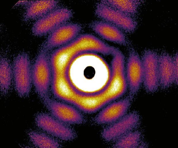
The researchers have provided an image showing their work,

Caption: This is a wide-angle X-ray diffraction image of a truncated twinned tetrahedra nanoparticle. Credit: Hannes Hartmann/University of Rostock

Caption: This is a wide-angle X-ray diffraction image of a truncated twinned tetrahedra nanoparticle.
Credit: Hannes Hartmann/University of Rostock

Here’s a link to and a citation for the paper,

The 3D-architecture of individual free ​silver nanoparticles captured by X-ray scattering by Ingo Barke, Hannes Hartmann, Daniela Rupp, Leonie Flückiger, Mario Sauppe, Marcus Adolph, Sebastian Schorb, Christoph Bostedt, Rolf Treusch, Christian Peltz, Stephan Bartling, Thomas Fennel, Karl-Heinz Meiwes-Broer, & Thomas Möller. Nature Communications 6, Article number: 6187 doi:10.1038/ncomms7187 Published 04 February 2015

This article is open access.

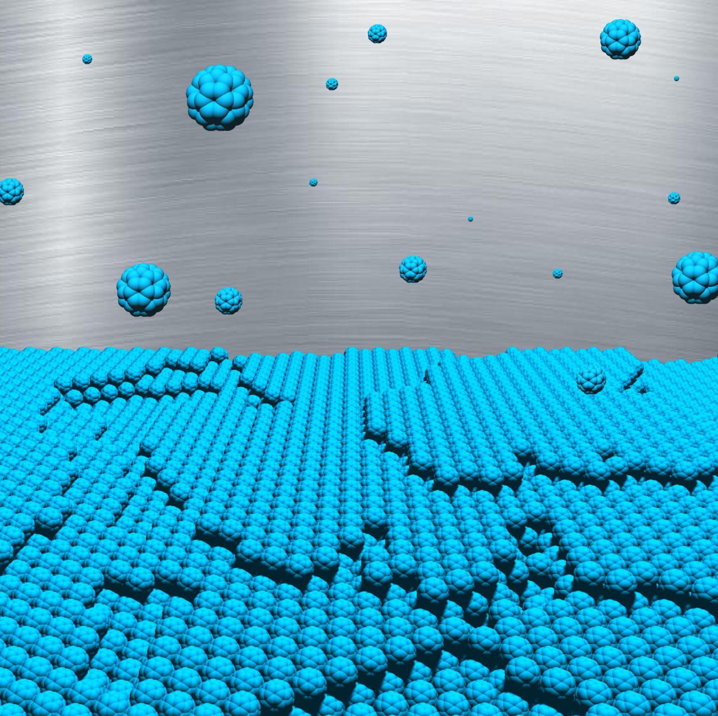
Watching buckyballs (buckminsterfullerenes) self-assemble in real-time

For the 5% or less of the world who need this explanation, the reference to a football later in this post is, in fact, a reference to a soccer ball. Moving on to a Nov. 5, 2014 news item on Nanowerk (Note: A link has been removed),

Using DESY’s ultrabright X-ray source PETRA III, researchers have observed in real-time how football-shaped carbon molecules arrange themselves into ultra-smooth layers. Together with theoretical simulations, the investigation reveals the fundamentals of this growth process for the first time in detail, as the team around Sebastian Bommel (DESY and Humboldt Universität zu Berlin) and Nicola Kleppmann (Technische Universität Berlin) reports in the scientific journal Nature Communications (“Unravelling the multilayer growth of the fullerene C60 in real-time”).

This knowledge will eventually enable scientists to tailor nanostructures from these carbon molecules for certain applications, which play an increasing role in the promising field of plastic electronics. The team consisted of scientists from Humboldt-Universität zu Berlin, Technische Universität Berlin, Universität Tübingen and DESY.

Here’s an image of the self-assembling materials,

Caption: This is an artist's impression of the multilayer growth of buckyballs. Credit: Nicola Kleppmann/TU Berlin

Caption: This is an artist’s impression of the multilayer growth of buckyballs.
Credit: Nicola Kleppmann/TU Berlin

A Nov. 5, 2014 DESY (Deutsches Elektronen-Synchrotron) press release (also on EurekAlert), describes the work further,

The scientists studied so called buckyballs. Buckyballs are spherical molecules, which consist of 60 carbon atoms (C60). Because they are reminiscent of American architect Richard Buckminster Fuller’s geodesic domes, they were christened buckminsterfullerenes or “buckyballs” for short. With their structure of alternating pentagons and hexagons, they also resemble tiny molecular footballs. [emphasis mine]

Using DESY’s X-ray source PETRA III, the researchers observed how buckyballs settle on a substrate from a molecular vapour. In fact, one layer after another, the carbon molecules grow predominantly in islands only one molecule high and barely form tower-like structures..“The first layer is 99% complete before 1% of the second layer is formed,” explains DESY researcher Bommel, who is completing his doctorate in Prof. Stefan Kowarik’s group at the Humboldt Universität zu Berlin. This is how extremely smooth layers form.

“To really observe the growth process in real-time, we needed to measure the surfaces on a molecular level faster than a single layer grows, which takes place in about a minute,” says co-author Dr. Stephan Roth, head of the P03 measuring station, where the experiments were carried out. “X-ray investigations are well suited, as they can trace the growth process in detail.”

“In order to understand the evolution of the surface morphology at the molecular level, we carried out extensive simulations in a non-equilibrium system. These describe the entire growth process of C60 molecules into a lattice structure,” explains Kleppmann, PhD student in Prof. Sabine Klapp’s group at the Institute of Theoretical Physics, Technische Universität Berlin. “Our results provide fundamental insights into the molecular growth processes of a system that forms an important link between the world of atoms and that of colloids.”

Through the combination of experimental observations and theoretical simulations, the scientists determined for the first time three major energy parameters simultaneously for such a system: the binding energy between the football molecules, the so-called “diffusion barrier,” which a molecule must overcome if it wants to move on the surface, and the Ehrlich-Schwoebel barrier, which a molecule must overcome if it lands on an island and wants to hop down from that island.

“With these values, we now really understand for the first time how such nanostructures come into existence,” stresses Bommel. “Using this knowledge, it is conceivable that these structures can selectively be grown in the future: How must I change my temperature and deposition rate parameters so that an island of a particular size will grow. This could, for example, be interesting for organic solar cells, which contain C60.” The researchers intend to explore the growth of other molecular systems in the future using the same methods.

Here’s a link to and a citation for the paper,

Unravelling the multilayer growth of the ​fullerene C60 in real time by S. Bommel, N. Kleppmann, C. Weber, H. Spranger, P. Schäfer, J. Novak, S.V. Roth, F. Schreiber, S.H.L. Klapp, & S. Kowarik. Nature Communications 5, Article number: 5388 doi:10.1038/ncomms6388 Published 05 November 2014

This article is open access.

I was not able to find any videos of these buckyballs assembling in real-time. Presumably, there are technical issues with recording the process, financial issues, or some combination thereof. Still, I can’t help but feel teased (tongue in cheek) by these scientists who give me an artist’s concept instead. Hopefully, budgets and/or technology will allow the rest of us to view this process at some time in the future.

Viewing a photosynthesis subsystem in a near-natural state

[downloaded from http://www.desy.de/infos__services/presse/pressemeldungen/@@news-view?id=9383]

Molecular structure of photosystem II, which arranges itself in rows. Credit: Martin Bommer/HU Berlin [downloaded from http://www.desy.de/infos__services/presse/pressemeldungen/@@news-view?id=9383]

Apparently, this image represents a near-natural state for a photosynthesis subsystem called, Photosynthesis II. Here’s more from a Nov. 4, 2014 news item on Nanowerk (Note: A link has been removed),

Photosynthesis is one of the most important processes in nature. The complex method with which all green plants harvest sunlight and thereby produce the oxygen in our air is, however, still not fully understood. Researchers using DESY’s X-ray light source PETRA III have examined a photosynthesis subsystem in a near-natural state. According to the scientists led by Privatdozentin Dr. Athina Zouni from the Humboldt University (HU) Berlin, the X-ray experiments on what is known as photosystem II reveal, for example, yet unknown structures. Their results are published in the scientific journal Structure (“Native-like Photosystem II Superstructure at 2.44 Å Resolution through Detergent Extraction from the Protein Crystal”). The technology utilised could also be of interest for analysing other biomolecules.

A Nov. 4, 2014 DESY (Deutsches Elektronen-Synchrotron) press release, which originated the news item, describes some of the issues with studying ‘photosynthetic machinery’,

Photosystem II forms part of the photosynthetic machinery where water, with the help of sunlight, is split into hydrogen and oxygen. As one of the membrane proteins, it sits in the cell membrane. Membrane proteins are a large and vital group of biomolecules that are, for example, important in addressing a variety of medical issues. In order to decode the protein structure and reveal details on how biomolecules function, researchers use the very bright and short-wave X-rays of PETRA III and other similar facilities. Small crystals, however, must initially be grown from these biomolecules.

“The structure of single molecules cannot be directly seen even with the brightest X-rays,” explains co-author and DESY researcher Dr. Anja Burkhardt of Measuring Station P11, where the experiments were carried out. “In a crystal, however, a multitude of these molecules are arranged in a highly symmetrical fashion. Thus the signal, resulting from X-ray diffraction of these molecules, is amplified. The molecular structure can then be calculated from the diffraction images.”

In addition to these difficulties the scientists were also grappling with this problem (from the press release),

Biomolecules – and especially membrane proteins – cannot easily be compelled into crystal form as it is contrary to their natural state. Preparing suitable samples is therefore a crucial step in the whole analysis process. For instance, photosystem II must be first separated from the membrane, where it is bound to numerous small fat molecules (lipids). Researchers use special detergents for this purpose, such as those also principally found in soap. The catch: instead of lipids, the biomolecules are now surrounded by detergents, which may make the crystals spongy under certain conditions, thus exacerbating the analysis.

“What we want is to come as close as possible to nature,” stresses Zouni. The closer the proteins in the crystal are to their natural state, the better the results.

The press release describes how the team solved the problem,

“The trick was to use a detergent that strongly differs from the lipids in composition and structure,” explains the researcher.

Before examining the biomolecular crystals using X-rays, a portion of the water is extracted and replaced by an anti-freeze. The crystals are usually frozen for the experiments because the high-energy X-ray doesn’t damage them so quickly in the frozen state. During this process, the researchers would like to avoid ice formation.

“The dehydration process removed not only the water in our samples, but also completely removed the detergent, something we didn’t expect,” says Zouni.“Our samples are closer to the natural state than what has been reported before.”

Consequently, the investigation’s spatial resolution increased from about 0.6 nanometres (a millionth of a millimetre) to 0.244 nanometres. This is not, in fact, the highest resolution ever achieved in a photosystem II study, but the analysis shows that the photosystem II proteins are arranged within the crystals as pairs of rows, something that also occurs in the natural environment.

This latest development builds on previous research according to the press release,

Electron microscope investigations by Professor Egbert Boekema’s group at the University of Groningen in the Netherlands had already shown the photosystems’ crystal like arrangement in the natural membrane — a kind of tiny solar cell. Electron microscopy could better recognize connections using direct observation of the native membrane while X-ray crystallography could reveal the smallest details.

The press release ends with how the latest work could have an impact on further research,

“We placed the structural data over the electron microscope images – they matched precisely,” says Zouni. The investigation also revealed structures that were invisible before. “We can see exactly where the bonds to the lipids are located,” the scientist explains. The more the researchers discover about photosystem II, the better they understand exactly how it functions.

The procedure of using a new detergent, however, is not only interesting in terms of photosystem II. “The method can potentially be applied to many membrane proteins,” stresses Zouni. In the future, many biomolecules could maybe examined in a more natural state than ever before.

Here’s a link to and a citation for the paper from Zouni’s team,

Native-like Photosystem II Superstructure at 2.44 Å Resolution through Detergent Extraction from the Protein Crystal by Julia Hellmich, Martin Bommer, Anja Burkhardt, Mohamed Ibrahim, Jan Kern, Alke Meents, Frank Müh, Holger Dobbek, and Athina Zouni. Structure Volume 22, Issue 11, p1607–1615, 4 November 2014  DOI: http://dx.doi.org/10.1016/j.str.2014.09.007

This paper is open access.

ETA Nov. 6, 2014: On the off chance the links to the Nanowerk news item or DESY press release do not yield results, you may be able to find the DESY Nov. 5, 2014 news release here on EurekAlert.

Hydrodynamic alignment and assembly of nano-fibrils results in cellulose fibers stronger than both aluminum and steel

A June 2, 2014 news item on Azonano describes the new fibres (which come from wood),

“Our filaments are stronger than both aluminium and steel per weight,” emphasizes lead author Prof. Fredrik Lundell from the Wallenberg Wood Science Center at the Royal Swedish Institute of Technology KTH in Stockholm. “The real challenge, however, is to make bio based materials with extreme stiffness that can be used in wind turbine blades, for example. With further improvements, in particular increased fibril alignment, this will be possible.”

The June 2, 2014 DESY ( one of the world’s leading accelerator centres) press release describes the research in detail,

A Swedish-German research team has successfully tested a new method for the production of ultra-strong cellulose fibres at DESY’s research light source PETRA III. The novel procedure spins extremely tough filaments from tiny cellulose fibrils by aligning them all in parallel during the production process. …

For their method, the researchers took tiny, nanometre-sized cellulose fibrils and fed them together with water through a small channel. Two additional water jets coming in perpendicular from left and right accelerate the fibril flow. “Following the acceleration, all nano fibrils align themselves more or less parallel with the flow,” explains co-author Dr. Stephan Roth from DESY, head of the experimental station P03 at PETRA III where the experiments took place. “Furthermore, salt is added to the outer streams. The salt makes the fibrils attach to each other, thereby locking the structure of the future filament.”

Finally, the wet filaments are left to dry in air where they shrink to form a strong fibre. “Drying takes a few minutes in air,” explains co-author Dr. Daniel Söderberg from KTH. “The resulting material is completely compatible with the biosphere, since the natural structure of the cellulose is maintained in the fibrils. Thus, it is biodegradable and compatible with human tissue.”

The bright X-ray light from PETRA III enabled the scientists to follow the process and check the configuration of the nano fibrils at various stages in the flow. “Research today is driven by cross-disciplanary collaborations,” underlines Söderberg. “Without the excellent competence and possibilities brought into the project by the team of DESY’s experimental station P03 this would not have been possible.”

As the scientists write, their fibres are much stronger than all other previously reported artificial filaments from cellulose nano fibrils. In fact, the artificial filaments can rival the strongest natural cellulose pulp fibres extracted from wood at the same degree of alignment of the nano fibrils. “In principle, we can make very long fibres,” says Lundell. “Up until now we have made samples that where ten centimetres long or so, but that is more of an equipment issue than a fundamental problem.”

For their experiments, the researchers have used nano fibrils extracted from fresh wood. “In principle, it should be possible to obtain fibrils from recycled paper also,” says Lundell. But he cautions: “The potential of recycled material in this context needs further investigations.”

Here’s a link to and a citation for the published paper,

Hydrodynamic alignment and assembly of nano-fibrils resulting in strong cellulose filaments by Karl M. O. Håkansson, Andreas B. Fall, Fredrik Lundell, Shun Yu, Christina Krywka, Stephan V. Roth, Gonzalo Santoro, Mathias Kvick, Lisa Prahl Wittberg, Lars Wågberg & L. Daniel Söderberg. Nature Communications, 2014; DOI: 10.1038/ncomms5018

This is an open access paper.

I posted a June 3, 2014 item on cellulose nanofibriil titled:  Doubling paper strength with nanofibrils; a nanocellulose.

All about time, metronomes, and attoseconds

Apparently there’s a metronome (the world’s most accurate) which makes it possible to get slow-motion videos/movies of atoms and molecules. The Jan. 16, 2012 news item on Nanowerk offers this,

The world’s most accurate metronome keeps stroke to an incredible 10 quintillionth of a second. The device enables slow-motion pictures from the world of molecules and atoms, scientists from the Center for Free-Electron Laser Science (CFEL) in Hamburg, Germany, and the Massachusetts Institute of Technology (MIT) report. The metronome, an ultrashort pulse laser, acting as an optical flywheel, is currently the most precise clock generator on short time scales, writes the research team headed by DESY scientist Prof. Franz X. Kärtner in the journal Nature Photonics (“Optical flywheels with attosecond jitter”). CFEL is a joint venture of DESY, the German Max Planck Society and the University of Hamburg.

I find this prospect gobsmacking (quite stunning), from the news item,

The accuracy of the laser beat is ten attoseconds (quintillionth of a second), or 0.000 000 000 000 000 01 seconds. [emphasis mine] Atomic clocks achieve a higher precision, yet on longer time scales. Only with this accurate laser beat it is possible to take motion pictures of the nanocosm, as the movement of electrons in molecules and atoms take place on time scales of some 100 attoseconds to femtoseconds. [emphasis mine] “That is about the time an electron needs for orbiting a hydrogen nucleus or for the electric charge to move through a molecule during photosynthesis,” Kärtner explains. With novel light sources, so-called free-electron lasers, researchers expect fundamental new insights into those processes.

I can hardly wait to see my first nanocosm in motion. There’s no word as to when this might be possible in either the news item on Nanowerk or on the Center for Free-Electron Laser Science (CFEL) announcement page.