Tag Archives: University of Massachusetts Medical School

Gene editing the lungs with nanoparticles

The nanoparticles in question are lipid nanoparticles designed for delivery to the lungs and they are somewhat similar to the ones in some of the COVID-19 vaccines (mRNA vaccines). From a March 30, 2023 news item on Nanowerk,

Engineers at MIT [Massachusetts Institute of Technology] and the University of Massachusetts Medical School have designed a new type of nanoparticle that can be administered to the lungs, where it can deliver messenger RNA encoding useful proteins.

With further development, these particles could offer an inhalable treatment for cystic fibrosis and other diseases of the lung, the researchers say.

“This is the first demonstration of highly efficient delivery of RNA to the lungs in mice. We are hopeful that it can be used to treat or repair a range of genetic diseases, including cystic fibrosis,” says Daniel Anderson, a professor in MIT’s Department of Chemical Engineering and a member of MIT’s Koch Institute for Integrative Cancer Research and Institute for Medical Engineering and Science (IMES).

In a study of mice, Anderson and his colleagues used the particles to deliver mRNA encoding the machinery needed for CRISPR/Cas9 gene editing. That could open the door to designing therapeutic nanoparticles that can snip out and replace disease-causing genes.
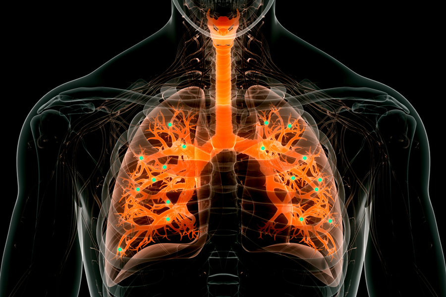
Engineers at MIT and the University of Massachusetts Medical School have designed a new type of nanoparticle that can be administered to the lungs, where it can deliver messenger RNA encoding useful proteins. Credits: Image: iStock, edited by MIT News

A March 30, 2023 MIT news release, also on EurekAlert, which originated the news item, describes the research in more detail,

Targeting the lungs

Messenger RNA holds great potential as a therapeutic for treating a variety of diseases caused by faulty genes. One obstacle to its deployment thus far has been difficulty in delivering it to the right part of the body, without off-target effects. Injected nanoparticles often accumulate in the liver, so several clinical trials evaluating potential mRNA treatments for diseases of the liver are now underway. RNA-based Covid-19 vaccines, which are injected directly into muscle tissue, have also proven effective. In many of those cases, mRNA is encapsulated in a lipid nanoparticle — a fatty sphere that protects mRNA from being broken down prematurely and helps it enter target cells. 

Several years ago, Anderson’s lab set out to design particles that would be better able to transfect the epithelial cells that make up most of the lining of the lungs. In 2019, his lab created nanoparticles that could deliver mRNA encoding a bioluminescent protein to lung cells. Those particles were made from polymers instead of lipids, which made them easier to aerosolize for inhalation into the lungs. However, more work is needed on those particles to increase their potency and maximize their usefulness. 

In their new study, the researchers set out to develop lipid nanoparticles that could target the lungs. The particles are made up of molecules that contain two parts: a positively charged headgroup and a long lipid tail. The positive charge of the headgroup helps the particles to interact with negatively charged mRNA, and it also help mRNA to escape from the cellular structures that engulf the particles once they enter cells. 

The lipid tail structure, meanwhile, helps the particles to pass through the cell membrane. The researchers came up with 10 different chemical structures for the lipid tails, along with 72 different headgroups. By screening different combinations of these structures in mice, the researchers were able to identify those that were most likely to reach the lungs. 

Efficient delivery

In further tests in mice, the researchers showed that they could use the particles to deliver mRNA encoding CRISPR/Cas9 components designed to cut out a stop signal that was genetically encoded into the animals’ lung cells. When that stop signal is removed, a gene for a fluorescent protein turns on. Measuring this fluorescent signal allows the researchers to determine what percentage of the cells successfully expressed the mRNA.

After one dose of mRNA, about 40 percent of lung epithelial cells were transfected, the researchers found. Two doses brought the level to more than 50 percent, and three doses up to 60 percent. The most important targets for treating lung disease are two types of epithelial cells called club cells and ciliated cells, and each of these was transfected at about 15 percent. 

“This means that the cells we were able to edit are really the cells of interest for lung disease,” Li says. “This lipid can enable us to deliver mRNA to the lung much more efficiently than any other delivery system that has been reported so far.”

The new particles also break down quickly, allowing them to be cleared from the lung within a few days and reducing the risk of inflammation. The particles could also be delivered multiple times to the same patient if repeat doses are needed. This gives them an advantage over another approach to delivering mRNA, which uses a modified version of harmless adenoviruses. Those viruses are very effective at delivering RNA but can’t be given repeatedly because they induce an immune response in the host.

To deliver the particles in this study, the researchers used a method called intratracheal instillation, which is often used as a way to model delivery of medication to the lungs. They are now working on making their nanoparticles more stable, so they could be aerosolized and inhaled using a nebulizer. 

The researchers also plan to test the particles to deliver mRNA that could correct the genetic mutation found in the gene that causes cystic fibrosis, in a mouse model of the disease. They also hope to develop treatments for other lung diseases, such as idiopathic pulmonary fibrosis, as well as mRNA vaccines that could be delivered directly to the lungs.

Here’s a link to and a citation from the paper,

Combinatorial design of nanoparticles for pulmonary mRNA delivery and genome editing by Bowen Li, Rajith Singh Manan, Shun-Qing Liang, Akiva Gordon, Allen Jiang, Andrew Varley, Guangping Gao, Robert Langer, Wen Xue & Daniel Anderson. Nature Biotechnology (2023) DOI: https://doi.org/10.1038/s41587-023-01679-x Published 30 March 2023

This paper is behind a paywall.
Counteracting chemotherapy resistance with nanoparticles that mimic salmonella

Given the reputation that salmonella (for those who don’t know, it’s a toxin you don’t want to find in your food) has, a nanoparticle which mimics its effects has a certain cachet. An Aug. 22, 2016 news item on Nanowerk,

Researchers at the University of Massachusetts Medical School have designed a nanoparticle that mimics the bacterium Salmonella and may help to counteract a major mechanism of chemotherapy resistance.

Working with mouse models of colon and breast cancer, Beth McCormick, Ph.D., and her colleagues demonstrated that when combined with chemotherapy, the nanoparticle reduced tumor growth substantially more than chemotherapy alone.

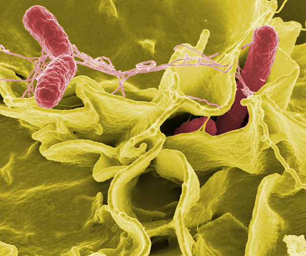
Credit: Rocky Mountain Laboratories,NIAID,NIHColor-enhanced scanning electron micrograph showing Salmonella typhimurium (red) invading cultured human cells.

Credit: Rocky Mountain Laboratories,NIAID,NIHColor-enhanced scanning electron micrograph showing Salmonella typhimurium (red) invading cultured human cells.

An Aug. 22, 2016 US National Institute of Cancer news release, which originated the news item, explains the research in more detail,

A membrane protein called P-glycoprotein (P-gp) acts like a garbage chute that pumps waste, foreign particles, and toxins out of cells. P-gp is a member of a large family of transporters, called ATP-binding cassette (ABC) transporters, that are active in normal cells but also have roles in cancer and other diseases. For instance, cancer cells can co-opt P-gp to rid themselves of chemotherapeutic agents, severely limiting the efficacy of these drugs.

In previous work, Dr. McCormick and her colleagues serendipitously discovered that Salmonella enterica, a bacterium that causes food poisoning, decreases the amount of P-gp on the surface of intestinal cells. Because Salmonella has the capacity to grow selectively in cancer cells, the researchers wondered whether there was a way to use the bacterium to counteract chemotherapy resistance caused by P-gp.

“While trying to understand how Salmonella invades the human host, we made this other observation that may be relevant to cancer therapeutics and multidrug resistance,” explained Dr. McCormick.

Salmonella and Cancer Cells

To determine the specific bacterial component responsible for reducing P-gp levels, the researchers engineered multiple Salmonella mutant strains and tested their effect on P-gp levels in colon cells. They found that a Salmonella strain lacking the bacterial protein SipA was unable to reduce P-gp levels in the colon of mice or in a human colon cancer cell line. Salmonella secretes SipA, along with other proteins, to help the bacterium invade human cells.

The researchers then showed that treatment with SipA protein alone decreased P-gp levels in cell lines of human colon cancer, breast cancer, bladder cancer, and lymphoma.

Because P-gp can pump drugs out of cells, the researchers next sought to determine whether SipA treatment would prevent cancer cells from expelling chemotherapy drugs.

When they treated human colon cancer cells with the chemotherapy agents doxorubicin or vinblastine, with or without SipA, they found that the addition of SipA increased drug retention inside the cells. SipA also increased the cancer cells’ sensitivity to both drugs, suggesting that it could possibly be used to enhance chemotherapy.

“Through millions of years of co-evolution, Salmonella has figured out a way to remove this transporter from the surface of intestinal cells to facilitate host infection,” said Dr. McCormick. “We capitalized on the organism’s ability to perform that function.”

A Nanoparticle Mimic

It would not be feasible to infect people with the bacterium, and SipA on its own will likely deteriorate quickly in the bloodstream, coauthor Gang Han, Ph.D., of the University of Massachusetts Medical School, explained in a press release. The researchers therefore fused SipA to gold nanoparticles, generating what they refer to as a nanoparticle mimic of Salmonella. They designed the nanoparticle to enhance the stability of SipA, while retaining its ability to interact with other proteins.

In an effort to target tumors without harming healthy tissues, the researchers used a nanoparticle of specific size that should only be able to access the tumor tissue due to its “leaky” architecture. “Because of this property, we are hoping to be able to avoid negative effects to healthy tissues,” said Dr. McCormick. Another benefit of this technology is that the nanoparticle can be modified to enhance tumor targeting and minimize the potential for side effects, she added.

The researchers showed that this nanoparticle was 100 times more effective than SipA protein alone at reducing P-gp levels in a human colon cancer cell line. The enhanced function of the nanoparticle is likely due to stabilization of SipA, explained the researchers.

The team then tested the nanoparticle in a mouse model of colon cancer, because this cancer type is known to express high levels of P-gp. When they treated tumor-bearing mice with the nanoparticle plus doxorubicin, P-gp levels dropped and the tumors grew substantially less than in mice treated with the nanoparticle or doxorubicin alone. The researchers observed similar results in a mouse model of human breast cancer.

There are concerns about the potential effect of nanoparticles on normal tissues. “P-gp has evolved as a defense mechanism” to rid healthy cells of toxic molecules, said Suresh Ambudkar, Ph.D., deputy chief of the Laboratory of Cell Biology in NCI’s Center for Cancer Research. It plays an important role in protecting cells of the blood-brain barrier, liver, testes, and kidney. “So when you try to interfere with that, you may create problems,” he said.

The researchers, however, found no evidence of nanoparticle accumulation in the brain, heart, kidney, or lungs of mice, nor did it appear to cause toxicity. They did observe that the nanoparticles accumulated in the liver and spleen, though this was expected because these organs filter the blood, said Dr. McCormick.

Moving Forward

The research team is moving forward with preclinical studies of the SipA nanoparticle to test its safety and toxicity, and to establish appropriate dosage levels.

However, Dr. Ambudkar noted, “the development of drug resistance in cancer cells is a multifactorial process. In addition to the ABC transporters, other phenomena are involved, such as drug metabolism.” And because there is a large family of ABC transporters, one transporter can compensate if another is blocked, he explained.

For the last 25 years, clinical trials with drugs that inhibit P-gp have failed to overcome chemotherapy resistance, Dr. Ambudkar said. Tackling the issue of multidrug resistance in cancer, he continued, “is not something that can be solved easily.”

Dr. McCormick and her team are also pursuing research to better characterize and understand the biology of SipA. “We are not naïve about the complexity of the problem,” she said. “However, if we know more about the biology, we believe we can ultimately make a better drug.”

Here’s a link to and a citation for the paper,

A Salmonella nanoparticle mimic overcomes multidrug resistance in tumours by Regino Mercado-Lubo, Yuanwei Zhang, Liang Zhao, Kyle Rossi, Xiang Wu, Yekui Zou, Antonio Castillo, Jack Leonard, Rita Bortell, Dale L. Greiner, Leonard D. Shultz, Gang Han, & Beth A. McCormick. Nature Communications 7, Article number: 12225  doi:10.1038/ncomms12225 Published 25 July 2016

This paper is open access.

A new nanoparticle—layered* like an onion

The new nanoparticle comes courtesy of an international collaboration (US, China, Sweden, and Russia. A Nov. 10, 2015 University of Buffalo news release (also on EurekAlert) by Charlotte Hu describes the particle and its properties,

A new, onion-like nanoparticle could open new frontiers in biomaging, solar energy harvesting and light-based security techniques.

The particle’s innovation lies in its layers: a coating of organic dye, a neodymium-containing shell, and a core that incorporates ytterbium and thulium. Together, these strata convert invisible near-infrared light to higher energy blue and UV light with record-high efficiency, a trick that could improve the performance of technologies ranging from deep-tissue imaging and light-induced therapy to security inks used for printing money.

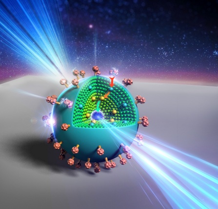
Here’s an artist’s representation of the new nanoparticle,

An artist’s rendering shows the layers of a new, onion-like nanoparticle whose specially crafted layers enable it to efficiently convert invisible near-infrared light to higher energy blue and UV light. Credit: Kaiheng Wei Courtesy: University of Buffalo

An artist’s rendering shows the layers of a new, onion-like nanoparticle whose specially crafted layers enable it to efficiently convert invisible near-infrared light to higher energy blue and UV light. Credit: Kaiheng Wei Courtesy: University of Buffalo

The news release goes on to describe technology in more detail,

When it comes to bioimaging, near-infrared light could be used to activate the light-emitting nanoparticles deep inside the body, providing high-contrast images of areas of interest. In the realm of security, nanoparticle-infused inks could be incorporated into currency designs; such ink would be invisible to the naked eye, but glow blue when hit by a low-energy laser pulse — a trait very difficult for counterfeiters to reproduce.

“It opens up multiple possibilities for the future,” says Tymish Ohulchanskyy, deputy director of photomedicine and research associate professor at the Institute for Lasers, Photonics, and Biophotonics (ILPB) at the University at Buffalo.

“By creating special layers that help transfer energy efficiently from the surface of the particle to the core, which emits blue and UV light, our design helps overcome some of the long-standing obstacles that previous technologies faced,” says Guanying Chen, professor of chemistry at Harbin Institute of Technology [China] and ILPB research associate professor.

“Our particle is about 100 times more efficient at ‘upconverting’ light than similar nanoparticles created in the past, making it much more practical,” says Jossana Damasco, a UB chemistry PhD student who played a key role in the project.

The research was published online in Nano Letters on Oct. 21 and led by the Institute for Lasers, Photonics, and Biophotonics at UB, and the Harbin Institute of Technology in China, with contributions from the Royal Institute of Technology in Sweden; Tomsk State University in Russia; and the University of Massachusetts Medical School.

The study’s senior author was Paras Prasad, ILPB executive director and SUNY [State University of New York] Distinguished Professor in chemistry, physics, medicine and electrical engineering at UB.

Peeling back the layers

Converting low-energy light to light of higher energies isn’t easy to do. The process involves capturing two or more tiny packets of light called “photons” from a low-energy light source, and combining their energy to form a single, higher-energy photon.

The onionesque nanoparticle performs this task beautifully. Each of its three layers fulfills a unique function:

  • The outermost layer is a coating of organic dye. This dye is adept at absorbing photons from low-energy near-infrared light sources. It acts as an “antenna” for the nanoparticle, harvesting light and transferring energy inside, Ohulchanskyy says.
  • The next layer is a neodymium-containing shell. This layer acts as a bridge, transferring energy from the dye to the particle’s light-emitting core.
  • Inside the light-emitting core, ytterbium and thulium ions work in concert. The ytterbium ions draw energy into the core and pass the energy on to the thulium ions, which have special properties that enable them to absorb the energy of three, four or five photons at once, and then emit a single higher-energy photon of blue and UV light.

So why not just use the core? Why add the dye and neodymium layer at all?

As Ohulchanskyy and Chen explain, the core itself is inefficient in absorbing photons from the outside world. That’s where the dye comes in.

Once you add the dye, the neodymium-containing layer is necessary for transferring energy efficiently from dye to core. Ohulchanskyy uses the analogy of a staircase to explain why this is: When molecules or ions in a material absorb a photon, they enter an “excited” state from which they can transfer energy to other molecules or ions. The most efficient transfer occurs between molecules or ions whose excited states require a similar amount of energy to obtain, but the dye and ytterbium ions have excited states with very different energies. So the team added neodymium — whose excited state is in between that of the dye and thulium’s — to act as a bridge between the two, creating a “staircase” for the energy to travel down to reach emitting thulium ions.

Here’s a link to and a citation for the paper,

Energy-Cascaded Upconversion in an Organic Dye-Sensitized Core/Shell Fluoride Nanocrystal by Guanying Chen, Jossana Damasco, Hailong Qiu, Wei Shao, Tymish Y. Ohulchanskyy, Rashid R. Valiev, Xiang Wu, Gang Han, Yan Wang, Chunhui Yang, Hans Ågren, and Paras N. Prasad. Nano Lett., 2015, 15 (11), pp 7400–7407 DOI: 10.1021/acs.nanolett.5b02830 Publication Date (Web): October 21, 2015

Copyright © 2015 American Chemical Society

This paper is behind a paywall.

Finally, there is a Nov. 11, 2015 article about the research by Jake Wilkinson for Azonano. He provides additional details such as this measurement,

Measuring approximately 50nm in diameter, the new nanoparticle features three differently designed layers. …

*’ayered’ changed to ‘layered’ on Nov. 11, 2015.

Wireless nano for remotely activating neurons

Every once in a while, there’s a piece of research that disconcerts me and this would be one of those pieces. From a May 22, 2014 news item on Nanowerk,

Yang Xiang, PhD, assistant professor of neurobiology at University of Massachusetts Medical School, has received a three-year, $900,000 grant from the Human Frontiers Science Program to lead an international team of scientists, including Gang Han, PhD, assistant professor of biochemistry & molecular pharmacology, in the development and implementation of a new optogenetic platform that can remotely activate neurons inside a free-moving organism.

Using a new class of nanoparticles developed by Dr. Han, Dr. Xiang and colleagues propose to selectively turn on non-image forming photoreceptors (NIFP) inside mice and Drosophila unencumbered by the fiber optic wires used in currently available optogenetic technologies. By wirelessly stimulating these photoreceptors, which are able to sense light even though they don’t generate vision, scientists can better understand their role in regulating physiological functions such as circadian rhythm, sleep and melatonin secretion. The hope is that this new technology can also be used to study the links between other types of neurons, physiology and behavior.

A May 22, 2014 University of Massachusetts Medical School news release by Jim Fessenden, which originated the news item, describes optogenetics and some of its challenges,

“Current optogenetic technologies are limited in their application because they require using ‘wired’ fiber optic implants to deliver blue light to activate neuron activities,” said Xiang. “This is a major technological problem that has become an obstacle to understanding the physiological role NIFP play in animal behavior. If we’re able to overcome this hurdle by using the nanoparticles developed by Dr. Han, it would open the door to more informed investigations of not only NIFP but a wide range of neurons and their effect on behavior.”

In use for only about a decade, optogenetic technology combines techniques from optics and genetics, allowing scientists to precisely control activities of individual neurons using light. By genetically inserting light-activated biological molecules such as channelrhodopsins, a family of proteins found in algae, into neurons, scientists can instantaneously turn them on using beams of blue light with millisecond precision.

A limiting factor to the wider application of this technology, however, is that blue wavelengths are unable to penetrate skin, bone and other tissues deep enough to activate the neurons inside free-moving animals. To overcome this obstacle, current techniques require the insertion of fiber optic wires close enough to the neurons so the light that activates them can be delivered. This technique restricts animal movement and makes it difficult to observe behavioral responses in natural conditions. This fiber optic approach further limits scientists’ ability to study behavior over longer periods of time as the effectiveness of light delivery is relatively short due to scarring.

The news release describes the new technique proposed by Xiang and his associates,

Han has developed an “upconversion nanoparticle” (UCNP) that has the potential to solve the limitations of wired optogenetic techniques. These nanoparticles are capable of absorbing infrared light that can’t be seen and converting it into visible blue light. In contrast to blue light, infrared light is capable of penetrating skin and tissue to a depth of several centimeters. Xiang and Han believe these nanoparticles, tuned to emit blue light, can be inserted into the brain and used as a substitute for traditional fiber optics to wirelessly activate neurons in animals.

The hope is that the nanoparticles will absorb infrared light that passes through the tissue, and convert it to blue light inside the animal. This blue light would then activate the NFIPs. If successful, Xiang and colleagues will be able to observe any changes in animal behavior brought about by activating these non-image forming photoreceptors.

“The nanoparticles act as a kind of relay station,” said Han. “They convert the low-energy red light into a high-energy blue light that can activate the neurons. This technique completely alleviates the need to use intrusive fiber optic wires. It vastly simplifies the technology and expands the potential uses for optogenetics.”

Xiang said, “In many ways, this is the perfect bridge between a technological advancement and an important biological question. With these nanoparticles it’s possible for us to begin answering fundamental neurobiological questions about NIFPs.

“More broadly, it would open up the possibility of using other model organisms, such as Drosophila, that can’t be used with the current wired optogenetic technologies, to investigate and answer important questions about how neural activities regulate behavior.”

Illogical as it is, the idea that neurons could be wirelessly and remotely activated by someone other the owner of those neurons disturbs me even though I know drugs are commonly used to do much the same thing in humans.

In any event, the news release provides this final paragraph about the funding,

HFSP [Human Frontiers Science Program] awards are given to highly innovative teams that demonstrate that they have developed and can test a paradigm-shifting idea that holds promise for the development of new approaches to problems in the life sciences with potential to advance the field of research significantly.

I looked up the HFSP online and found this on the About Us page on the HFSP website,

The Human Frontier Science Program is a program of funding for frontier research in the life sciences. It is implemented by the International Human Frontier Science Program Organization (HFSPO) with its office in Strasbourg.

The members of the HFSPO, the so-called Management Supporting Parties (MSPs) are the contributing countries and the European Union, which contributes on behalf of the non-G7 EU members.

The current MSPs are Australia, Canada, France, Germany, India, Italy, Japan, Republic of Korea, Norway, New Zealand, Switzerland the United Kingdom, the United States of America and the European Union. [emphasis mine]

I was not expecting to find Canada on that list.