Tag Archives: James Weaver

Answer to why Roman concrete was so durable

Roman concrete lasts for millenia while our ‘modern’ concrete doesn’t and that’s what makes the Roman stuff so fascinating. There’s a very good January 6, 2023 Massachusetts Institute of Technology (MIT) news release (also on EurekAlert) which may provide an answer the mystery of the this material’s longevity,

The ancient Romans were masters of engineering, constructing vast networks of roads, aqueducts, ports, and massive buildings, whose remains have survived for two millennia. Many of these structures were built with concrete: Rome’s famed Pantheon, which has the world’s largest unreinforced concrete dome and was dedicated in A.D. 128, is still intact, and some ancient Roman aqueducts still deliver water to Rome today. Meanwhile, many modern concrete structures have crumbled after a few decades.

Researchers have spent decades trying to figure out the secret of this ultradurable ancient construction material, particularly in structures that endured especially harsh conditions, such as docks, sewers, and seawalls, or those constructed in seismically active locations.

Now, a team of investigators from MIT, Harvard University, and laboratories in Italy and Switzerland, has made progress in this field, discovering ancient concrete-manufacturing strategies that incorporated several key self-healing functionalities. The findings are published in the journal Science Advances, in a paper by MIT professor of civil and environmental engineering Admir Masic, former doctoral student Linda Seymour, and four others.

For many years, researchers have assumed that the key to the ancient concrete’s durability was based on one ingredient: pozzolanic material such as volcanic ash from the area of Pozzuoli, on the Bay of Naples. [emphasis mine] This specific kind of ash was even shipped all across the vast Roman empire to be used in construction, and was described as a key ingredient for concrete in accounts by architects and historians at the time.

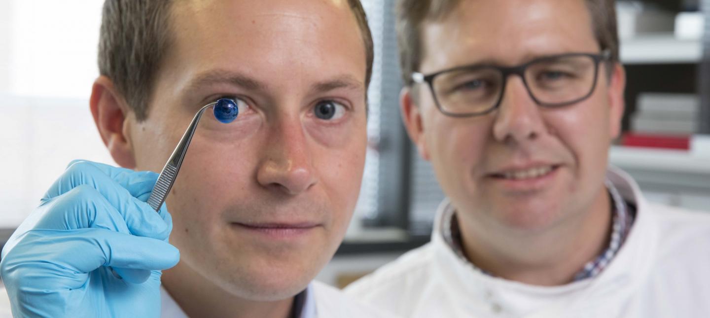
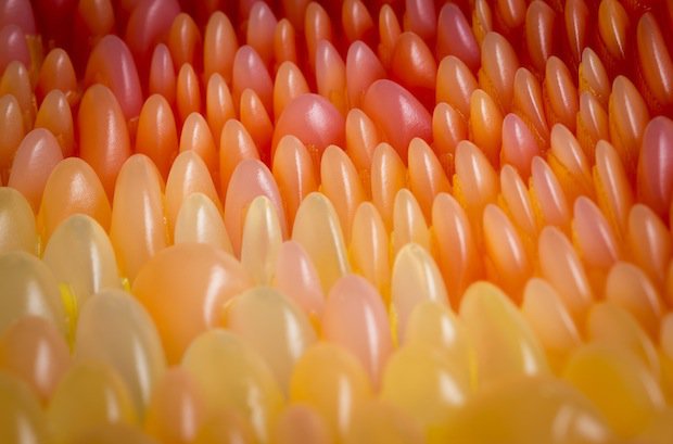
Under closer examination, these ancient samples also contain small, distinctive, millimeter-scale bright white mineral features, which have been long recognized as a ubiquitous component of Roman concretes. These white chunks, often referred to as “lime clasts,” originate from lime, another key component of the ancient concrete mix. “Ever since I first began working with ancient Roman concrete, I’ve always been fascinated by these features,” says Masic. “These are not found in modern concrete formulations, so why are they present in these ancient materials?”

Previously disregarded as merely evidence of sloppy mixing practices, or poor-quality raw materials, the new study suggests that these tiny lime clasts gave the concrete a previously unrecognized self-healing capability. [emphasis mine] “The idea that the presence of these lime clasts was simply attributed to low quality control always bothered me,” says Masic. “If the Romans put so much effort into making an outstanding construction material, following all of the detailed recipes that had been optimized over the course of many centuries, why would they put so little effort into ensuring the production of a well-mixed final product? There has to be more to this story.”

Upon further characterization of these lime clasts, using high-resolution multiscale imaging and chemical mapping techniques pioneered in Masic’s research lab, the researchers gained new insights into the potential functionality of these lime clasts.

Historically, it had been assumed that when lime was incorporated into Roman concrete, it was first combined with water to form a highly reactive paste-like material, in a process known as slaking. But this process alone could not account for the presence of the lime clasts. Masic wondered: “Was it possible that the Romans might have actually directly used lime in its more reactive form, known as quicklime?”

Studying samples of this ancient concrete, he and his team determined that the white inclusions were, indeed, made out of various forms of calcium carbonate. And spectroscopic examination provided clues that these had been formed at extreme temperatures, as would be expected from the exothermic reaction produced by using quicklime instead of, or in addition to, the slaked lime in the mixture. Hot mixing, the team has now concluded, was actually the key to the super-durable nature.

“The benefits of hot mixing are twofold,” Masic says. “First, when the overall concrete is heated to high temperatures, it allows chemistries that are not possible if you only used slaked lime, producing high-temperature-associated compounds that would not otherwise form. Second, this increased temperature significantly reduces curing and setting times since all the reactions are accelerated, allowing for much faster construction.”

During the hot mixing process, the lime clasts develop a characteristically brittle nanoparticulate architecture, creating an easily fractured and reactive calcium source, which, as the team proposed, could provide a critical self-healing functionality. As soon as tiny cracks start to form within the concrete, they can preferentially travel through the high-surface-area lime clasts. This material can then react with water, creating a calcium-saturated solution, which can recrystallize as calcium carbonate and quickly fill the crack, or react with pozzolanic materials to further strengthen the composite material. These reactions take place spontaneously and therefore automatically heal the cracks before they spread. Previous support for this hypothesis was found through the examination of other Roman concrete samples that exhibited calcite-filled cracks.

To prove that this was indeed the mechanism responsible for the durability of the Roman concrete, the team produced samples of hot-mixed concrete that incorporated both ancient and modern formulations, deliberately cracked them, and then ran water through the cracks. Sure enough: Within two weeks the cracks had completely healed and the water could no longer flow. An identical chunk of concrete made without quicklime never healed, and the water just kept flowing through the sample. As a result of these successful tests, the team is working to commercialize this modified cement material.

“It’s exciting to think about how these more durable concrete formulations could expand not only the service life of these materials, but also how it could improve the durability of 3D-printed concrete formulations,” says Masic.

Through the extended functional lifespan and the development of lighter-weight concrete forms, he hopes that these efforts could help reduce the environmental impact of cement production, which currently accounts for about 8 percent of global greenhouse gas emissions. Along with other new formulations, such as concrete that can actually absorb carbon dioxide from the air, another current research focus of the Masic lab, these improvements could help to reduce concrete’s global climate impact.

The research team included Janille Maragh at MIT, Paolo Sabatini at DMAT in Italy, Michel Di Tommaso at the Instituto Meccanica dei Materiali, in Switzerland, and James Weaver at the Wyss Institute for Biologically Inspired Engineering at Harvard University. The work was carried out with the assistance of the archeological museum of Priverno, Italy.

I remember the excitement over volcanic ash (it’s mentioned in my June 3, 2016 posting titled: “Making better concrete by looking to nature for inspiration” and my February 17, 2021 posting “Nuclear power plants take a cue from Roman concrete“). As for something being ignored as unimportant or being a result poor practice when it’s not, that’s one of my favourite kinds of science story.

For the really curious, Jennifer Ouellette’s January 6, 2023 article (Ancient Roman concrete could self-heal thanks to “hot mixing” with quicklime) for Ars Technica provides a little more detail.

Here’s a link to and a citation for the latest paper,

Hot mixing: Mechanistic insights into the durability of ancient Roman concrete by Linda M. Seymour, Janille Maragh, Paolo Sabatini, Michel Di Tommaso, James C. Weaver, and Admir Masic. Science Advances 6 Jan 2023 Vol 9, Issue 1 DOI: 10.1126/sciadv.add1602

This paper is open access.

One last note, DMAT is listed as Paolo Sabatini’s home institution. It is a company for which Sabatini is a co-founder and CEO (chief executive officer). DMAT has this on its About page, “Our mission is to develop breakthrough innovations in construction materials at a global scale. DMAT is at the helm of concrete’s innovation.”

A 3D printed eye cornea and a 3D printed copy of your brain (also: a Brad Pitt connection)

Sometimes it’s hard to keep up with 3D tissue printing news. I have two news bits, one concerning eyes and another concerning brains.

3D printed human corneas

A May 29, 2018 news item on ScienceDaily trumpets the news,

The first human corneas have been 3D printed by scientists at Newcastle University, UK.

It means the technique could be used in the future to ensure an unlimited supply of corneas.

As the outermost layer of the human eye, the cornea has an important role in focusing vision.

Yet there is a significant shortage of corneas available to transplant, with 10 million people worldwide requiring surgery to prevent corneal blindness as a result of diseases such as trachoma, an infectious eye disorder.

In addition, almost 5 million people suffer total blindness due to corneal scarring caused by burns, lacerations, abrasion or disease.

The proof-of-concept research, published today [May 29, 2018] in Experimental Eye Research, reports how stem cells (human corneal stromal cells) from a healthy donor cornea were mixed together with alginate and collagen to create a solution that could be printed, a ‘bio-ink’.

Here are the proud researchers with their cornea,

Caption: Dr. Steve Swioklo and Professor Che Connon with a dyed cornea. Credit: Newcastle University, UK

A May 30,2018 Newcastle University press release (also on EurekAlert but published on May 29, 2018), which originated the news item, adds more details,

Using a simple low-cost 3D bio-printer, the bio-ink was successfully extruded in concentric circles to form the shape of a human cornea. It took less than 10 minutes to print.

The stem cells were then shown to culture – or grow.

Che Connon, Professor of Tissue Engineering at Newcastle University, who led the work, said: “Many teams across the world have been chasing the ideal bio-ink to make this process feasible.

“Our unique gel – a combination of alginate and collagen – keeps the stem cells alive whilst producing a material which is stiff enough to hold its shape but soft enough to be squeezed out the nozzle of a 3D printer.

“This builds upon our previous work in which we kept cells alive for weeks at room temperature within a similar hydrogel. Now we have a ready to use bio-ink containing stem cells allowing users to start printing tissues without having to worry about growing the cells separately.”

The scientists, including first author and PhD student Ms Abigail Isaacson from the Institute of Genetic Medicine, Newcastle University, also demonstrated that they could build a cornea to match a patient’s unique specifications.

The dimensions of the printed tissue were originally taken from an actual cornea. By scanning a patient’s eye, they could use the data to rapidly print a cornea which matched the size and shape.

Professor Connon added: “Our 3D printed corneas will now have to undergo further testing and it will be several years before we could be in the position where we are using them for transplants.

“However, what we have shown is that it is feasible to print corneas using coordinates taken from a patient eye and that this approach has potential to combat the world-wide shortage.”

Here’s a link to and a citation for the paper,

3D bioprinting of a corneal stroma equivalent by Abigail Isaacson, Stephen Swioklo, Che J. Connon. Experimental Eye Research Volume 173, August 2018, Pages 188–193 and 2018 May 14 pii: S0014-4835(18)30212-4. doi: 10.1016/j.exer.2018.05.010. [Epub ahead of print]

This paper is behind a paywall.

A 3D printed copy of your brain

I love the title for this May 30, 2018 Wyss Institute for Biologically Inspired Engineering news release: Creating piece of mind by Lindsay Brownell (also on EurekAlert),

What if you could hold a physical model of your own brain in your hands, accurate down to its every unique fold? That’s just a normal part of life for Steven Keating, Ph.D., who had a baseball-sized tumor removed from his brain at age 26 while he was a graduate student in the MIT Media Lab’s Mediated Matter group. Curious to see what his brain actually looked like before the tumor was removed, and with the goal of better understanding his diagnosis and treatment options, Keating collected his medical data and began 3D printing his MRI [magnetic resonance imaging] and CT [computed tomography] scans, but was frustrated that existing methods were prohibitively time-intensive, cumbersome, and failed to accurately reveal important features of interest. Keating reached out to some of his group’s collaborators, including members of the Wyss Institute at Harvard University, who were exploring a new method for 3D printing biological samples.

“It never occurred to us to use this approach for human anatomy until Steve came to us and said, ‘Guys, here’s my data, what can we do?” says Ahmed Hosny, who was a Research Fellow with at the Wyss Institute at the time and is now a machine learning engineer at the Dana-Farber Cancer Institute. The result of that impromptu collaboration – which grew to involve James Weaver, Ph.D., Senior Research Scientist at the Wyss Institute; Neri Oxman, [emphasis mine] Ph.D., Director of the MIT Media Lab’s Mediated Matter group and Associate Professor of Media Arts and Sciences; and a team of researchers and physicians at several other academic and medical centers in the US and Germany – is a new technique that allows images from MRI, CT, and other medical scans to be easily and quickly converted into physical models with unprecedented detail. The research is reported in 3D Printing and Additive Manufacturing.

“I nearly jumped out of my chair when I saw what this technology is able to do,” says Beth Ripley, M.D. Ph.D., an Assistant Professor of Radiology at the University of Washington and clinical radiologist at the Seattle VA, and co-author of the paper. “It creates exquisitely detailed 3D-printed medical models with a fraction of the manual labor currently required, making 3D printing more accessible to the medical field as a tool for research and diagnosis.”

Imaging technologies like MRI and CT scans produce high-resolution images as a series of “slices” that reveal the details of structures inside the human body, making them an invaluable resource for evaluating and diagnosing medical conditions. Most 3D printers build physical models in a layer-by-layer process, so feeding them layers of medical images to create a solid structure is an obvious synergy between the two technologies.

However, there is a problem: MRI and CT scans produce images with so much detail that the object(s) of interest need to be isolated from surrounding tissue and converted into surface meshes in order to be printed. This is achieved via either a very time-intensive process called “segmentation” where a radiologist manually traces the desired object on every single image slice (sometimes hundreds of images for a single sample), or an automatic “thresholding” process in which a computer program quickly converts areas that contain grayscale pixels into either solid black or solid white pixels, based on a shade of gray that is chosen to be the threshold between black and white. However, medical imaging data sets often contain objects that are irregularly shaped and lack clear, well-defined borders; as a result, auto-thresholding (or even manual segmentation) often over- or under-exaggerates the size of a feature of interest and washes out critical detail.

The new method described by the paper’s authors gives medical professionals the best of both worlds, offering a fast and highly accurate method for converting complex images into a format that can be easily 3D printed. The key lies in printing with dithered bitmaps, a digital file format in which each pixel of a grayscale image is converted into a series of black and white pixels, and the density of the black pixels is what defines the different shades of gray rather than the pixels themselves varying in color.

Similar to the way images in black-and-white newsprint use varying sizes of black ink dots to convey shading, the more black pixels that are present in a given area, the darker it appears. By simplifying all pixels from various shades of gray into a mixture of black or white pixels, dithered bitmaps allow a 3D printer to print complex medical images using two different materials that preserve all the subtle variations of the original data with much greater accuracy and speed.

The team of researchers used bitmap-based 3D printing to create models of Keating’s brain and tumor that faithfully preserved all of the gradations of detail present in the raw MRI data down to a resolution that is on par with what the human eye can distinguish from about 9-10 inches away. Using this same approach, they were also able to print a variable stiffness model of a human heart valve using different materials for the valve tissue versus the mineral plaques that had formed within the valve, resulting in a model that exhibited mechanical property gradients and provided new insights into the actual effects of the plaques on valve function.

“Our approach not only allows for high levels of detail to be preserved and printed into medical models, but it also saves a tremendous amount of time and money,” says Weaver, who is the corresponding author of the paper. “Manually segmenting a CT scan of a healthy human foot, with all its internal bone structure, bone marrow, tendons, muscles, soft tissue, and skin, for example, can take more than 30 hours, even by a trained professional – we were able to do it in less than an hour.”

The researchers hope that their method will help make 3D printing a more viable tool for routine exams and diagnoses, patient education, and understanding the human body. “Right now, it’s just too expensive for hospitals to employ a team of specialists to go in and hand-segment image data sets for 3D printing, except in extremely high-risk or high-profile cases. We’re hoping to change that,” says Hosny.

In order for that to happen, some entrenched elements of the medical field need to change as well. Most patients’ data are compressed to save space on hospital servers, so it’s often difficult to get the raw MRI or CT scan files needed for high-resolution 3D printing. Additionally, the team’s research was facilitated through a joint collaboration with leading 3D printer manufacturer Stratasys, which allowed access to their 3D printer’s intrinsic bitmap printing capabilities. New software packages also still need to be developed to better leverage these capabilities and make them more accessible to medical professionals.

Despite these hurdles, the researchers are confident that their achievements present a significant value to the medical community. “I imagine that sometime within the next 5 years, the day could come when any patient that goes into a doctor’s office for a routine or non-routine CT or MRI scan will be able to get a 3D-printed model of their patient-specific data within a few days,” says Weaver.

Keating, who has become a passionate advocate of efforts to enable patients to access their own medical data, still 3D prints his MRI scans to see how his skull is healing post-surgery and check on his brain to make sure his tumor isn’t coming back. “The ability to understand what’s happening inside of you, to actually hold it in your hands and see the effects of treatment, is incredibly empowering,” he says.

“Curiosity is one of the biggest drivers of innovation and change for the greater good, especially when it involves exploring questions across disciplines and institutions. The Wyss Institute is proud to be a space where this kind of cross-field innovation can flourish,” says Wyss Institute Founding Director Donald Ingber, M.D., Ph.D., who is also the Judah Folkman Professor of Vascular Biology at Harvard Medical School (HMS) and the Vascular Biology Program at Boston Children’s Hospital, as well as Professor of Bioengineering at Harvard’s John A. Paulson School of Engineering and Applied Sciences (SEAS).

Here’s an image illustrating the work,

Caption: This 3D-printed model of Steven Keating’s skull and brain clearly shows his brain tumor and other fine details thanks to the new data processing method pioneered by the study’s authors. Credit: Wyss Institute at Harvard University

Here’s a link to and a citation for the paper,

From Improved Diagnostics to Presurgical Planning: High-Resolution Functionally Graded Multimaterial 3D Printing of Biomedical Tomographic Data Sets by Ahmed Hosny , Steven J. Keating, Joshua D. Dilley, Beth Ripley, Tatiana Kelil, Steve Pieper, Dominik Kolb, Christoph Bader, Anne-Marie Pobloth, Molly Griffin, Reza Nezafat, Georg Duda, Ennio A. Chiocca, James R.. Stone, James S. Michaelson, Mason N. Dean, Neri Oxman, and James C. Weaver. 3D Printing and Additive Manufacturing http://doi.org/10.1089/3dp.2017.0140 Online Ahead of Print:May 29, 2018

This paper appears to be open access.

A tangential Brad Pitt connection

It’s a bit of Hollywood gossip. There was some speculation in April 2018 that Brad Pitt was dating Dr. Neri Oxman highlighted in the Wyss Institute news release. Here’s a sample of an April 13, 2018 posting on Laineygossip (Note: A link has been removed),

It took him a long time to date, but he is now,” the insider tells PEOPLE. “He likes women who challenge him in every way, especially in the intellect department. Brad has seen how happy and different Amal has made his friend (George Clooney). It has given him something to think about.”

While a Pitt source has maintained he and Oxman are “just friends,” they’ve met up a few times since the fall and the insider notes Pitt has been flying frequently to the East Coast. He dropped by one of Oxman’s classes last fall and was spotted at MIT again a few weeks ago.

Pitt and Oxman got to know each other through an architecture project at MIT, where she works as a professor of media arts and sciences at the school’s Media Lab. Pitt has always been interested in architecture and founded the Make It Right Foundation, which builds affordable and environmentally friendly homes in New Orleans for people in need.

“One of the things Brad has said all along is that he wants to do more architecture and design work,” another source says. “He loves this, has found the furniture design and New Orleans developing work fulfilling, and knows he has a talent for it.”

It’s only been a week since Page Six first broke the news that Brad and Dr Oxman have been spending time together.

I’m fascinated by Oxman’s (and her colleagues’) furniture. Rose Brook writes about one particular Oxman piece in her March 27, 2014 posting for TCT magazine (Note: Links have been removed),

MIT Professor and 3D printing forerunner Neri Oxman has unveiled her striking acoustic chaise longue, which was made using Stratasys 3D printing technology.

Oxman collaborated with Professor W Craig Carter and Composer and fellow MIT Professor Tod Machover to explore material properties and their spatial arrangement to form the acoustic piece.

Christened Gemini, the two-part chaise was produced using a Stratasys Objet500 Connex3 multi-colour, multi-material 3D printer as well as traditional furniture-making techniques and it will be on display at the Vocal Vibrations exhibition at Le Laboratoire in Paris from March 28th 2014.

An Architect, Designer and Professor of Media, Arts and Science at MIT, Oxman’s creation aims to convey the relationship of twins in the womb through material properties and their arrangement. It was made using both subtractive and additive manufacturing and is part of Oxman’s ongoing exploration of what Stratasys’ ground-breaking multi-colour, multi-material 3D printer can do.

Brook goes on to explain how the chaise was made and the inspiration that led to it. Finally, it’s interesting to note that Oxman was working with Stratasys in 2014 and that this 2018 brain project is being developed in a joint collaboration with Statasys.

That’s it for 3D printing today.