Monthly Archives: July 2012

Ethical nano in Second Life

Isn’t Second Life dead? Apparently not.

While you won’t be able to attend the live event online, there will be free access to the nano and ethics discussion held on July 20, 2012, from 1 pm to 4 pm EDT at the Terasem Island Conference Center in Second Life. The question and speakers were (from the July 20, 2012 event posting on the Kurzweil Accelerating Intelligence website,

What should be the ethical constraints on nanotechnology?

Speakers include:

  • Martine Rothblatt, Ph.D. — “Geoethical Rules for Nanotechnological Advances”
  • Peter Wicks — “Nanotechnology and the Environment: Enemies or Allies?”
  • Alex Wissner-Gross, Ph.D. — “Physically Programmable Surfaces”

The workshop is an exchange of scholarly views on uses of lifesaving nanotechnologies, including the impacts on people, accessibility, monitoring compliance with ethical norms.

I think if you check out the Terasem Island Conference Center in Second Life (SLURL), you will be able to access the archived discussion.

Pop up event based on European Commission’s Science: It’s a girl thing on July 27, 2012 in Vancouver (Canada)

The Society for Canadian Women in Science and Technology (SCWIST) will be holding a free ‘pop up’ event at Joey’s on Broadway (1424 W. Broadway at Hemlock St.) on Friday, July 27, 2012 from 6 pm – 8 pm.This event is a local outcome of the international discussion taking place about the European Commissions’ Science: It’s a Girl Thing campaign video (first mentioned in my July 6, 2012 posting and then in my July 18, 2012 posting).

Here’s more about the Vancouver topic and the event (from the July 20, 2012 posting on the Westcoast Women in Engineering, Science, and Technology (WWEST) blog on the University of British Columbia website),

Topic: It’s a girl thing: How do we get more girls to pursue STEM [Science, Technology, Engineering, and Mathematics] careers?

What is a SCWIST Pop-Up Discussion? A casual evening of networking, socializing, and discussion on current and relevant media topics held at a local restaurant! It’s a chance to get out and chat and network with like-minded people!

There’s also information abut th4 event on the SCWIST  Facebook page.

Zombies, brains, collapsing boundaries, and entanglements at the 4th annual S.NET conference

My proposal, Zombies, brains, collapsing boundaries, and entanglements, for the 4th annual S.NET (Society for the Study of Nanoscience and Emerging Technologies) conference was accepted. Mentioned in my Feb. 9, 2012 posting, the conference will be held at the University of Twente (Netherlands) from Oct. 22 – 25, 2012.

Here’s the abstract I provided,

The convergence between popular culture’s current fascination with zombies and their appetite for human brains (first established in the 1985 movie, Night of the Living Dead) and an extraordinarily high level of engagement in brain research by various medical and engineering groups around the world is no coincidence

Amongst other recent discoveries, the memristor (a concept from nanoelectronics) is collapsing the boundaries between humans and machines/robots and ushering in an age where humanistic discourse must grapple with cognitive entanglements.

Perceptible only at the level of molecular electronics (nanoelectronics), the memristor was a theoretical concept until 2008. Traditionally in electrical engineering, there are three circuit elements: resistors, inductors, and capacitors. The new circuit element, the memristor, was postulated in a paper by Dr. Leon Chua in 1971 to account for anomalies that had been experienced and described in the literature since the 1950s.

According to Chua’s theory and confirmed by the research team headed by R. Stanley Williams, the memristor remembers how much and when current has been flowing. The memristor is capable of an in-between state similar to certain brain states and this capacity lends itself to learning. As some have described it, the memristor is a synapse on a chip making neural computing a reality and/or the possibility of repairing brains stricken with neurological conditions. In other words, with post-human engineering exploiting discoveries such as the memristor we will have machines/robots that can learn and think and human brains that could incorporate machines.

As Jacques Derrida used the zombie to describe a state that this is neither life nor death as undecidable, the memristor can be described as an agent of transformation conferring robots with the ability to learn (a human trait) thereby rendering them as undecidable, i.e., neither machine nor life. Mirroring its transformative agency in robots, the memristor could also confer the human brain with machine/robot status and undecidability when used for repair or enhancement.

The memristor moves us past Jacques Derrida’s notion of undecidability as largely theoretical to a world where we confront this reality in a type of cognitive entanglement on a daily basis.

You can find the preliminary programme here.  My talk is scheduled for Thursday, Oct. 25, 2012 in one of the last sessions for the conference, 11 – 12:30 pm in the Tracing Transhuman Narratives strand.

I do see a few names I recognize, Wickson, Pat (Roy)  Mooney and Youtie. I believe Wickson is Fern Wickson from the University of Bergen last mentioned here in a Jul;y 7, 2010 posting about nature, nanotechnology, and metaphors. Pat Roy Mooney is from The ETC Group (an activist or civil society group) and was last mentioned here in my Oct. 7, 2011 posting), and I believe Youtie is Jan Youtie who wss mentioned in my March 29, 2012 posting about nanotechnology, economic impacts, and full life cycle assessments.

Nanopore instruments, femtomolar concentrations, Ireland, and New Zealand

It was the word femtomolar that did it for me. While I have somehow managed to conceptualize the nanoscale, the other scales (pico, femto, atto, zetto, and yocto) continue to  elude me. If my experience with the ‘nanoscale ‘ is any guide, the only solution will be to find as much information as I can on these other ones and immerse myself in them. With that said, here’s more from the July 19, 2012 Izon press release,

Researchers at the Lee Bionanosciences Laboratory at UCD [University College Dublin] School of Chemistry and Chemical Biology in Dublin have demonstrated the detection and measurement of biological analytes down to femtomolar concentration levels using an off the shelf qNano instrument. This ultra low level biodetection capability has implications for biomedical research and clinical development as trace amounts of a biological substance in a sample can now be detected amd quantfied using standard commercially available equipment.

Platt [Dr Mark Platt] and colleagues’ [Professor Gil Lee and Dr Geoff Willmott] method for femtomolar-level detection uses magnetic particle systems and can be applied to any biological particle or protein for which specific aptamers or antibodies exist. Resistive pulse sensing, the underlying technology of the qNano [Izon product], was used to monitor individual and aggregated rod-shaped nanoparticles as they move through tunable pores in elastomeric membranes.

Dr Platt says, “The strength of using the qNano is the ability to interrogate individual particles through a nanopore. This allowed us to establish a very sensitive measurement of concentration because we could detect the interactions occurring down to individual particle level.

”The unique and technically innovative approach of the authors was to detect a molecule’s presence by a process that results in end on end or side by side aggregation of rod shaped nickel-gold particles. The rods were designed so that the aptamers could be attached to one end only.

“By comparing particles of similar dimensions we demonstrated that the resistive pulse signal is fundamentally different for rod and sphere-shaped particles, and for rod shaped particles of different lengths. We could exploit these differences in a new agglutina¬tion assay to achieve these low detection levels,” says Dr Platt.

In the agglutination assay particles with a particular aspect ratio can be distinguished by two measurements: the measured drop in current as particles traverse the pore (∆ip), which reveals the particle’s size; and the full width at half maximum (FWHM) duration of the resistive pulse, which relates to the particle’s speed and length. Limits of detection down to femtomolar levels were thus able to be demonstrated.

I’m a little unclear as to what qNano actually is. I did find this description on the qNano product page,

qNano uses unique nanopore-based detection to enable the physical properties of a wide range of particle types to be measured with unsurpassed accuracy.

Detailed Multi-Parameter Analysis.

Particle-by-particle measurement through qNano enables detailed determination of:

Nanopore-based detection allows thousands of particles to be measured individually, providing far greater detail and accuracy than light-based techniques.

Applications & Particle Types

A wide range of biological and synthetic particle types, spanning 50 nm – 10 μm, can be measured, across a broad range of research fields.

qNano Package

qNano is sold as a full system ready for use including the base instrument, variable pressure module, fluid cell and a starter kit of nanopores, buffer solution and standard particle sets.

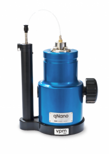
Here’s what the product looks like,

qNano (from the Izon website)

As for what this all might mean to those of us who exist at the macroscale (from the Izon press release),

Izon Science will continue to work with Dr Platt at Loughborough, and with University College Dublin and various customers to develop a series of diagnostic kits that can be used with the qNano to identify and measure biomolecules, viruses, and microvesicles.“This is a real milestone for Izon’s technology as being able to measure biomolecules down to these extremely low levels opens up new bio-analysis options for researchers. 10 femtomolar was achieved, which is the equivalent dilution to 1 gram in 3.3 billion litres, or 1 gram in 1300 Olympic sized swimming pools,” says Hans van der Voorn, Executive Chairman of Izon Science.

For those interested in finding out about nanopores, these were mentioned in my July 18, 2012 posting while aptamers were discussed in my interview (Oct. 25, 2011 posting) with Dr. Maria DeRosa who researches them in her Carleton University laboratory (Ottawa, Canada).

RNA (ribonucleic acid) video game

I am a great fan of  Foldit, a protein-folding game I have mentioned several times here (my first posting about Foldit was Aug. 6, 2010) and now via the Foresight Insitute’s July 16, 2012 blog posting, I have discovered an RNA video game (Note: I have removed links),

As we pointed out a few months ago, the greater complexity of folding rules for RNA compared to its chemical cousin DNA gives RNA a greater variety of compact, three-dimensional shapes and a different set of potential functions than is the case with DNA, and this gives RNA nanotechnology a different set of advantages compared to DNA nanotechnology … Proteins have even more complex folding rules and an even greater variety of structures and functions. We also noted here that online gamers playing Foldit topped scientists in redesigning a protein to achieve a novel enzymatic activity that might be especially useful in developing molecular building blocks for molecular manufacturing. Now KurzweilAI.net brings news of an online game that allows players to design RNA molecules …

Here’s more from the KurzwelAI.net June 26, 3012 posting about the new RNA game EteRNA,

EteRNA, an online game with more than 38,000 registered users, allows players to design molecules of ribonucleic acid — RNA — that have the power to build proteins or regulate genes.

EteRNA players manipulate nucleotides, the fundamental building blocks of RNA, to coax molecules into shapes specified by the game.

Those shapes represent how RNA appears in nature while it goes about its work as one of life’s most essential ingredients.

EteRNA was developed by scientists at Stanford and Carnegie Mellon universities, who use the designs created by players to decipher how real RNA works. The game is a direct descendant of Foldit — another science crowdsourcing tool disguised as entertainment — which gets players to help figure out the folding structures of proteins.

Here’s how the EteRNA folks describe this game (from the About EteRNA page),

By playing EteRNA, you will participate in creating the first large-scale library of synthetic RNA designs. Your efforts will help reveal new principles for designing RNA-based switches and nanomachines — new systems for seeking and eventually controlling living cells and disease-causing viruses. By interacting with thousands of players and learning from real experimental feedback, you will be pioneering a completely new way to do science. Join the global laboratory!

The About EteRNA webpage also offers a discussion about RNA,

RNA is often called the “Dark Matter of Biology.” While originally thought to be an unstable cousin of DNA, recent discoveries have shown that RNA can do amazing things. They play key roles in the fundamental processes of life and disease, from protein synthesis and HIV replication, to cellular control. However, the full biological and medical implications of these discoveries is still being worked out.

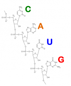
RNA is made of four nucleotides (A, C,G,and U, which stand for adenine, cytosine, guanine, and uracil). Chemically, each of these building blocks is made of atoms of carbon, oxygen, nitrogen, phosphorus, and hydrogen. When you design RNAs with EteRNA, you’re really creating a chain of these nucleotides.

RNA Nucleotides (from the About EteRNA webpage)

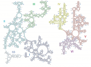
Scientists do not yet understand all of RNA’s roles, but we already know about a large collection of RNAs that are critical for life: (see the Thermus Thermophilus image representing following points)

  1. mRNAs are short copies of a cell’s DNA genome that gets cut up, pasted, spliced, and otherwise remixed before getting translated into proteins.
  1. rRNA forms the core machinery of an ancient machine, the ribosome. This machine synthesizes the proteins of your cells and all living cells, and is the target of most antibiotics.
  2. miRNAs (microRNAs) are short molecules (about 22-letters) that are used by all complex cells as commands for silencing genes and appear to have roles in cancer, heart disease, and other medical problems.
  3. Riboswitches are ubiquitous in bacteria. They sense all sorts of small molecules that could be food or signals from other bacteria, and turn on or off genes by changing their shapes. These are interesting targets for new antibiotics.
  4. Ribozymes are RNAs that can act as enzymes. They catalyze chemical reactions like protein synthesis and RNA splicing, and provide evidence of RNA’s dominance in a primordial stage of Life’s evolution.
  5. Retroviruses, like Hepatitis C, poliovirus, and HIV, are very large RNAs coated with proteins.
  6. And much much more… shRNA, piRNA, snRNA, and other new classes of important RNAs are being discovered every year.

Thermus Thermophilus – Large Subunit Ribosomal RNA
Source: Center for Molecular Biology (downloaded from the About EteRNA webpage)

I do wonder about the wordplay EteRNA/eternal. Are these scientists trying to tell us something?

Science should it be ‘in’ or ‘and’ society?

The European Science  Foundation (ESF) has just launched a report on science ‘in’ society as opposed to the more common phrasing science ‘and’ society. I’m not sure changing  from science ‘and’ society to science ‘in’ society is going to be noticed all that much. (In marketing circles it was known as that the company’s correct name was The Gap but no one ever called it that and it was rarely written correctly by anyone other than company officials. Despite strenuous effort, it remained the Gap.)

From the July 16, 2012 news release on EurekAlert,

At the ESOF 2012 conference The European Science Foundation’s (ESF) dedicated Member Organisation Forum (MO Forum) on ‘Science in Society Relationships’ has released its latest report: “Science in Society: a Challenging Frontier for Science Policy”. The report has called for a strengthening of ‘Science in Society’ (SiS) activities in a time of ambiguity for science.

The latest report aims to highlight the role of science in society, to raise awareness of how scientific knowledge is translated into society and to encourage better practice in the relationship between science and society. In order to achieve a better society and increase the quality of research and innovation, the MO Forum offers several recommendations:

  • A clear commitment to SiS in MO science policy and strategy has to be enhanced
  • Transparent SiS processes must be put in place within the organisational structures of Member Organisations and other research funding and performing bodies. SiS processes must also be seen as an essential and central part of a researcher’s work
  • Researchers and research groups must be properly rewarded for their work in this area
  • More experiments concerning instruments, activities and methods should be encouraged
  • Sharing experience and best practice through networks for exchange within Europe on a regular basis would increase efficiency in SiS

Networks to jointly develop systems for indicators, evaluations and measurements are needed. There is a need to coordinate efforts for greater impact. Organisations need the instruments to do this and this involves ensuring that SiS activities are formally evaluated, which is not the case today

You can download the report from here. Don’t worry about using the ‘shopping cart’, all I had to do was click on the ‘download’ button. I liked this graphic (from p. 11 of the PDF of “Science in Society: a Challenging Frontier for Science Policy”), which illustrates a changing approach to the topic,

Given that my primary interest is the communication of science (in whatever direction it occurs), I was most interested in these recommendations (on pp. 21-2 of the PDF),

4.2.1 Key recommendations for Research Funding Organisations (RFOs)

RFOs can be small or large, public or private, typically employing from 20 to 200 administrative staff. The main focus for organisations of this sort should be to ensure that there is sufficient funding for SiS activities. RFOs also have a responsibility to evaluate the quantity, quality and impact of SiS activities and to use this evaluation to reward researchers and research groups accordingly. Measures appropriate for RFOs are:

• Start by surveying the current situation. Is the present model working and does it fit with the mission statement with regard to SiS? Analyse if the funding situation with regard to SiS activities is optimal for the task. Is it within the research grant, or is there a separate fund for SiS, or a mixture of the two?

• Identify how to monitor what activities researchers funded by your MO currently participate in. Consider the audience being targeted, the impact, the quality, the cost (in terms of time and money). In what ways can you as a funding body influence this?

• Experiment, explore and learn ways to increase and enhance researchers’ participation in SiS relations. There should be inclusiveness across sciences and research groups; it should not be mandatory for every individual researcher but it is imperative that the dialogue is not confined to just a few selected researchers and research groups.

• Identify gaps in SiS activities, capacities and expertise where extra funding or support, initiated through the funding scheme, could improve things; for example through training and workshops.

4.2.2 Key recommendations for Research Performing Organisations (RPOs)

RPOs are often large organisations with tens, hundreds or thousands of researchers. This type of organisation, therefore, has the capacity to heavily influence the decisions and motivations of researchers when they consider involvement in SiS activities. Thus RPOs should ensure that there are sufficient resources allocated for SiS activities. RPOs also have the potential to include SiS as a consideration in promotions and pay rises. They will also have the power and the facilities to coordinate, organise or simply participate in training and workshops around SiS.

• Start by surveying current practices of society/ public interaction. Identify what activities go on now, the audience reached, the impact, the quality, the cost (in terms of time and money). Do they fit with the mission statement?

• Define and compare their qualities, publics, efficiency, etc. Select which seem to be more efficient and describe them in their context. Identify areas of strength and of weakness.

• Survey funding available to research groups for dissemination of research results (e.g. budgets provided for the EU-funded projects). Could the funds be used more efficiently provided that RPOs offer professional support to researchers both in performing SiS activities and in drafting new projects?

• Identify ways to increase and enhance scientists’ participation in SiS. There should be inclusiveness across sciences and research groups; it should not be mandatory for every individual researcher but it is imperative that the dialogue is not confined to just a few selected researchers and research groups.

• Identify gaps in funding, capacity and expertise and make plans to provide the necessary funding, training and support by professional staff for researchers and research groups.

• Integrate the scientific education of researchers with skills for science communication and dissemination.

• Raise awareness of stakeholders and decision makers, as well as of public groups of different kinds.

• Set up or participate in infrastructures for public engagement activities and communication arenas of all kinds: forums, dialogues, training, competences, etc.

I think the really important recommendation (if you want to see action from the researchers) is this one (from p. 27 of the PDF),

4.6 Make evaluation of SiS part of research funding schemes

By and large, there is need for change in the culture of scientific organisations. This is a clear conclusion from the work of the MO Forum on SiS relations. SiS activities should not represent an obstacle to researchers’ career progress. One effective way for RFOs and RPOs to show that they value SiS is to consider rewarding researchers for their SiS work, particularly by means of funding and merits.

The MO Forum recommends that RFOs and RPOs consider the following measures as a first step towards linking SiS activities with research funding.

(a) Introduce evaluation methods and indicators: [emphasis mine]

• Activities and time spent

• Resources – budget and human resources

• Income

• Develop impact measurements

• Indicators should be simple, transparent, easy to collect, generally accepted

(b) Make SiS an intrinsic part of funding and merits:

• Introduce SiS requirements at grant application stage – for instance, a plan of SiS activities at the grant application stage in order to prompt researchers to think about SiS issues

• In peer review decisions, use SiS as a differentiator when projects score equally on scientific excellence

• Collect data on SiS and enable researchers to report their SiS activities within current grant monitoring systems (annual, interim, end-of-grant, evaluation reports)

• When awarding grants, allocate a percentage of time to be spent on SiS activities

• Allocate funds for specific SiS-promoting activities

I believe this is where some previous programmes have fallen short. Including a section on funding applications about ‘public engagement’ or ‘science in society’ activities is all very well but it’s meaningless unless there is some overview and evaluation of the activities undertaken. As well, I think that at least one more element should be introduced and that’s substantive encouragement and recognition for the efforts from academic institutions in the form of career credit.  The unwritten rule in academe (in Canada anyway) is still that you’re better off with research credits than teaching credits despite all the chat about the importance of students and their experience in the classroom.

One last note, I was quite intrigued by the definition of science used in the report (from p. 9 of the PDF),

Science may be considered as a broad field which includes a body of publicly proven knowledge that is separated into specific fields (disciplines). Research may be defined as the exploration of new fields or new questions, as exploratory activities by scientists in search of new approaches which contribute to our understanding of the world as well as to influence the world.

In this report, we took into consideration both aspects; so ‘science’ refers to science or research, and covers both abstract and practical activities, and encompasses all sciences, including humanities and social sciences as well as natural sciences, medicine and engineering.

It’s the first time I’ve seen the humanities included.
Repairing joints with nanoscale scaffolds and stem cells

Cartilage damage is a major problem for millions of people and chondroitin supplements are widely used to counteract the pain and damage since cartilage does not regrow. Until now.

Researchers at Johns Hopkins University have used chondroitin sulfate to create nanoscaffolds for growing new cartilage. From the July 17, 2012 news release on EurekAlert,

Unlike skin, cartilage can’t repair itself when damaged. For the last decade, Elisseeff’s [Jennifer Elisseeff, Ph.D., Jules Stein Professor of Ophthalmology and director of the Translational Tissue Engineering Center at the Johns Hopkins University School of Medicine] team has been trying to better understand the development and growth of cartilage cells called chondrocytes, while also trying to build scaffolding that mimics the cartilage cell environment and generates new cartilage tissue. This environment is a 3-dimensional mix of protein fibers and gel that provides support to connective tissue throughout the body, as well as physical and biological cues for cells to grow and differentiate.

In the laboratory, the researchers created a nanofiber-based network using a process called electrospinning, which entails shooting a polymer stream onto a charged platform, and added chondroitin sulfate—a compound commonly found in many joint supplements—to serve as a growth trigger. After characterizing the fibers, they made a number of different scaffolds from either spun polymer or spun polymer plus chondroitin. They then used goat bone marrow-derived stem cells (a widely used model) and seeded them in various scaffolds to see how stem cells responded to the material.

Elisseeff  and her team watched the cells grow and found that compared to cells growing without scaffold, these cells developed into more voluminous, cartilage-like tissue. “The nanofibers provided a platform where a larger volume of tissue could be produced,” says Elisseeff, adding that 3-dimensional nanofiber scaffolds were more useful than the more common nanofiber sheets for studying cartilage defects in humans.

They’ve also experimented with animal models,

The investigators then tested their system in an animal model. They implanted the nanofiber scaffolds into damaged cartilage in the knees of rats, and compared the results to damaged cartilage in knees left alone.

They found that the use of the nanofiber scaffolds improved tissue development and repair as measured by the production of collagen, a component of cartilage. The nanofiber scaffolds resulted in greater production of a more durable type of collagen, which is usually lacking in surgically repaired cartilage tissue. In rats, for example, they found that the limbs with damaged cartilage treated with nanofiber scaffolds generated a higher percentage of the more durable collagen (type 2) than those damaged areas that were left untreated.

“Whereas scaffolds are generally pretty good at regenerating cartilage protein components in cartilage repair, there is often a lot of scar tissue-related type 1 collagen produced, which isn’t as strong,” says Elisseeff. “We found that our system generated more type 2 collagen, which ensures that cartilage lasts longer.”

“Creating a nanofiber network that enables us to more equally distribute cells and more closely mirror the actual cartilage extracellular environment are important advances in our work and in the field. These results are very promising,” she says.

I wouldn’t rush out yet for new cartilage . It’s likely to be several years before this is available to people.

Intelligent tablet (pill) packaging for medication regimes

Most people who take a lot of pills/medications either own elaborate pill boxes or have their medications prepared in special blister packages so they can track their use. After an initial period of hypervigilance, it can be easy to lose track of whether or not you took your pill two hours ago.

Holst Centre (a Belgian/Dutch collaborative, independent, and open innovation R&D [research and development] centre) and Qolpac (a pharmaceutical packaging company) are developing a new, intelligent type of packaging for medications. Here’s what the July 17, 2012 news item on Nanowerk has to say about it,

Smart blisters are pharmaceutical packages capable of monitoring when a pill is taken out of its packaging. Qolpac and Holst Centre have jointly developed a technology that integrates an ultra-low-power processor and radio into a thin plastic foil that could replace the standard backing foil of a blister package. Drawing on Qolpac’s expertise in therapy adherence solutions, the two partners will further develop this technology for mass-market use. This includes finding suppliers and manufacturers capable of supporting a high-volume application.

I can certainly understand how helpful it would be to know when the pill was removed from the package (e.g., knowing a pill was removed two hours ago tells me I took it the pill at 10 am so I can now take my 12 pm pill with confidence). Still, this is one more piece of life being monitored and, sometimes, it seems as if every activity (breathing, sweating, blinking, etc.)  will be. It reminds of the koan, if a tree falls in the forest and no one is there to hear, does it make a sound? But, I’m changing it to this:: if we start monitoring all our activities and no one is there to notice, do we really exist?

‘Girly’ girls aren’t motivated to study science by ‘girly’ scientists

Liz Else in a June 22, 2012 article for New Scientist discusses, in light of the recent  ‘Science: It’s a Girl Thing’ campaign video/debacle (mentioned in my July 6, 2012 posting), some recent research which suggests that ‘girly’ or ‘feminine’ scientist role models are demotivating (Note: I have removed links),

But the team really should have done some background before launching the teaser video for the initiative (above). If they had, they would have probably come across some recent research by University of Michigan psychologists Diana Betz and Denise Sekaquaptewa that would have stopped them dead in their tracks.

Betz and Sekaquaptewa recruited 142 girls aged 11 to 13 and showed them mocked-up magazine articles about three female university students who were either described as doing well in science, engineering, technology or mathematics (STEM), or as rising stars in unspecified fields. The three also either displayed overtly feminine characteristics or gender-neutral traits.

Oddly, the researchers found that girls who read about the feminine science students decreased their self-rated interest in maths ability and short-term expectations of success. [emphasis mine]

Else’s article describes other related outcomes and provides a link to the research article (which is behind a paywall).

This research contrasts with the response from the Australian teen science bloggers (in my July 6, 2012 posting) who were very enthusiastic about this more girly approach.

In conjunction with the material in my previous posting on this topic,  it seems this whole incident has sparked an extraordinary conversation taking place internationally and across various social media. For those on Twitter, I recommend the #ScienceGirlThing discussion. Locally (Vancouver, British Columbia, Canada), I believe the Society for Canadian Women in Science and Technology (SCWIST) is considering an event focused on the ‘Science: It’s a Girl Thing’. I’ll let you know more as this evolves.

Thanks to @CarlsonEngineer for the link to article by Else.

Self-assembling, size-specific nanopores or nanotubes mimic nature

I guess you can call this biomimicry or biomimetics as it’s also known. From the  State University of New York at Buffalo  July 17, 2012 news releaseby Charlotte Hsu,

Inspired by nature, an international research team has created synthetic pores that mimic the activity of cellular ion channels, which play a vital role in human health by severely restricting the types of materials allowed to enter cells.

The pores the scientists built are permeable to potassium ions and water, but not to other ions such as sodium and lithium ions.

This kind of extreme selectivity, while prominent in nature, is unprecedented for a synthetic structure, said University at Buffalo chemistry professor Bing Gong, PhD, who led the study.

Here’s how they did it (from the news release),

To create the synthetic pores, the researchers developed a method to force donut-shaped molecules called rigid macrocycles to pile on top of one another. [emphasis mine] The scientists then stitched these stacks of molecules together using hydrogen bonding. The resulting structure was a nanotube with a pore less than a nanometer in diameter.

The July 17, 2012 media advisory by Tona Kunz from the Argonne National Laboratory (one of the partners in this research) describes why creating consistently sized nanopores/nanotubes has been so difficult and offers more information about the macrocycles,

Nanopores and their rolled up version, nanotubes, consist of atoms bonded to each other in a hexagonal pattern to create an array of nanometer-scale openings or channels. This structure creates a filter that can be sized to select which molecules and ions pass into drinking water or into a cell. The same filter technique can limit the release of chemical by-products from industrial processes.

Successes in making synthetic nanotubes from various materials have been reported previously, but their use has been limited because they degrade in water, the pore size of water-resistant carbon nanotubes is difficult to control, and, more critically, the inability to assemble them into appropriate filters.

An international team of researchers, with help of the Advanced Photon Source at Argonne National Laboratory, have succeeded in overcoming these hurdles by building self-assembling, size-specific nanopores. This new capability enables them to engineer nanotubes for specific functions and use pore size to selectively block specific molecules and ions.

Scientists used groupings of atoms called ridged macrocycles that share a planar hexahenylene ethynylene core that bears six amide side chains. Through a cellular self-assembly process, the macrocycles stack cofacially, or atom on top of atom. Each layer of the macrocycle is held together by bonding among hydrogen atoms in the amide side chains. This alignment creates a uniform pore size regardless of the length of the nanotube. A slight misalignment of even a few macrocycles can alter the pore size and greatly compromise the nanotube’s functionality.

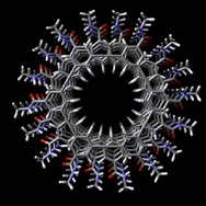
Here’s an image of the macrocycles supplied by the Agronne National Labortory,

A snapshot of a helical stack of macryocycles generated in the computer simulation.

The size specificity is  important if  nanopores/nanotubes are going to be used in medical applications,

The pore sizes can be adjusted to filter molecules and ions according to their size by changing the macroycle size, akin to the way a space can be put into a wedding ring to make it fit tighter. The channels are permeable to water, which aids in the fast transmission of intercellular information. The synthetic nanopores mimic the activity of cellular ion channels used in the human body. The research lays the foundation for an array of exciting new technology, such as new ways to deliver directly into cells proteins or medicines to fight diseases.

The research group’s paper has appeared in Nature Communications as of July 17, 2012, from Hsu’s news release,

The study’s lead authors are Xibin Zhou of Beijing Normal University; Guande Liu of Shanghai Jiao Tong University; Kazuhiro Yamato, postdoctoral scientist at UB; and Yi Shen of Shanghai Jiao Tong University and the Shanghai Institute of Applied Physics, Chinese Academy of Sciences. Other institutions that contributed to the work include the University of Nebraska-Lincoln and Argonne National Laboratory. Frank Bright, a SUNY Distinguished Professor of chemistry at UB, assisted with spectroscopic studies.