Tag Archives: bacteria

Hybrid bacterial genes and virus shell combined to create ‘nano reactor’ for hydrogen biofuel

Turning water into fuel may seem like an almost biblical project (e.g., Jesus turning water to wine in the New Testament) but scientists at Indiana University are hopeful they are halfway to their goal. From a Jan. 4, 2016 news item on ScienceDaily,

Scientists at Indiana University have created a highly efficient biomaterial that catalyzes the formation of hydrogen — one half of the “holy grail” of splitting H2O to make hydrogen and oxygen for fueling cheap and efficient cars that run on water.

A Jan. 4, 2016 Indiana University (IU) news release (also on EurekAlert*), which originated the news item, explains further (Note: Links have been removed),

A modified enzyme that gains strength from being protected within the protein shell — or “capsid” — of a bacterial virus, this new material is 150 times more efficient than the unaltered form of the enzyme.

“Essentially, we’ve taken a virus’s ability to self-assemble myriad genetic building blocks and incorporated a very fragile and sensitive enzyme with the remarkable property of taking in protons and spitting out hydrogen gas,” said Trevor Douglas, the Earl Blough Professor of Chemistry in the IU Bloomington College of Arts and Sciences’ Department of Chemistry, who led the study. “The end result is a virus-like particle that behaves the same as a highly sophisticated material that catalyzes the production of hydrogen.”

The genetic material used to create the enzyme, hydrogenase, is produced by two genes from the common bacteria Escherichia coli, inserted inside the protective capsid using methods previously developed by these IU scientists. The genes, hyaA and hyaB, are two genes in E. coli that encode key subunits of the hydrogenase enzyme. The capsid comes from the bacterial virus known as bacteriophage P22.

The resulting biomaterial, called “P22-Hyd,” is not only more efficient than the unaltered enzyme but also is produced through a simple fermentation process at room temperature.

The material is potentially far less expensive and more environmentally friendly to produce than other materials currently used to create fuel cells. The costly and rare metal platinum, for example, is commonly used to catalyze hydrogen as fuel in products such as high-end concept cars.

“This material is comparable to platinum, except it’s truly renewable,” Douglas said. “You don’t need to mine it; you can create it at room temperature on a massive scale using fermentation technology; it’s biodegradable. It’s a very green process to make a very high-end sustainable material.”

In addition, P22-Hyd both breaks the chemical bonds of water to create hydrogen and also works in reverse to recombine hydrogen and oxygen to generate power. “The reaction runs both ways — it can be used either as a hydrogen production catalyst or as a fuel cell catalyst,” Douglas said.

The form of hydrogenase is one of three occurring in nature: di-iron (FeFe)-, iron-only (Fe-only)- and nitrogen-iron (NiFe)-hydrogenase. The third form was selected for the new material due to its ability to easily integrate into biomaterials and tolerate exposure to oxygen.

NiFe-hydrogenase also gains significantly greater resistance upon encapsulation to breakdown from chemicals in the environment, and it retains the ability to catalyze at room temperature. Unaltered NiFe-hydrogenase, by contrast, is highly susceptible to destruction from chemicals in the environment and breaks down at temperatures above room temperature — both of which make the unprotected enzyme a poor choice for use in manufacturing and commercial products such as cars.

These sensitivities are “some of the key reasons enzymes haven’t previously lived up to their promise in technology,” Douglas said. Another is their difficulty to produce.

“No one’s ever had a way to create a large enough amount of this hydrogenase despite its incredible potential for biofuel production. But now we’ve got a method to stabilize and produce high quantities of the material — and enormous increases in efficiency,” he said.

The development is highly significant according to Seung-Wuk Lee, professor of bioengineering at the University of California-Berkeley, who was not a part of the study.

“Douglas’ group has been leading protein- or virus-based nanomaterial development for the last two decades. This is a new pioneering work to produce green and clean fuels to tackle the real-world energy problem that we face today and make an immediate impact in our life in the near future,” said Lee, whose work has been cited in a U.S. Congressional report on the use of viruses in manufacturing.

Beyond the new study, Douglas and his colleagues continue to craft P22-Hyd into an ideal ingredient for hydrogen power by investigating ways to activate a catalytic reaction with sunlight, as opposed to introducing elections using laboratory methods.

“Incorporating this material into a solar-powered system is the next step,” Douglas said.

Here’s a link to and a citation for the paper,

Self-assembling biomolecular catalysts for hydrogen production by Paul C. Jordan, Dustin P. Patterson, Kendall N. Saboda, Ethan J. Edwards, Heini M. Miettinen, Gautam Basu, Megan C. Thielges, & Trevor Douglas. Nature Chemistry (2015) doi:10.1038/nchem.2416 Published online 21 December 2015

This paper is behind a paywall.

*(also on EurekAlert) added on Jan. 5, 2016 at 1550 PST.
Touchless displays with 2D nanosheets and sweat

Swiping touchscreens with your finger has become a dominant means of accessing information in many applications but there is at least one problem associated with this action. From an Oct. 2, 2015 news item on phys.org,

While touchscreens are practical, touchless displays would be even more so. That’s because, despite touchscreens having enabled the smartphone’s advance into our lives and being essential for us to be able to use cash dispensers or ticket machines, they do have certain disadvantages. Touchscreens suffer from mechanical wear over time and are a transmission path for bacteria and viruses. To avoid these problems, scientists at Stuttgart’s Max Planck Institute for Solid State Research and LMU Munich have now developed nanostructures that change their electrical and even their optical properties as soon as a finger comes anywhere near them.

Here’s what a touchless screen looks like when tracking,

Touchless colour change: A nanostructure containing alternating layers of phosphatoantimonate nanosheets and oxide ... [more] © Advanced Materials 2015/MPI for Solid State Research

Touchless colour change: A nanostructure containing alternating layers of phosphatoantimonate nanosheets and oxide … [more]
© Advanced Materials 2015/MPI for Solid State Research

An Oct. 1, 2015 Max Planck Institute press release, which originated the news item, gives technical details,

A touchless display may be able to capitalize on a human trait which is of vital importance, although sometimes unwanted: This is the fact that our body sweats – and is constantly emitting water molecules through tiny pores in the skin. Scientists of the Nanochemistry group led by Bettina Lotsch at the Max Planck Institute for Solid State Research in Stuttgart and the LMU Munich have now been able to visualize the transpiration of a finger with a special moisture sensor which reacts as soon as an object – like an index finger – approaches its surface, without touching it. The increasing humidity is converted into an electrical signal or translated into a colour change, thus enabling it to be measured.

Phosphatoantimonic acid is what enables it to do this. This acid is a crystalline solid at room temperature with a structure made up of antimony, phosphorous, oxygen and hydrogen atoms. “It’s long been known to scientists that this material is able to take up water and swells considerably in the process,” explained Pirmin Ganter, doctoral student at the Max Planck Institute for Solid State Research and the Chemistry Department at LMU Munich. This water uptake also changes the properties of the material. For instance, its electrical conductivity increases as the number of stored water molecules rises. This is what enables it to serve as a measure of ambient moisture.

A sandwich nanomaterial structure exposed to moisture also changes its colour

However, the scientists aren’t so interested in developing a new moisture sensor. What they really want is to use it in touchless displays. “Because these sensors react in a very local manner to any increase in moisture, it is quite conceivable that this sort of material with moisture-dependent properties could also be used for touchless displays and monitors,” said Ganter. Touchless screens of this kind would require nothing more than a finger to get near the display to change their electrical or optical properties – and with them the input signal – at a specific point on the display.

Taking phosphatoantimonate nanosheets as their basis, the Stuttgart scientists then developed a photonic nanostructure which reacts to the moisture by changing colour. “If this was built into a monitor, the users would then receive visible feedback to  their finger motion” explained Katalin Szendrei, also a doctoral student in Bettina Lotsch’s group. To this end, the scientists created a multilayer sandwich material with alternating layers of ultrathin phosphatoantimonate nanosheets and silicon dioxide (SiO2) or titanium dioxide nanoparticles (TiO2). Comprising more than ten layers, the stack ultimately reached a height of little more than one millionth of a metre.

For one thing, the colour of the sandwich material can be set via the thickness of the layers. And for another, the colour of the sandwich changes if the scientists increase the relative humidity in the immediate surroundings of the material, for instance by moving a finger towards the screen. “The reason for this lies in the storage of water molecules between the phosphatoantimonate layers, which makes the layers swell considerably,” explained Katalin Szendrei. “A change in the thickness of the layers in this process is accompanied by a change in the colour of the sensor – produced in a similar way to what gives colour to a butterfly wing or in mother-of-pearl.”

The material reacts to the humidity change within a few milliseconds

This is a property that is fundamentally well known and characteristic of so-called photonic crystals. But scientists had never before observed such a large colour change as they now have in the lab in Stuttgart. “The colour of the nanostructure turns from blue to red when a finger gets near, for example. In this way, the colour can be tuned through the whole of the visible spectrum depending on the amount of water vapour taken up,” stressed Bettina Lotsch.

The scientists’ new approach is not only captivating because of the striking colour change. What’s also important is the fact that the material reacts to the change in humidity within a few milliseconds – literally in the blink of an eye. Previously reported materials normally took several seconds or more to respond. That is much too slow for practical applications. And there’s another thing that other materials couldn’t always do: The sandwich structure consisting of phosphatoantimonate nanosheets and oxide nanoparticles is highly stable from a chemical perspective and responds selectively to water vapour.

A layer protecting against chemical influences has to let moisture through

The scientists can imagine their materials being used in much more than just future generations of smartphones, tablets or notebooks. “Ultimately, we could see touchless displays also being deployed in many places where people currently have to touch monitors to navigate,” said Bettina Lotsch. For instance in cash dispensers or ticket machines, or even at the weighing scales in the supermarket’s vegetable aisle. Displays in public placesthat are used by many different people would have distinct hygiene benefits if they were touchless.

But before we see them being used in such places, the scientists have a few more challenges to overcome. It’s important, for example, that the nanostructures can be produced economically. To minimize wear, the structures still need to be coated with a protective layer if they’re going to be used in anything like a display. And that, again, has to meet not one but two different requirements: It must protect the moisture-sensitive layers against chemical and mechanical influences. And it must, of course, let the moisture pass through. But the Stuttgart scientists have an idea for how to achieve that already. An idea they are currently starting to put into practice with an additional cooperation partner on board.

Dexter Johnson’s Oct. 2, 2015 posting on his Nanoclast blog (on the IEEE [Institute of Electrical and Electronics Engineers] website) provides some additional context for this research (Note: A link has been removed),

In a world where the “swipe” has become a dominant computer interface method along with moving and clicking the mouse, the question becomes what’s next? For researchers at Stuttgart’s Max Planck Institute for Solid State Research and LMU Munich, Germany, the answer continues to be a swipe, but one in which you don’t actually need to touch the screen with your finger. Researchers call these no-contact computer screens touchless positioning interfaces (TPI).

Here’s a link to and a citation for the paper,

Touchless Optical Finger Motion Tracking Based on 2D Nanosheets with Giant Moisture Responsiveness by Katalin Szendrei, Pirmin Ganter, Olalla Sànchez-Sobrado, Roland Eger, Alexander Kuhn, and Bettina V. Lotsch. Advanced Materials DOI: 10.1002/adma.201503463 Article first published online: 22 SEP 2015

© 2015 WILEY-VCH Verlag GmbH & Co. KGaA, Weinheim

This paper is behind a paywall.

Greening silver nanoparticles with lignin

A July 13, 2015 news item on phys.org highlights a new approach to making silver nanoparticles safer in the environment,

North Carolina State University researchers have developed an effective and environmentally benign method to combat bacteria by engineering nanoscale particles that add the antimicrobial potency of silver to a core of lignin, a ubiquitous substance found in all plant cells. The findings introduce ideas for better, greener and safer nanotechnology and could lead to enhanced efficiency of antimicrobial products used in agriculture and personal care.

A July 13, 2015 North Carolina State University (NCSU) news release (also on EurekAlert), which originated the news item, adds a bit more information,

As the nanoparticles wipe out the targeted bacteria, they become depleted of silver. The remaining particles degrade easily after disposal because of their biocompatible lignin core, limiting the risk to the environment.

“People have been interested in using silver nanoparticles for antimicrobial purposes, but there are lingering concerns about their environmental impact due to the long-term effects of the used metal nanoparticles released in the environment,” said Velev, INVISTA Professor of Chemical and Biomolecular Engineering at NC State and the paper’s corresponding author. “We show here an inexpensive and environmentally responsible method to make effective antimicrobials with biomaterial cores.”

The researchers used the nanoparticles to attack E. coli, a bacterium that causes food poisoning; Pseudomonas aeruginosa, a common disease-causing bacterium; Ralstonia, a genus of bacteria containing numerous soil-borne pathogen species; and Staphylococcus epidermis, a bacterium that can cause harmful biofilms on plastics – like catheters – in the human body. The nanoparticles were effective against all the bacteria.

The method allows researchers the flexibility to change the nanoparticle recipe in order to target specific microbes. Alexander Richter, the paper’s first author and an NC State Ph.D. candidate who won a 2015 Lemelson-MIT prize, says that the particles could be the basis for reduced risk pesticide products with reduced cost and minimized environmental impact.

“We expect this method to have a broad impact,” Richter said. “We may include less of the antimicrobial ingredient without losing effectiveness while at the same time using an inexpensive technique that has a lower environmental burden. We are now working to scale up the process to synthesize the particles under continuous flow conditions.”

I don’t quite understand how the silver nanoparticles/ions are rendered greener. I gather the lignin is harmless but where do the silver nanoparticles/ions go after they’ve been stripped of their lignin cover and have killed the bacteria? I did try reading the paper’s abstract (not much use for someone with my science level),

Silver nanoparticles have antibacterial properties, but their use has been a cause for concern because they persist in the environment. Here, we show that lignin nanoparticles infused with silver ions and coated with a cationic polyelectrolyte layer form a biodegradable and green alternative to silver nanoparticles. The polyelectrolyte layer promotes the adhesion of the particles to bacterial cell membranes and, together with silver ions, can kill a broad spectrum of bacteria, including Escherichia coli, Pseudomonas aeruginosa and quaternary-amine-resistant Ralstonia sp. Ion depletion studies have shown that the bioactivity of these nanoparticles is time-limited because of the desorption of silver ions. High-throughput bioactivity screening did not reveal increased toxicity of the particles when compared to an equivalent mass of metallic silver nanoparticles or silver nitrate solution. Our results demonstrate that the application of green chemistry principles may allow the synthesis of nanoparticles with biodegradable cores that have higher antimicrobial activity and smaller environmental impact than metallic silver nanoparticles.

If you can explain what happens to the silver nanoparticles, please let me know.

Meanwhile, here’s a link to and a citation for the paper,

An environmentally benign antimicrobial nanoparticle based on a silver-infused lignin core by Alexander P. Richter, Joseph S. Brown, Bhuvnesh Bharti, Amy Wang, Sumit Gangwal, Keith Houck, Elaine A. Cohen Hubal, Vesselin N. Paunov, Simeon D. Stoyanov, & Orlin D. Velev. Nature Nanotechnology (2015) doi:10.1038/nnano.2015.141 Published online 13 July 2015

This paper is behind a paywall.

Customizing bacteria (E. coli) into squares, circles, triangles, etc.

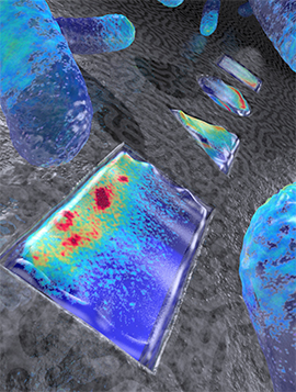
The academic paper for this latest research from Delft University of Technology (TU Delft, Netherlands), uses the term ‘bacterial sculptures,’ an intriguing idea that seems to have influenced the artistic illustration accompanying the research announcement.

Artistic rendering live E.coli bacteria that have been shaped into a rectangle, triangle, circle, and square (from front to back). Colors indicate the density of the Min proteins that represent a snapshot in time (based on actual data), as these proteins oscillate back and forth within the bacterium, to determine the mid plane of the cell for cellular division. Image credit:  ‘Image Cees Dekker lab TU Delft / Tremani’

Artistic rendering live E.coli bacteria that have been shaped into a rectangle, triangle, circle, and square (from front to back). Colors indicate the density of the Min proteins that represent a snapshot in time (based on actual data), as these proteins oscillate back and forth within the bacterium, to determine the mid plane of the cell for cellular division.
Image credit: ‘Image Cees Dekker lab TU Delft / Tremani’

A June 22, 2015 news item on Nanowerk provides more insight into the research (Note: A link has been removed),

The E.coli bacterium, a very common resident of people’s intestines, is shaped as a tiny rod about 3 micrometers long. For the first time, scientists from the Kavli Institute of Nanoscience at Delft University have found a way to use nanotechnology to grow living E.coli bacteria into very different shapes: squares, triangles, circles, and even as letters spelling out ‘TU Delft’. They also managed to grow supersized E.coli with a volume thirty times larger than normal. These living oddly-shaped bacteria allow studies of the internal distribution of proteins and DNA in entirely new ways.

In this week’s Nature Nanotechnology (“Symmetry and scale orient Min protein patterns in shaped bacterial sculptures”), the scientists describe how these custom-designed bacteria still manage to perfectly locate ‘the middle of themselves’ for their cell division. They are found to do so using proteins that sense the cell shape, based on a mathematical principle proposed by computer pioneer Alan Turing in 1953.

A June 22, 2015 TU Delft press release, which originated the news item, expands on the theme,

Cell division

“If cells can’t divide properly, biological life wouldn’t be possible. Cells need to distribute their cell volume and genetic materials equally into their daughter cells to proliferate.”, says prof. Cees Dekker, “It is fascinating that even a unicellular organism knows how to divide very precisely. The distribution of certain proteins in the cell is key to regulating this, but how exactly do those proteins get that done?”

Turing

As the work of the Delft scientist exemplifies, the key here is a process discovered by the famous Alan Turing in 1953. Although Turing is mostly known for his role in deciphering the Enigma coding machine and the Turing Test, the impact of his ‘reaction-diffusion theory’ on biology might be even more spectacular. He predicted how patterns in space and time emerge as the result of only two molecular interactions – explaining for instance how a zebra gets its stripes, or how an embryo hand develops five fingers.

MinD and MinE

Such a Turing process also acts with proteins within a single cell, to regulate cell division. An E.coli cell uses two types of proteins, known as MinD and MinE, that bind and unbind again and again at the inner surface of the bacterium, thus oscillating back and forth from pole to pole within the bacterium every minute. “This results in a low average concentration of the protein in the middle and high concentrations at the ends, which drives the division machinery to the cell center”, says PhD-student Fabai Wu, who ran the experiments. “As our experiments show, the Turing patterns allow the bacterium to determine its symmetry axes and its center. This applies to many bacterial cell shapes that we custom-designed, such as squares, triangles and rectangles of many sizes. For fun, we even made ‘TUDelft’ and ‘TURING’ letters. Using computer simulations, we uncovered that the shape-sensing abilities are caused by simple Turing-type interactions between the proteins.”

Actual data for live E.coli bacteria that have been shaped into the letters TUDELFT.
The red color shows the cytosol contents of the cell, while the green color shows the density of the Min proteins, representing a snapshot in time, as these proteins oscillate back and forth within the bacterium to determine the mid plane of the cell for cellular division. The letters are about 5 micron high.
Image credit:  ‘Fabai Wu, Cees Dekker lab at TU Delft’

Spatial control for building synthetic cells

“Discovering this process is not only vital for our understanding of bacterial cell division – which is important in developing new strategies for antibiotics. But the approach will likely also be fruitful to figuring out how cells distribute other vital systems within a cell, such as chromosomes”, says Cees Dekker. “The ultimate goal in our research is to be able to completely build a living cell from artificial components, as that is the only way to really understand how life works. Understanding cell division – both the process that actually pinches off the cell into two daughters and the part that spatially regulates that machinery – is a major part of that.”

Here’s a link to and a citation for the paper,

Symmetry and scale orient Min protein patterns in shaped bacterial sculptures by Fabai Wu, Bas G. C. van Schie, Juan E. Keymer, & Cees Dekker. Nature Nanotechnology (2015) doi:10.1038/nnano.2015.126 Published online 22 June 2015

This paper is behind a paywall but there does seem to be another link (in the excerpt below) which gives you a free preview via ReadCube Access (according to the TU Delft press release),

The DOI for this paper will be 10.1038/nnano.2015.126. Once the paper is published electronically, the DOI can be used to retrieve the abstract and full text by adding it to the following url: http://dx.doi.org/

Enjoy!

Metal nanoparticles and gut microbiomes

What happens when you eat or drink nanoparticles, metallic or otherwise? No one really knows. Part of the problem with doing research now is there are no benchmarks for the quantity we’ve been ingesting over the centuries. Nanoparticles do occur naturally, as well, people who’ve eaten with utensils made of or coated with silver or gold have ingested silver or gold nanoparticles that were shed by those very utensils. In short, we’ve been ingesting any number of nanoparticles through our food, drink, and utensils in addition to the engineered nanoparticles that are found in consumer products. So, part of what researchers need to determine is the point at which we need to be concerned about nanoparticles. That’s trickier than it might seem since we ingest our nanoparticles and recycle them into the environment (air, water, soil) to reingest (inhale, drink, eat, etc.) them at a later date. The endeavour to understand what impact engineered nanoparticles in particular will have on us as more are used in our products is akin to assembling a puzzle.

There’s a May 5, 2015 news item on Azonano which describes research into the effects that metallic nanoparticles have on the micriobiome (bacteria) in our guts,

Exposure of a model human colon to metal oxide nanoparticles, at levels that could be present in foods, consumer goods, or treated drinking water, led to multiple, measurable differences in the normal microbial community that inhabits the human gut. The changes observed in microbial metabolism and the gut microenvironment with exposure to nanoparticles could have implications for overall human health, as discussed in an article published in Environmental Engineering Science, a peer-reviewed journal from Mary Ann Liebert, Inc., publishers. The article is available free on the Environmental Engineering Science website until June 1, 2015.

A May 4, 2015 Mary Ann Liebert publisher news release on EurekAlert, which originated the news item, describes the research in more detail (Note: A link has been removed),

Alicia Taylor, Ian Marcus, Risa Guysi, and Sharon Walker, University of California, Riverside, individually introduced three different nanoparticles–zinc oxide, cerium dioxide, and titanium dioxide–commonly used in products such as toothpastes, cosmetics, sunscreens, coatings, and paints, into a model of the human colon. The model colon mimics the normal gut environment and contains the microorganisms typically present in the human microbiome.

In the article “Metal Oxide Nanoparticles Induce Minimal Phenotypic Changes in a Model Colon Gut Microbiota” the researchers described changes in both specific characteristics of the microbial community and of the gut microenvironment after exposure to the nanoparticles.

Here’s a link to and a citation for the paper,

Metal Oxide Nanoparticles Induce Minimal Phenotypic Changes in a Model Colon Gut Microbiota by Alicia A. Taylor, Ian M. Marcus Ian, Risa L., Guysi, and Sharon L. Walker. Environmental Engineering Science. DOI:10.1089/ees.2014.0518 Online Ahead of Print: April 24, 2015

I’ve taken a quick look at the research while it’s still open access (till June 1, 2015) to highlight the bits I consider interesting. There’s this about the nanoparticle (NP) quantities used in the study (Note: Links have been removed),

Environmentally relevant NP concentrations were chosen to emulate human exposures to NPs through ingestion of both food and drinking water at 0.01 μg/L ZnO NP, 0.01 μg/L CeO2 NP, and 3 mg/L TiO2 NP (Gottschalk et al., 2009; Kiser et al., 2009, 2013; Weir et al., 2012; Keller and Lazareva, 2013). Recent work has also indicated that adults in the USA ingest 5 mg TiO2 per day, half of which is in the nano-size range (Lomer et al., 2000; Powell et al., 2010). Exposure routes and reliable dosing information of NPs that are embedded in solid matrices are difficult to predict, and this is often a limitation of analytical techniques (Nowack et al., 2012; Yang and Westerhoff, 2014). The exposure levels used in this study were predominately selected from literature values that give predictions on amount of NPs in water and food sources (Gottschalk et al., 2009; Kiser et al., 2009; Weir et al., 2012; Keller and Lazareva, 2013; Keller et al., 2013).

For anyone unfamiliar with chemical notations, ZnO NP is zinc oxide nanoparticle, 0.01 μg/L is one/one hundredth of a microgram per litre,  CeO2 is cesisum dioxide NP, and TiO2 is titanium dioxide NP while 3 mg/L, is 3 milligrams per litre.

After assuring the quantities used in the study are the same as they expect humans to be ingesting on a regular basis, the researchers describe some of the factors which may affect the interaction between the tested nanoparticles and the bacteria (Note: Links have been removed),

It is essential to note that interactions between NPs and bacteria in the intestines may be dependent on numerous factors: the surface charge of the NPs and bacteria, the chemical composition and surface charge of the digested food, and variability in diet. These factors may ultimately correlate to effects seen in humans on an individual basis. In fact, similar work has demonstrated that exposing common NPs found in food to stomach-like conditions will change their surface chemistry from negative to neutral or positive, causing the NPs to interact with negatively charged mucus proteins in the gastrointestinal tract and, in turn, affecting the transport of NPs within the intestine (McCracken et al., 2013). The purpose of this work was to measure responses of the microbial community during the NP exposures. Based on previous research, it is anticipated that the NPs altered by stomach-like conditions would also cause changes in the gut environment (McCracken et al., 2013).

Here’s some of what they discovered,

Our initial hypothesis, that NPs induce phenotypic changes in a gut microbial community, was affirmed through significant measurable effects seen in the data. Tests that supported that NPs caused changes in the phenotype included hydrophobicity, EPM, sugar content of the EPS, cell size, conductivity, and SFCA (specifically butyric acid) production. Data for cell concentration and the protein content of the EPS demonstrated no significant results. Data were inconclusive for pH. With such a complex biological system, it is very likely that the phenotypic and biochemical changes observed are combinations of responses happening in parallel. The effects seen may be attributed to both changes induced by the NPs and natural phenomena associated with microbial community activity and other metabolic processes in a multifaceted environment such as the gut. Some examples of natural processes that could also influence the phenotypic and biochemical parameters are osmolarity, active metabolites, and electrolyte concentrations (Miller and Wood, 1996; Record et al., 1998).

Here’s the concluding sentence from the abstract,

Overall, the NPs caused nonlethal, significant changes to the microbial community’s phenotype, which may be related to overall health effects. [emphasis mine]

This research like the work I featured in a June 27, 2013 posting points to some issues with researching the impact that nanoparticles may have on our bodies. There was no cause for immediate alarm in 2013 and it appears that is still the case in 2015. However, that assumes quantities being ingested don’t increase significantly.

Essential oil nanoemulsions for foodborne bacteria

An April 30, 2015 news item on Azonano describes essential oil research at Wayne State University (Detroit, Michigan, US),

Nearly half of foodborne illnesses in the U.S. from 1998 through 2008 have been attributed to contaminated fresh produce. Prevention and control of bacterial contamination on fresh produce is critical to ensure food safety. The current strategy remains industrial washing of the product in water containing chlorine. However, due to sanitizer ineffectiveness there is an urgent need to identify alternative antimicrobials, particularly those of natural origin, for the produce industry.

A team of researchers at Wayne State University have been exploring natural, safe and alternative antimicrobials to reduce bacterial contamination. Plant essential oils such as those from thyme, oregano and clove are known to have a strong antimicrobial effect, but currently their use in food protection is limited due to their low solubility in water. The team, led by Yifan Zhang, Ph.D., assistant professor of nutrition and food science in the College of Liberal Arts and Sciences, explored ways to formulate oil nanoemulsions to increase the solubility and stability of essential oils, and consequently, enhance their antimicrobial activity.

An April 29, 2015 Wayne State University news release (also on EurekAlert), which originated the news item, expands on the theme,

“Much of the research on the antimicrobial efficacy of essential oils has been conducted using products made by mixing immiscible oils in water or phosphate buffered saline,” said Zhang. “However, because of the hydrophobic nature of essential oils, organic compounds from produce may interfere with reducing the sanitizing effect or duration of the effectiveness of these essential oils. Our team set out to find a new approach to inhibit these bacteria with the use of oregano oil, one of the most effective plant essential oils with antimicrobial effect.”

Zhang, and then-Ph.D. student, Kanika Bhargava, currently assistant professor of human environmental sciences at the University of Central Oklahoma, approached Sandro da Rocha, Ph.D., associate professor of chemical engineering and materials science in the College of Engineering at Wayne State, to explore options.

“In our research, we discovered that oregano oil was able to inhibit common foodborne bacteria, such as E. coli O157, Salmonella and Listeria, in artificially contaminated fresh lettuce” said Zhang. “We wanted to explore the possibility of a nanodelivery system for the oil, which is an area of expertise of Dr. da Rocha.”

The team initially considered the use of solid polymeric nanoparticles for the delivery of the oil, but da Rocha suggested the use of nanoemulsions.

“My team felt the use of nanoemulsions would improve the rate of release compared to other nanoformulations, and the ability of the food grade surfactant to wet the surface of the produce,” said da Rocha. “We were able to reduce L. monocytogenes, S. Typhimurium, and E. coli O157 on fresh lettuce. Former Ph.D. student Denise S. Conti, now at the Generics Division of the FDA, helped design the nanocarriers and characterize them.”

The team added that while there is still work to be done, their study suggests promise for the use of essential oil nanoemulsions as a natural alternative to chemicals for safety controls in produce.

“Our future research aims to investigate the antimicrobial effects of essential oil nanoemulsions in various combinations, as well as formulate the best proportions of each ingredient at the lowest possible necessary levels needed for food application, which ultimately will aid in maintaining the taste of the produce.”

Here’s a link to and a citation for the paper,

Application of an oregano oil nanoemulsion to the control of foodborne bacteria on fresh lettuce by Kanika Bhargava, Denise S. Conti, Sandro R.P. da Rocha, and Yifan Zhang. Food Microbiology Volume 47, May 2015, Pages 69–73, DOI: 10.1016/j.fm.2014.11.007

This paper is behind a paywall.

And the bacteria shall save us—nanobiobots

A March 24, 2015 University of Illinois at Chicago news release (also on EurekAlert) describes the NERD, a Nano-Electro-Robotic Device which employs bacteria and graphene quantum dots,

As nanotechnology makes possible a world of machines too tiny to see, researchers are finding ways to combine living organisms with nonliving machinery to solve a variety of problems.

Like other first-generation bio-robots, the new nanobot engineered at the University of Illinois at Chicago [UIC] is a far cry from Robocop. It’s a robotic germ.

UIC researchers created an electromechanical device–a humidity sensor–on a bacterial spore. They call it NERD, for Nano-Electro-Robotic Device. …

“We’ve taken a spore from a bacteria, and put graphene quantum dots on its surface–and then attached two electrodes on either side of the spore,” said Vikas Berry, UIC associate professor of chemical engineering and principal investigator on the study.

“Then we change the humidity around the spore,” he said.

When the humidity drops, the spore shrinks as water is pushed out. As it shrinks, the quantum dots come closer together, increasing their conductivity, as measured by the electrodes.

“We get a very clean response–a very sharp change the moment we change humidity,” Berry said. The response was 10 times faster, he said, than a sensor made with the most advanced man-made water-absorbing polymers.

There was also better sensitivity in extreme low-pressure, low-humidity situations.

“We can go all the way down to a vacuum and see a response,” said Berry, which is important in applications where humidity must be kept low, for example, to prevent corrosion or food spoilage. “It’s also important in space applications, where any change in humidity could signal a leak,” he said.

Currently available sensors increase in sensitivity as humidity rises, Berry said. NERD’s sensitivity is actually higher at low humidity.

“This is a fascinating device,” Berry said. “Here we have a biological entity. We’ve made the sensor on the surface of these spores, with the spore a very active complement to this device. The biological complement is actually working towards responding to stimuli and providing information.”

Interesting, yes? Here’s a link to and a citation for the research paper,

Graphene Quantum Dots Interfaced with Single Bacterial Spore for Bio-Electromechanical Devices: A Graphene Cytobot by T. S. Sreeprasad, Phong Nguyen, Ahmed Alshogeathri, Luke Hibbeler, Fabian Martinez, Nolan McNeil, & Vikas Berry. Scientific Reports 5, Article number: 9138 doi:10.1038/srep09138 Published 16 March 2015

This paper is open access.

Dexter Johnson provides more context for this research in a March 26, 2015 post on his Nanoclast blog (on the IEEE [institute of Electrical and Electronics Engineers]) where he notes,

Recently, James Tours’ group at Rice University, who were the first to develop GQCs [graphene quantum dots] in 2013, created an improved way to manufacture them that promised to open them up to a new range of applications in optics.

Dexter’s insights make for worthwhile reading.

New molecular ruler could help with developing antibiotics

Researchers at the University of Utah have developed a molecular ruler which could help to determine the length at which a nanoscale needle is effective. From a March 17, 2015 news item on Azonano,

When a salmonella bacterium attacks a cell, it uses a nanoscopic needle to inject it with proteins to aid the infection. If the needle is too short, the cell won’t be infected. Too long, and the needle breaks. Now, University of Utah biologists report how a disposable molecular ruler or tape measure determines the length of the bacterial needle so it is just right.

The findings have potential long-term applications for developing new antibiotics against salmonella and certain other disease-causing bacteria, for designing bacteria that could inject cancer cells with chemotherapy drugs, and for helping people how to design machines at the nanoscopic or molecular scale.

A March 16, 2015 University of Utah news release, which originated the news item, provides some insight from the researchers,

“If you look at important pathogens – the bubonic plague bacterium, salmonella, shigella and plant pathogens like fire blight – they all use hypodermic-like needles to inject proteins that facilitate disease processes,” Hughes [University of Utah biology professor Kelly Hughes] says.

“Our work says that there is one mechanism – the molecular ruler – to explain how the lengths are controlled for needles in gram-negative bacteria and for hooks on flagella [the U-joints in propellers bacteria use to move] in all bacteria,” he adds.

In their study, Wee [University of Utah doctoral student Daniel Wee] and Hughes found that as a bacterial needle or “injectisome” grows, a molecular ruler – really, more like a gooey tape measure – is secreted from within the needle’s base. It oozes up through the tube-like needle, and when the bottom end of the ruler reaches the bottom end of the needle, the needle stops growing and begins to inject proteins into the target cell to help the infection process.

The biologists say the [US] National Institutes of Health-funded study refutes other theories for how salmonella and some other disease bacteria determine needle lengths.

The news release also explains how this finding could be made useful,

“What we understand from bacteria can help us build nanomachines and nanobots,” Hughes says, noting that bacterial flagella – the nanoscopic motor-and-propeller system they use to swim to dinner or to targets – are “the most sophisticated nanomachines in the universe.”

In one example, Swiss scientists are using the design of bacterial flagella as the basis for a nanobot that will be put inside the eye to do nanoscale surgery, he adds.

In addition to flagella, a number of disease-causing bacteria also have injectisomes, which also are built of proteins, as are most structures in living organisms.

“In the case of the needle, you have a structure that extends from the surface of the bacterium like a hypodermic,” Hughes says. “These needles are fragile. If one is too long, it will break off and be useless. If you make it too short, then it can’t get past the surface proteins on cells it needs to invade.”

By understanding how bacteria determine the length for their needles, it someday may be possible to engineer bacteria to inject chemotherapy drugs right into cancer cells.

“People would like to design bacteria that can get to cancer cells and inject poisons into just those cells and kill them, and not harm the rest of us,” Hughes says.

And by understanding how certain disease-causing bacteria build their injectisomes, new antibiotics might be developed in a decade or so to target and destroy the needles and thus deter bacterial infections. The rulers that help build flagella also might be attacked by drugs to prevent bacteria from reaching target cells, “so you can kill two birds with one stone by hitting the two machines at the same time,” Hughes says.

He says that approach might work against injectisome-equipped bacteria such as salmonella species that cause typhoid fever and food poisoning; shigella species that cause dysentery; the bubonic plague bacterium Yersinia pestis; disease-causing E. coli; sexually transmitted Chlamydia trachomatis; many plant pathogens; and Pseudomonas aeruginosa, which often infects burn patients and the lungs of cystic fibrosis patients.

Not usually my kind of thing, I find this quite fascinating (from the news release),

Bacteria secrete a molecular ruler to measure needle length

Bacterial injectisomes are incredibly small, measuring only 20 to 100 nanometers long. A nanometer is one billionth of a meter, and a meter is about 39 inches long. The width of a typical human hair often is given as 100 microns, so the maximum length of a bacterial needle, 100 nanometers, is one-thousandth of the width of a human hair.

Gram-negative, disease-causing bacteria “are very closely related species, so how do they subtly control the various needle lengths to be perfect?” Hughes asks. “In one case it might be 40 nanometers versus 55 nanometers. These are small sizes. So to do this, the bacteria developed molecular rulers to differentiate needles of different lengths.”

(Gram-negative bacteria are those with membranes lining both the inside and outside of their cell wall, while gram-positive bacteria have only an inner membrane.)

Like any cell, a bacterium is encased in a cell wall. So bacteria developed all kinds of secretions to make contact with and infect other cells: flagellar propellers to swim to food or target cells, docking structures to help bacteria stick to targets, and injectisomes to inject infection-promoting proteins into targets.

When a bacterium builds a needle, it first builds a base. “A series of proteins form a doughnut, and inside the doughnut hole, the actual secretion machine gets constructed,” Hughes says. “It’s the same for the flagella as it is for these needles.”

Next, proteins start assembling to form the needle or injectisome.

The new study demonstrated that in salmonella, the ruler or tape measure is secreted slowly through the channel of the growing needle. Once amino acids at the bottom end of the ruler pass through the base of the needle, they tell the bacterium that the needle is long enough and to stop growing. They also tell the needle to injecting virulence proteins into the target cell, and the molecular ruler is ejected, Wee says.

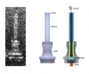
Here’s an image of what the injectisome looks like,

On the left is an electron microscope image of an injectisome, the nanoscopic needle that salmonella and certain other bacteria use to inject proteins into target cells as part of the infection process. The illustration at center depicts the exterior of the needle and its base. The cross-section at right shows the string-like molecular ruler that determines the length of salmonella’s bacteria needle, according to a new University of Utah study by doctoral student Daniel Wee and biology professor Kelly Hughes. Credit: Daniel Wee, University of Utah

On the left is an electron microscope image of an injectisome, the nanoscopic needle that salmonella and certain other bacteria use to inject proteins into target cells as part of the infection process. The illustration at center depicts the exterior of the needle and its base. The cross-section at right shows the string-like molecular ruler that determines the length of salmonella’s bacteria needle, according to a new University of Utah study by doctoral student Daniel Wee and biology professor Kelly Hughes. Credit: Daniel Wee, University of Utah

The news release also offers some specific details about the research,

How the study was performed

The new study used the Typhimurium strain of Salmonella enterica, which causes food poisoning. The researchers proved the molecular ruler determines needle length in salmonella by inserting amino acids from the plague bacterium’s molecular ruler genes into genes for salmonella’s molecular ruler, making rulers with seven different lengths.

Genetically engineered salmonella with seven ruler lengths were grown in a flask, their needles isolated, and the needle lengths measured under an electron microscope.

Wee found the ruler lengths correlated precisely with the lengths of the resulting needles or injectisomes, with each amino acid added to the ruler gene making the resulting needle 0.2 nanometers longer.

Previous studies found the molecular ruler determines the length of the hook or U-joint that helps turn flagella or propellers in many bacteria. Research also found the molecular ruler determines the length of both the flagellar hook and the needle in plague bacteria. But some researchers argued salmonella needle’s length was determined by some other mechanism:

– One theory holds that a molecular measuring cup in the needle’s base sends a cupful of needle components to assemble the needle, and the length of the needle is determined by the size of the cup. The new study disproved that by genetically removing the cup and showing that the injectisomes or needles still grew to correct lengths.

– Another theory says that as needle components assemble outside the needle’s base, a rod-shaped structure assembles inside the base to link the base and needle, and that when the rod is complete, needle assembly stops, thus determining needle length. But the Utah study found the rod and needle components are not made simultaneously, but compete with each other, so as more rod parts are made, fewer needle parts are made, giving an illusion that rod completion controls needle length.

Here’s a link to and a citation for the paper,

Molecular ruler determines needle length for the Salmonella Spi-1 injectisome by Daniel H. Wee and Kelly T. Hughes. Published online before print March 16, 2015, doi: 10.1073/pnas.1423492112 PNAS March 16, 2015

This paper is behind a paywall.


Bacteria is shocked, I tell you, shocked

Casablanca (1942, black and white, Hollywood movie) lovers may recognize the paraphrase of just one of the many famous lines in the movie. However, this ‘shocking’ news has more to do with preventing bacteria from congregating on surfaces according to a Jan. 12, 2014 news item by Alexander Chilton on Azonano (Note: Links have been removed),

Researchers at Rensselaer Polytechnic Institute and Cornell University have devised a new nanoscale surface which uses an electrochemical anodization process in order to prevent the surface attachment of bacteria.

The research published in the Biofouling journal focuses on the formation of nanoscale pores which alter the surface energy and electrical charge of a metal surface. A repulsive force is exerted on the bacterial cells, which prevents the attachment of bacteria and the formation of a biofilm. The size of the nanoscale pores formed can be as small as 15 nanometers.

The application of the anodization process to aluminum created a nanoporous surface, known as alumina. This surface proved effective in preventing the attachment of two popular bacterial species: Listeria monocytogenes and Escherichia coli O157:H7.

Krishna Ramanujan’s Jan. 9, 2015 article for Cornell University’s Chronicle explains why the scientists are excited about the anodization technique,

“It’s probably one of the lowest-cost possibilities to manufacture a nanostructure on a metallic surface,” said Carmen Moraru, associate professor of food science and the paper’s senior author. …

Finding low-cost solutions to limiting bacterial attachments is key, especially in biomedical and food processing applications.  …

Anodized metals could be used to prevent buildups of biofilms – slick communities of bacteria that adhere to surfaces and are tricky to remove – in biomedical clean rooms and in equipment parts that are hard to reach or clean, Moraru said.

There are other strategies for limiting bacterial attachment to surfaces, including chemicals and bactericides, but these have limited applications, especially when it comes to food processing, Moraru said. With food processing, surfaces must meet food safety guidelines and be inert to food that they may contact.

Here’s a link to and a citation for the paper,

Alumina surfaces with nanoscale topography reduce attachment and biofilm formation by Escherichia coli and Listeria spp. by Guoping Feng, Yifan Cheng, Shu-Yi Wang, Lillian C. Hsu, Yazmin Feliz, Diana A. Borca-Tasciuc, Randy W. Worobo, & Carmen I. Moraru. Biofouling: The Journal of Bioadhesion and Biofilm Research Volume 30, Issue 10, 2014 pages 1253-1268 DOI: 10.1080/08927014.2014.976561 Published online: 27 Nov 2014

This article is open access.

Futuristic fashion with Biocouture and other future-focused clothing companies

Suzanne Lee and her ‘green tea’ couture are being featured in a May 20, 2014 article by Adele Peters about futuristic fashion and a new documentary, ‘The Next Black’, for Fast Company,

Fabric grown from bacteria. T-shirt designs that “refresh” themselves. Or how about a new way to dye fabrics without water or pollution? These are ideas for the future of fashion that blend style and sustainability.

Biocouture is growing new fabric from bacteria using a process more like brewing beer than making any other textiles. The company hopes that eventually clothing could be grown directly on dress forms, creating zero waste. …

Studio XO, a company pioneering interactive digital fashion, shares their vision for a “Tumblr for the body”–a subscription service for clothing that could automatically refresh itself as you wear it (picture a T-shirt with an ever-evolving print curated by designers or your friends). …

You can find the full 45 min. documentary embedded in the Peters article. You can also find additional information about Suzanne Lee’s work in my June 8, 2012 post titled, Material changes, which also features other designers.